纺织学报 ›› 2022, Vol. 43 ›› Issue (05): 1-6.doi: 10.13475/j.fzxb.20211109306
• 特约专栏:第十一届中国纺织学术年会专家观点 • 下一篇
顾张弘1,2, 姚响1,2, 王锦思1,2, 张耀鹏1,2( )
)
GU Zhanghong1,2, YAO Xiang1,2, WANG Jinsi1,2, ZHANG Yaopeng1,2()
摘要:
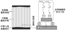
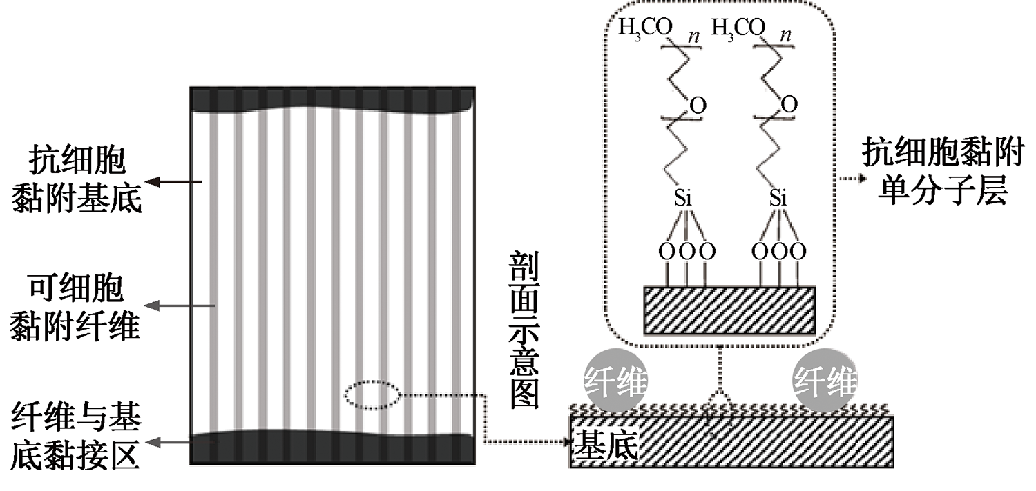
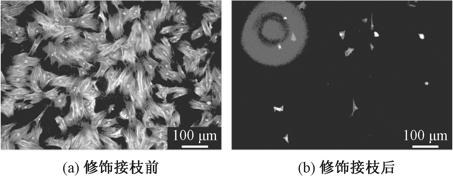
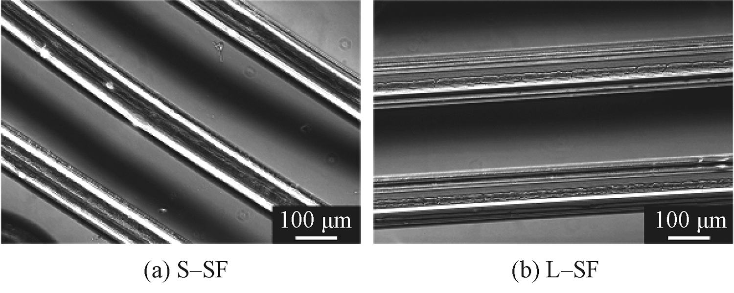
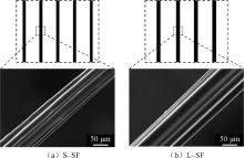
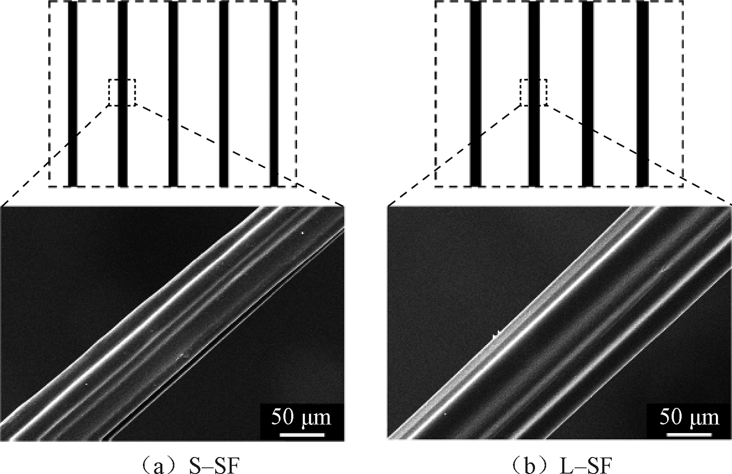
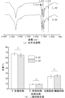
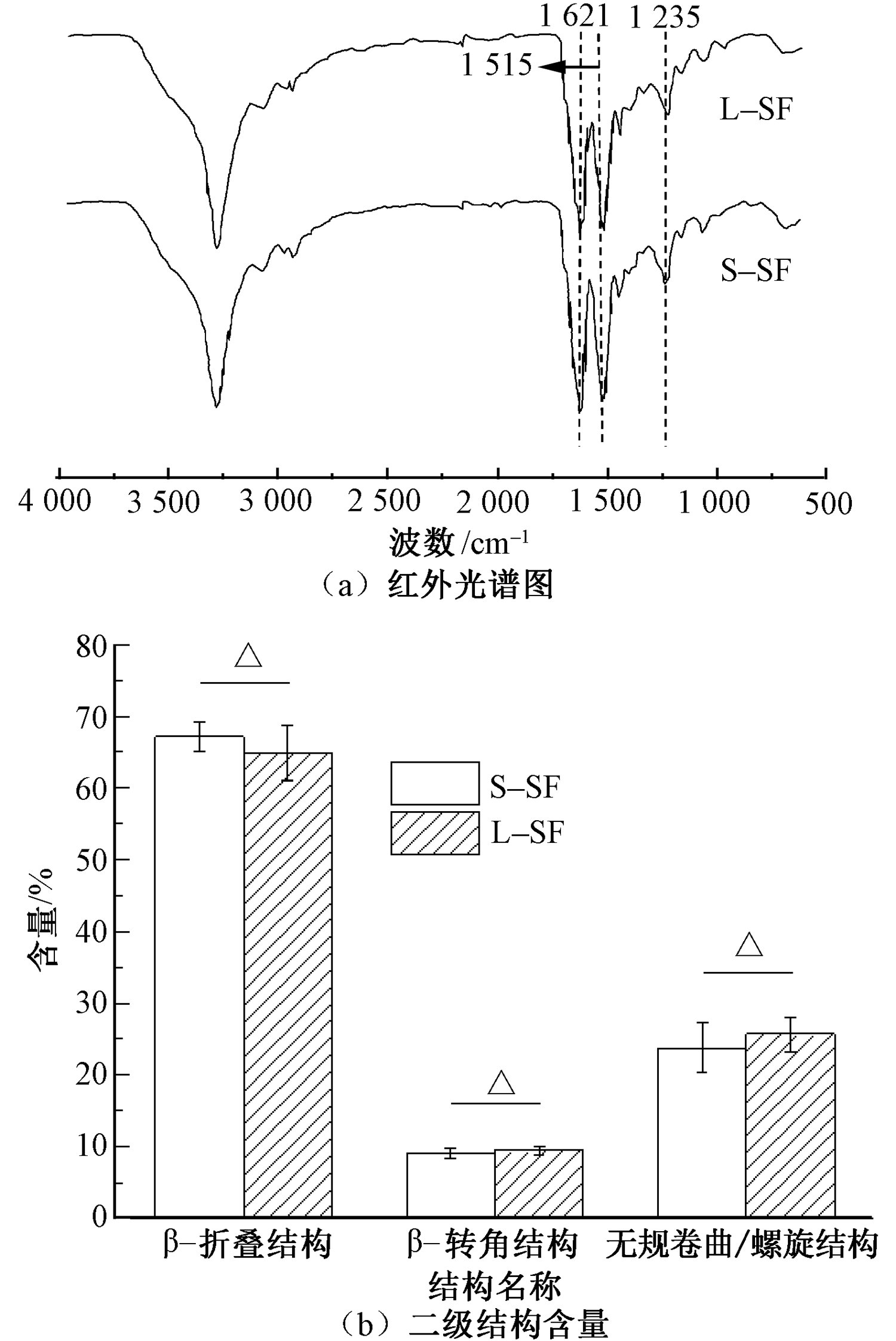
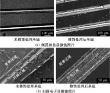
针对在分析纤维自身特征对细胞行为的影响时各类背景黏附产生干扰的问题,首先将寡聚乙二醇硅烷化试剂接枝在玻片表面得到具有抗细胞黏附特征的基底材料,然后利用湿法纺丝技术在基底表面制备可细胞黏附的单层丝素蛋白(SF)平行纤维,最后利用聚二甲基硅氧烷将SF纤维两端固定于基底上制备得到单层平行纤维图案,分析了纤维图案中纤维的直径、纤维间距、表面形貌和结构组成等特性,并以L929成纤维细胞为例检验该单层平行纤维图案的细胞黏附反差功能。结果表明:单层平行纤维图案中不同直径的SF纤维具有类似的外观形貌和二级结构组成特征,L929成纤维细胞仅可黏附在纤维表面,而不会黏附到基底背景区域,展现出优秀的细胞黏附反差特性。
中图分类号:
| [1] |
CIPITRIA A, SKELTON A, DARGAVILLE T R, et al. Design, fabrication and characterization of PCL electrospun scaffolds:a review[J]. Journal of Materials Chemistry, 2011, 21(26): 9419-9453.
doi: 10.1039/c0jm04502k |
| [2] |
DING J X, ZHANG J, LI J N, et al. Electrospun polymer biomaterials[J]. Progress in Polymer Science, 2019, 90: 1-34.
doi: 10.1016/j.progpolymsci.2019.01.002 |
| [3] | CHEN W M, XU Y, LIU Y Q, et al. Three-dimensional printed electrospun fiber-based scaffold for cartilage regeneration[J]. Materials & Design, 2019, 179: 107886. |
| [4] |
YAO X, PENG R, DING J D. Cell-material interactions revealed via material techniques of surface patterning[J]. Advanced Materials, 2013, 25(37): 5257-5286.
doi: 10.1002/adma.201301762 |
| [5] |
LIU M, ZENG X, MA C, et al. Injectable hydrogels for cartilage and bone tissue engineering[J]. Bone Research, 2017, 5: 17014.
doi: 10.1038/boneres.2017.14 |
| [6] |
LU L, FAN S N, GENG L H, et al. Flow analysis of regenerated silk fibroin/cellulose nanofiber suspensions via a bioinspired microfluidic chip[J]. Advanced Materials Technologies, 2021, 6(10): 2100124.
doi: 10.1002/admt.202100124 |
| [7] |
BADAMI A S, KREKE M R, THOMPSON M S, et al. Effect of fiber diameter on spreading, proliferation, and differentiation of osteoblastic cells on electrospun poly(lactic acid) substrates[J]. Biomaterials, 2006, 27(4): 596-606.
doi: 10.1016/j.biomaterials.2005.05.084 |
| [8] |
GHOBEIRA R, PHILIPS C, LIEFOOGHE L, et al. Synergetic effect of electrospun PCL fiber size, orientation and plasma-modified surface chemistry on stem cell behavior[J]. Applied Surface Science, 2019, 485: 204-221.
doi: 10.1016/j.apsusc.2019.04.109 |
| [9] |
ZOU S Z, WANG X R, FAN S N, et al. Electrospun regenerated Antheraea pernyi silk fibroin scaffolds with improved pore size, mechanical properties and cytocompatibility using mesh collectors[J]. Journal of Materials Chemistry B, 2021, 9(27): 5514-5527.
doi: 10.1039/D1TB00944C |
| [10] |
YANG S, ZHU J, LU C, et al. Aligned fibrin/functionalized self-assembling peptide interpenetrating nanofiber hydrogel presenting multi-cues promotes peripheral nerve functional recovery[J]. Bioactive Materials, 2022, 8: 529-544.
doi: 10.1016/j.bioactmat.2021.05.056 |
| [11] | SILVA J C, UDANGAWA R N, CHEN J L, et al. Kartogenin-loaded coaxial PGS/PCL aligned nanofibers for cartilage tissue engineering[J]. Materials Science & Engineering C: Materials for Biological Applications, 2020, 107: 12. |
| [12] | LI Z B, LIU Q Q, WANG H S, et al. Bladder acellular matrix graft reinforced silk fibroin composite scaffolds loaded VEGF with aligned electrospun fibers in multiple layers[J]. ACS Biomaterials Science & Engineering, 2015, 1(4): 238-246. |
| [13] |
YAO X, WANG X L, DING J D. Exploration of possible cell chirality using material techniques of surface patterning[J]. Acta Biomaterialia, 2021, 126: 92-108.
doi: 10.1016/j.actbio.2021.02.032 |
| [14] | YAO X, DING J D. Effects of microstripe geometry on guided cell migration[J]. ACS Applied Materials & Interfaces, 2020, 12(25): 27971-27983. |
| [15] | YAO X, LIU R, LIANG X, et al. Critical areas of proli-feration of single cells on micropatterned surfaces and corresponding cell type dependence[J]. ACS Applied Materials & Interfaces, 2019, 11(17): 15366-15380. |
| [16] |
HA M, ATHIRASALA A, TAHAYERI A, et al. Micropatterned hydrogels and cell alignment enhance the odontogenic potential of stem cells from apical papilla in-vitro[J]. Dental Materials, 2020, 36(1): 88-96.
doi: 10.1016/j.dental.2019.10.013 |
| [17] |
ANTMEN E, DEMIRCI U, HASIRCI V. Amplification of nuclear deformation of breast cancer cells by seeding on micropatterned surfaces to better distinguish their malignancies[J]. Colloids and Surfaces B: Biointerfaces, 2019, 183: 110402.
doi: 10.1016/j.colsurfb.2019.110402 |
| [18] | 姚响. 基于材料表面图案化技术研究细胞形状和表面手性特征对干细胞黏附与分化的影响[D]. 上海: 复旦大学, 2014: 1-194. |
| YAO Xiang. Effects of cell shape and surface chirality on adhesion and differentiation of stem cells revealed via material techniques of surface patterning[D]. Shanghai: Fudan University, 2014: 1-194. | |
| [19] |
CHEN J, ZHUANG A, SHAO H, et al. Robust silk fibroin/bacterial cellulose nanoribbon composite scaffolds with radial lamellae and intercalation structure for bone regeneration[J]. Journal of Materials Chemistry B, 2017, 5(20): 3640-3650.
doi: 10.1039/C7TB00485K |
| [20] |
LEE J H, KOPECEK J, ANDRADE J D. Protein-resistant surfaces prepared by PEO-containing block copolymer surfactants[J]. Journal of Biomedical Materials Research, 1989, 23(3): 351-368.
doi: 10.1002/jbm.820230306 |
| [21] |
LIU V A, JASTROMB W E, BHATIA S N. Engineering protein and cell adhesivity using PEO-terminated triblock polymers[J]. Journal of Biomedical Materials Research, 2002, 60(1): 126-134.
doi: 10.1002/jbm.10005 |
| [22] | ZARKOOB S, RENEKER D H, EBY R K, et al. Structure and morphology of nano electrospun silk fibers[J]. Abstracts of Papers of the American Chemical Society, 1998, 216(3): 122. |
| [23] | HUANG L, HUANG J, SHAO H, et al. Silk scaffolds with gradient pore structure and improved cell infiltration performance[J]. Materials Science & Engineering C: Materials for Biological Applications, 2019, 94: 179-189. |
| [24] |
ZHU J X, ZHANG Y P, SHAO H L, et al. Electrospinning and rheology of regenerated Bombyx mori silk fibroin aqueous solutions: the effects of pH and concentration[J]. Polymer, 2008, 49(12): 2880-2885.
doi: 10.1016/j.polymer.2008.04.049 |
| [25] |
ZHU Z H, OHGO K, ASAKURA T. Preparation and characterization of regenerated Bombyx mori silk fibroin fiber with high strength[J]. Express Polymer Letters, 2008, 2(12): 885-889.
doi: 10.3144/expresspolymlett.2008.103 |
| [26] |
LU Y, JIANG J W, PARK S, et al. Wet-spinning fabrication of flexible conductive composite fibers from silver nanowires and fibroin[J]. Bulletin of the Korean Chemical Society, 2020, 41(2): 162-169.
doi: 10.1002/bkcs.11945 |
| [27] |
YAO X, PENG R, DING J. Effects of aspect ratios of stem cells on lineage commitments with and without induction media[J]. Biomaterials, 2013, 34(4): 930-939.
doi: 10.1016/j.biomaterials.2012.10.052 |
| [28] | DORISHETTY P, BALU R, ATHUKORALALAGE S S, et al. Tunable biomimetic hydrogels from silk fibroin and nanocellulose[J]. ACS Sustainable Chemistry & Engineering, 2020, 8(6): 2375-2389. |
| [29] |
HU X, KAPLAN D, CEBE P. Determining beta-sheet crystallinity in fibrous proteins by thermal analysis and infrared spectroscopy[J]. Macromolecules, 2006, 39(18): 6161-6170.
doi: 10.1021/ma0610109 |
| [30] | 刘明. FTIR对丝素蛋白构象的研究[D]. 杭州: 浙江大学, 2006:1-68. |
| LIU Ming. Studies on the conformation of silk fibroin by FTIR[D]. Hangzhou: Zhejiang University, 2006: 1-68. |
| [1] | 孙光武, 李杰聪, 辛三法, 王新厚. 基于非牛顿流体本构方程的熔喷纤维直径预测[J]. 纺织学报, 2019, 40(11): 20-25. |
| [2] | 吴惠英. 脱胶工艺对蚕丝溶解及再生丝素蛋白纤维性能的影响[J]. 纺织学报, 2017, 38(08): 75-80. |
| [3] | 谢胜 韩万里. 熔喷过程中纤维直径再次变大的模拟与验证[J]. 纺织学报, 2017, 38(04): 17-21. |
| [4] | 梁超 胡春艳 阎克路 朱晓敏 THOMAS Helga. 影响熔融静电纺聚丙烯纤维直径的工艺因素[J]. 纺织学报, 2016, 37(11): 14-18. |
| [5] | 杨喜爱 肖爱平 冷鹃 程毅 廖丽萍. 亚麻纤维线密度与直径回归相关模型的构建及验证[J]. 纺织学报, 2014, 35(8): 21-0. |
| [6] | 庄昌明 孟晓华 曾泳春. 静电纺丝接收装置的大小对电场分布和纤维接收的影响[J]. 纺织学报, 2014, 35(6): 7-0. |
| [7] | 李志民;孙亚峰;王新厚. 静电纺丝拉伸模型与实验[J]. 纺织学报, 2008, 29(10): 5-8. |
| [8] | 潘志娟;徐安长;夏艳杰. 聚酰胺含量对静电纺丝素纤维结构和性能的影响[J]. 纺织学报, 2007, 28(6): 23-27. |
| [9] | 鲍韡韡;张幼珠;尹桂波;虞青亮. 丝素与明胶共混静电纺丝[J]. 纺织学报, 2007, 28(3): 1-4. |
| [10] | 白秀娥;陈国强;孙雪梅;盛新华. 甲基丙烯酸甲酯对丝素无引发剂接枝增重研究[J]. 纺织学报, 1999, 20(06): 45-47. |
|
||
 京公网安备11010502044800号