纺织学报 ›› 2022, Vol. 43 ›› Issue (07): 193-199.doi: 10.13475/j.fzxb.20201207107
宋子钰1, 赵居阳1, 秦玥1, 黄小睿1, 高晶1,2( ), 王璐1,2
SONG Ziyu1, ZHAO Juyang1, QIN Yue1, HUANG Xiaorui1, GAO Jing1,2(), WANG Lu1,2
摘要:
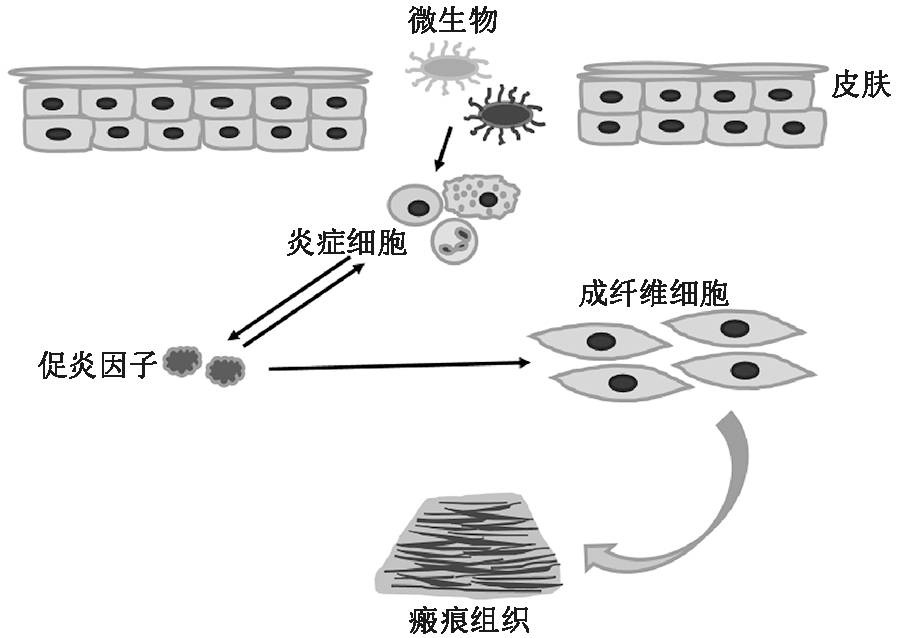
为实现皮肤伤口的无疤愈合,针对功能敷料在皮肤伤口愈合中所起到的抑制瘢痕作用,对国内外的相关研究进行概括总结。以伤口愈合过程为切入点,从瘢痕形成的影响因素,即外部环境和内部生物信号调控入手,系统分析了伤口愈合不同阶段的不同生理环境和需求下对抑制瘢痕敷料的性能要求。在炎症期、增殖期和重塑期3个阶段中,功能敷料分别以降低炎症反应、调节信号传导和促进组织再生的作用机制来抑制瘢痕的产生。认为瘢痕形成是一个动态连续而又复杂的过程,该过程涉及炎症细胞、角质形成细胞、成纤维细胞以及各类生长因子等物质,指出用于抑制瘢痕的功能敷料要想达到更好的效果,需要注重新型给药系统的研究和敷料结构的优化设计。
中图分类号:
| [1] |
PROFYRIS C, TZIOTZIOS C, DOVALE I. Cutaneous scarring: pathophysiology, molecular mechanisms, and scar reduction therapeutics: part I: the molecular basis of scar formation[J]. Journal of the American Academy of Dermatology, 2012, 66(1): 1-10.
doi: 10.1016/j.jaad.2011.05.055 |
| [2] |
KONDO T, ISHIDA Y. Molecular pathology of wound healing[J]. Forensic Science International, 2010, 203(1-3): 93-98.
doi: 10.1016/j.forsciint.2010.07.004 |
| [3] |
COENTRO J Q, PUGLIESE E, HANLEY G, et al. Current and upcoming therapies to modulate skin scarring and fibrosis[J]. Advanced Drug Delivery Reviews, 2019, 146: 37-59.
doi: 10.1016/j.addr.2018.08.009 |
| [4] |
PRATSINIS H, MAVROGONATOU E, KLETSAS D. Scarless wound healing: from development to senesc-ence[J]. Advanced Drug Delivery Reviews, 2019, 146: 325-343.
doi: 10.1016/j.addr.2018.04.011 |
| [5] |
ALMINE J F, WISE S G, WEISS A S. Elastin signaling in wound repair[J]. Birth defects research Part C: Embryo Today : Reviews, 2012, 96(3): 248-257.
doi: 10.1002/bdrc.21016 |
| [6] |
ZHAO Yuqian, LI Xueyong, XU Xiaoli, et al. Lumican alleviates hypertrophic scarring by suppressing integrin-FAK signaling[J]. Biochemical and Biophysical Research Communications, 2016, 480(2): 153-159.
doi: S0006-291X(16)31633-3 pmid: 27693693 |
| [7] | 李玉林. 病理学[M]. 8版. 北京: 人民卫生出版社, 2013: 27-43. |
| LI Yulin. Pathology[M]. 8th ed. Beijing: People's Medical Publishing House, 2013: 27-43. | |
| [8] |
LORDEN E R, MILLER K J, IBRAHIM M, et al. Biostable electrospun microfibrous scaffolds mitigate hypertrophic scar contraction in an immune-competent murine model[J]. Acta Biomaterialia, 2016, 32: 100-109.
doi: 10.1016/j.actbio.2015.12.025 |
| [9] |
LORDEN E R, MILLER K J, BASHIROV L, et al. Mitigation of hypertrophic scar contraction via an elastomeric biodegradable scaffold[J]. Biomaterials, 2015, 43: 61-70.
doi: 10.1016/j.biomaterials.2014.12.003 |
| [10] |
CHENG Liying, SUN Xiaoming, HU Changmin, et al. In vivo inhibition of hypertrophic scars by implantable ginsenoside-Rg3-loaded electrospun fibrous membr-anes[J]. Acta Biomaterialia, 2013, 9(12): 9461-9473.
doi: 10.1016/j.actbio.2013.07.040 pmid: 23938200 |
| [11] |
HU Xiaolong, LI Na, TAO Ke, et al. Effects of integrin ανβ3 on differentiation and collagen synthesis inducedby connective tissue growth factor in human hypertrophic scar fibroblasts[J]. International Journal of Molecular Medicine, 2014, 34(5): 1323-1334.
doi: 10.3892/ijmm.2014.1912 pmid: 25174803 |
| [12] | MONSTREY S, MIDDELKOOP E, JEROEN J, et al. Updated scar management practical guidelines: non-invasive and invasive measures[J]. Journal of Plastic, Reconstructive & Aesthetic Surgery, 2014, 67(8): 1017-1025. |
| [13] |
LIN Shiqi, QUAN Guilan, HOU Ailin, et al. Strategy for hypertrophic scar therapy: improved delivery of triamcinolone acetonide using mechanically robust tip-concentrated dissolving microneedle array[J]. Journal of Controlled Release, 2019, 306: 69-82.
doi: S0168-3659(19)30302-5 pmid: 31145948 |
| [14] |
DAVID C Y, SHARON W T C, XU Chenjie. Polymeric biomaterials for management of pathological scarring[J]. ACS Applied Polymer Materials, 2019, 1: 612-624.
doi: 10.1021/acsapm.8b00203 |
| [15] | WILGUS Traci A. Inflammation as an orchestrator of cutaneous scar formation: a review of the literature[J]. Plastic and aesthetic research, 2020, 7: 54. |
| [16] |
TRACI A W, VALERIE K B, KATHLEEN L T, et al. The Impact of cyclooxygenase-2 mediated inflammation on scarless fetal wound healing[J]. The American Journal of Pathology, 2004, 165(3): 753-761.
doi: 10.1016/S0002-9440(10)63338-X |
| [17] |
WU Qianli, FOURCAUDOT A B, YAMANE K, et al. Exacerbated and prolonged inflammation impairs wound healing and increases scarring[J]. Wound Repair and Regeneration, 2016, 24(1): 26-34.
doi: 10.1111/wrr.12381 |
| [18] | 邓雨萌, 雷霞. 炎性反应在瘢痕疙瘩发生发展中的作用及机制研究[J]. 中国美容医学, 2020, 29(4): 167-169. |
| DENG Yumeng, LEI Xia. The role and mechanism of inflammatory responses in the development of keloid[J]. Chinese Journal of Aesthetic Medicine, 2020, 29(4): 167-169. | |
| [19] |
SADIYA A, ANHA A, ALAM M S, et al. Development of antimicrobial and scar preventive chitosan hydrogel wound dressingsmicrobial and scar preventive chitosan hydrogel wound dressings[J]. International Journal of Pharmaceutics, 2016, 508(1/2): 92-101.
doi: 10.1016/j.ijpharm.2016.05.013 |
| [20] |
VIVEK K P, GUFRAN A, SIDDH N U, et al. Nano-fibrous scaffold with curcumin for anti-scar wound healing[J]. International Journal of Pharmaceutics, 2020. DOI: 10.1016/j.ijpharm.2020.119858.
doi: 10.1016/j.ijpharm.2020.119858 |
| [21] | 王佳琪, 王国栋, 吴洋. 活性氧在创伤愈合中作用的研究[J]. 现代生物医学进展, 2013, 13(31): 6194-6196. |
| WANG Jiaqi, WANG Guodong, WU Yang. The review on role of reactive oxygen species in wound healing[J]. Progress in Modern Biomedicine, 2013, 13(31): 6194-6196. | |
| [22] | DESMOULIERE A, BONTÉ F, LAVERDET B, et al. Fibroblasts and myofibroblasts in wound healing[J]. Clinical, Cosmetic and Investigational Dermatology, 2014, 7: 301-311. |
| [23] |
XU Wei, HONG Seokjong, ZEITCHEK M, et al. Hydration status regulates sodium flux and inflammatory pathways through epithelial sodium channel (ENaC) in the skin[J]. The Journal of Investigative Dermatology, 2015, 135(3): 796-806.
doi: 10.1038/jid.2014.477 |
| [24] | ZHANG Dongmei, CAI Guanke, MUKHERJEE S, et al. Elastic, persistently moisture-retentive, and wearable biomimetic film inspired by fetal scarless repair for promoting skin wound healing[J]. ACS applied Materials & Interfaces, 2020, 12(5): 5542-5556. |
| [25] |
ZHAO Jingling, YU Jianxing, XU Yingbin, et al. Epidermal HMGB1 activates dermal fibroblasts and causes hypertrophic scar formation in reduced hydration[J]. The Journal of Investigative Dermatology, 2018, 138(11): 2322-2332.
doi: 10.1016/j.jid.2018.04.036 |
| [26] |
ZHAO Jingling, ZHONG Aimei, FRIEDRICH E, et al. S100A12 induced in the epidermis by reduced hydration activates dermal fibroblasts and causes dermal fibrosis[J]. Journal of Investigative Dermatology, 2017, 137(3): 650-659.
doi: S0022-202X(16)32638-0 pmid: 27840235 |
| [27] | 陈晓洁, 吕爱凤, 高晶, 等. 功能敷料的“伤口湿润环境愈合”理论与实践[J]. 生物医学工程学进展, 2013, 34(1): 31-34. |
| CHEN Xiaojie, LV Aifeng, GAO Jing, et al. The theory and practice of moisture wound healing on functional dressings[J]. Progress in Biomedical Engineering, 2013, 34(1): 31-34. | |
| [28] | 何贵东, 李政, 华嘉川, 等. 水凝胶在医学领域应用研究进展[J]. 化工新型材料, 2017, 45(5): 223-225. |
| HE Guidong, LI Zheng, HUA Jiachuan, et al. Research and application progress of hydrogel in medical field[J]. New Chemical Materials, 2017, 45(5): 223-225. | |
| [29] | HUANG Xin, ZHANG Yaqing, ZHANG Xiangmei, et al. Influence of radiation crosslinked carboxymethyl-chitosan/gelatin hydrogel on cutaneous wound healing[J]. Materials Science & Engineering C, 2013, 33(8): 4816-4824. |
| [30] |
DINESH K S, ALOK R R. Biomedical applications of chitin, chitosan, and their derivatives[J]. Journal of Macromolecular Science: Part C, 2000, 40(1): 69-83.
doi: 10.1081/MC-100100579 |
| [31] |
WANG Dong, ZHANG Nihui, MENG Guolong, et al. The effect of form of carboxymethyl-chitosan dressings on biological properties in wound healing[J]. Colloids and Surfaces B: Biointerfaces, 2020. DOI: 10.1016/j.colsurfb.2020.111191.
doi: 10.1016/j.colsurfb.2020.111191. |
| [32] |
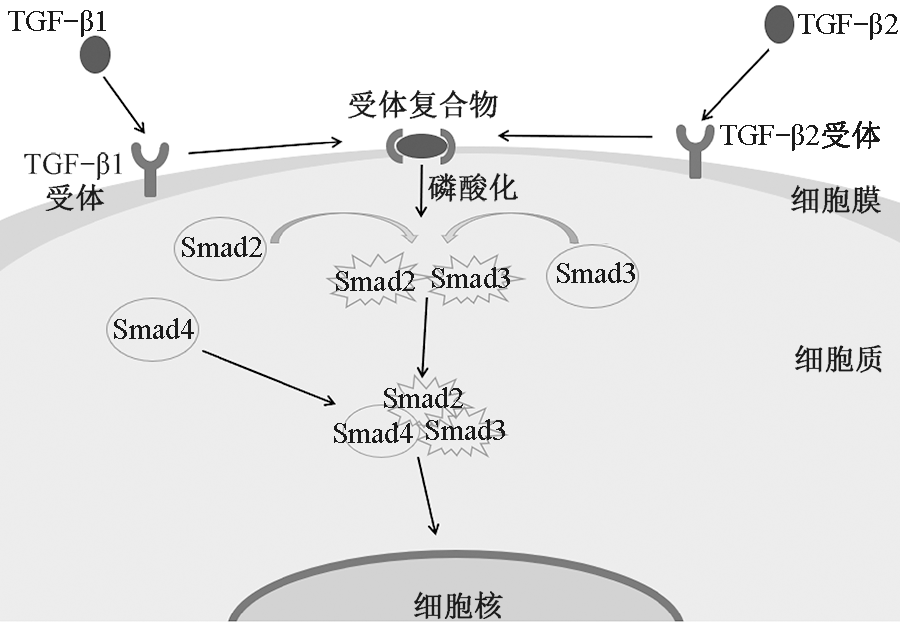
ZHANG Tao, WANG Xiaofeng, WANG Zhengcai, et al. Current potential therapeutic strategies targeting the TGF-β/Smad signaling pathway to attenuate keloid and hypertrophic scar formation[J]. Biomedicine & Pharmacotherapy, 2020. DOI: 10.1016/j.biopha.2020.110287.
doi: 10.1016/j.biopha.2020.110287. |
| [33] | WANG Le, YANG Junchuan, RAN Bei, et al. Small molecular TGF-β1-inhibitor-loaded electrospun fibrous scaffolds for preventing hypertrophic scars[J]. ACS Applied Materials & Interfaces, 2017, 9(38): 32545-32553. |
| [34] |
PADMANABAN M, SATHIYAMOORTHY S, RAMYA M, et al. Molecular signaling of ginsenosides Rb1, Rg1, and Rg3 and their mode of actions[J]. Journal of Ginseng Research, 2018, 42(2): 123-132.
doi: 10.1016/j.jgr.2017.01.008 |
| [35] |
CUI Wenguo, CHENG Liying, HU Changmin, et al. Electrospun poly(L-Lactide) fiber with ginsenoside Rg3 for inhibiting scar hyperplasia of skin[J]. PLOS ONE, 2013. DOI: 10.1371/journal.pone.0068771.
doi: 10.1371/journal.pone.0068771. |
| [36] |
FARAZ C, TAHEREH M, ALI H R, et al. Design, fabrication, and optimization of a dual function three-layer scaffold for controlled release of metformin hydrochloride to alleviate fibrosis and accelerate wound healing[J]. Acta Biomaterialia, 2020, 113: 144-163.
doi: 10.1016/j.actbio.2020.06.031 |
| [37] |
OLIVEIRA G V, HAWKINS H K, CHINKES D, et al. Hypertrophic versus non hypertrophic scars compared by immunohistochemistry and laser confocal microscopy: type I and III collagens[J]. International Wound Journal, 2009, 6(6): 445-451.
doi: 10.1111/j.1742-481X.2009.00638.x |
| [38] | PRIYA G, LESLIE T, SNEHAL S, et al. CD44-dependent inflammation, fibrogenesis, and collagenolysis regulates extracellular matrix remodeling and tensile strength during cutaneous wound healing[J]. Matrix Biology, 2018, 75: 314-330. |
| [39] | LI Haichang, DUANN P, LIN Peihui, et al. MG53 promotes wound healing and reduces scar formation by facilitating cell membrane repair and controlling myofibroblast differentiation[J]. Biophysical Journal, 2016, 110(3): 589. |
| [40] |
LI Haichang, DUANN P, LIN Peihui, et al. Modulation of wound healing and scar formation by MG53 protein-mediated cell membrane repair[J]. Journal of Biological Chemistry, 2015, 290(40): 24592-24603.
doi: 10.1074/jbc.M115.680074 pmid: 26306047 |
| [41] | 刘青武, 何秀娟, 张金超, 等. 细胞外基质在皮肤组织创面修复中的研究进展[J]. 医学研究杂志, 2019, 48(9): 21-24. |
| LIU Qingwu, HE Xiujuan, ZHANG Jinchao, et al. Research progress of extracellular matrix in skin tissue wound repair[J]. Journal of Medical Research, 2019, 48(9): 21-24. | |
| [42] | 王成, 荣艳华, 沈余明, 等. 正常皮肤与增生性瘢痕中Ⅰ型和Ⅲ型胶原的含量与比例[J]. 山东大学学报(医学版), 2016, 54(11): 64-67. |
| WANG Cheng, RONG Yanhua, SHEN Yuming, et al. Contents and ratios of typeⅠand type Ⅲ collagens in normal skin and hypertrophic scar in people of different ages[J]. Journal of Shandong University(Health Sciences), 2016, 54(11): 64-67. | |
| [43] |
YALMAN V, NELISA T L. Development of humic acid and alginate-based wound dressing and evaluation on inflammation[J]. Materials Technology, 2019, 34(12): 705-717.
doi: 10.1080/10667857.2019.1619961 |
| [44] |
COELHO N M, WANG A, MCCULLOCH C A. Discoidin domain receptor 1 interactions with myosin motors contribute to collagen remodeling and tissue fibrosis[J]. Biochimica et Biophysica Acta: Molecular Cell Research, 2019. DOI: 10.1016/j.bbamcr.2019.07.005.
doi: 10.1016/j.bbamcr.2019.07.005. |
| [45] |
ZHANG Nihui, GAO Tao, WANG Yao, et al. Modulating cationicity of chitosan hydrogel to prevent hypertrophic scar formation during wound healing[J]. International Journal of Biological Macromolecules, 2020, 154: 835-843.
doi: S0141-8130(20)32065-1 pmid: 32194120 |
| [46] |
CHEN Xi, PENG Lihua, LI Ni, et al. The healing and anti-scar effects of astragaloside IV on the wound repair in vitro and in vivo[J]. Journal of Ethnopharmacology, 2012, 139(3): 721-727.
doi: 10.1016/j.jep.2011.11.035 |
| [47] |
CHEN Xi, PENG Lihua, SHAN Yinghui, et al. Astragaloside IV-loaded nanoparticle-enriched hydrogel induces wound healing and anti-scar activity through topical delivery[J]. International Journal of Pharmaceutics, 2013, 447(1/2): 171-181.
doi: 10.1016/j.ijpharm.2013.02.054 |
| [48] |
SHAN Yinghui, PENG Lihua, LIU Xin, et al. Silk fibroin/gelatin electrospun nanofibrous dressing functionalized with astragaloside IV induces healing and anti-scar effects on burn wound[J]. International Journal of Pharmaceutics, 2015, 479(2): 291-301.
doi: 10.1016/j.ijpharm.2014.12.067 pmid: 25556053 |
| [49] |
SHI Hongxue, LIN Cai, LIN Beibei, et al. The anti-scar effects of basic fibroblast growth factor on the wound repair in vitro and in vivo[J]. PLOS ONE, 2013. DOI: 10.1371/journal.pone.0059966.
doi: 10.1371/journal.pone.0059966. |
| [50] |
CHENG Liying, SUN Xiaoming, ZHAO Xin, et al. Surface biofunctional drug-loaded electrospun fibrous scaffolds for comprehensive repairing hypertrophic scars[J]. Biomaterials, 2016, 83: 169-181.
doi: 10.1016/j.biomaterials.2016.01.002 pmid: 26774564 |
| [1] | 岳程飞, 丁长坤, 李璐, 程博闻. 碳化二亚胺/羟基丁二酰亚胺交联改性胶原蛋白纤维制备及其性能[J]. 纺织学报, 2020, 41(03): 1-7. |
| [2] | 吕婷婷, 安瑛, 李好义, 刘宇健, 焦志伟. 静电纺动物蛋白纳米纤维研究进展[J]. 纺织学报, 2019, 40(12): 140-145. |
| [3] | 贾琳 王西贤 张海霞 覃小红. 聚乳酸/胶原蛋白取向纳米纤维支架的性能[J]. 纺织学报, 2016, 37(11): 8-13. |
| [4] | 陈晓盟 王树根 薛晨. 山羊绒夹杂肤皮屑的组成及其结构[J]. 纺织学报, 2015, 36(06): 24-29. |
| [5] | 王响 靳向煜. 再生牛皮胶原蛋白复合纤维的性能[J]. 纺织学报, 2015, 36(04): 1-6. |
| [6] | 于金泽 陈莹 杨庆斌 唐劲天. 丙烯酸和胶原蛋白对PET的表面改性研究进展[J]. 纺织学报, 2014, 35(7): 152-0. |
| [7] | 许云辉;黄晨;陈宇岳;林红. 棉纤维经胶原蛋白涂覆处理后的结构[J]. 纺织学报, 2007, 28(6): 1-4. |
| [8] | 许云辉;陈宇岳;黄晨. 胶原蛋白涂覆棉纤维的研究[J]. 纺织学报, 2007, 28(5): 23-27. |
| [9] | 王雪娟;唐屹;吴炜誉;徐建军;叶光斗. 戊二醛交联胶原蛋白/PVA 复合纤维的结构与性能[J]. 纺织学报, 2007, 28(11): 13-16. |
| [10] | 姚理荣;林红;陈宇岳. 胶原蛋白纤维的性能与应用[J]. 纺织学报, 2006, 27(9): 105-107. |
|
 京公网安备11010502044800号