Journal of Textile Research ›› 2026, Vol. 47 ›› Issue (05): 45-55.doi: 10.13475/j.fzxb.20250707801

• Fiber Materials • Previous Articles     Next Articles

Study on anticoagulant properties and endothelialization promotion of polytetrafluoroethylene vascular stent membranes by airflow-assisted electrospinning

CHENG Ersu1, WANG Haojie1, LIU Yuqing1,2, MENG Kai1,2, ZHAO Huijing1,2()   

  1. 1 College of Textiles and Clothing Engineering, Soochow University, Suzhou, Jiangsu 215021, China
    2 National Engineering Laboratory for Modern Silk(Suzhou), Soochow University, Suzhou, Jiangsu 215123, China
  • Received:2025-07-30 Revised:2026-04-01 Online:2026-05-15 Published:2026-07-10
  • Contact: ZHAO Huijing E-mail:zhhj@suda.edu.cn

Abstract:

Objective Cardiovascular diseases (CVDs) are a class of clinical syndromes characterized by dysfunction of the heart and vascular system, and have become the leading cause of death globally. In clinical practice, stent grafts are widely used for interventional treatment, where the graft membrane serves as a physical barrier and plays a critical role in cases such as aneurysm occlusion, vascular perforation repair, and arterial stenosis management. Currently, expanded polytetrafluoroethylene (ePTFE) has become one of the commonly used membrane materials in clinical practice by virtue of its excellent biocompatibility, stable mechanical properties, and chemical inertness. However, commercial ePTFE grafts often suffer from poor long-term patency due to their chemical inertness, which hinders endothelialization and triggers thrombogenic responses. While electrospinning offers a promising alternative for mimicking the natural extracellular matrix (ECM), conventional electrospinning is severely limited by low production efficiency and jet instability, hindering industrial-scale manufacturing. Therefore, this study aims to address these dual challenges by developing a high-throughput airflow-assisted electrospinning strategy to fabricate PTFE vascular stent membranes. Furthermore, to overcome the bio-inert nature of PTFE, a surface functionalization strategy is proposed to simultaneously endow the grafts with potent anticoagulant properties and the capacity to promote rapid endothelialization, thereby enhancing their clinical translation potential.

Method A novel airflow-assisted electrospinning system utilized a high-velocity air stream to manipulate the polymer jet trajectory and enhance solvent evaporation. The influences of key processing parameters, particularly extrusion rate, on fiber morphology and deposition efficiency were systematically optimized. In order to functionalize the chemically inert PTFE surface, a multi-step modification protocol was employed. First, the membranes underwent air plasma activation to introduce initial reactive groups. Subsequently, a bio-adhesive intermediate layer was constructed by co-depositing dopamine (DA) and polyethyleneimine (PEI) under mild alkaline conditions (Tris-HCl, pH=8.5). Finally, heparin (HEP) was covalently immobilized onto the amine-rich DA/PEI layer by EDC/NHS activation chemistry. Physicochemical properties were characterized using scanning electron microscopy (SEM), Fourier transform infrared spectroscopy(FT-IR), energy dispersive spectroscopy(EDS), and contact angle measurements. Biological performance was evaluated through in vitro heparin release profiles, hemolysis assays, blood clotting index (BBCI) tests, platelet adhesion studies, and endothelial cell (ECs) proliferation assays using CCK-8 and fluorescence imaging.

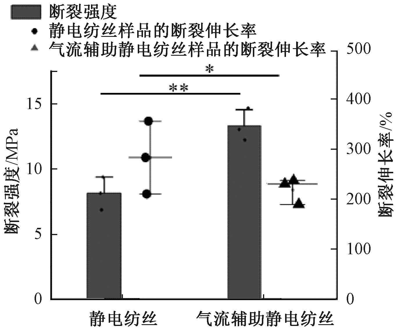
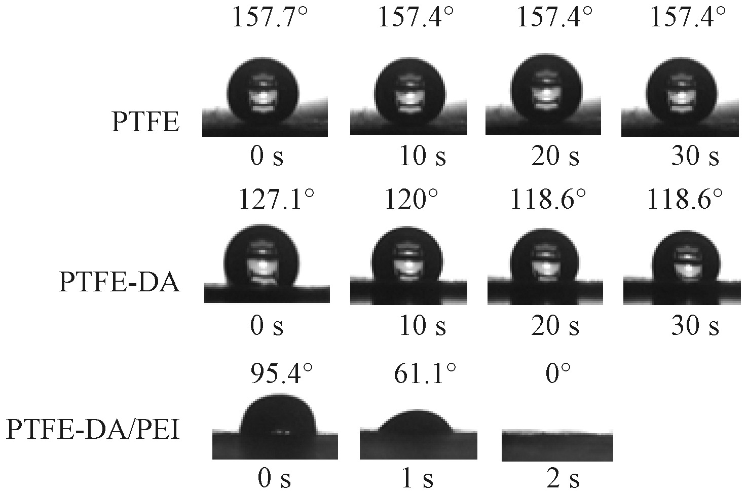
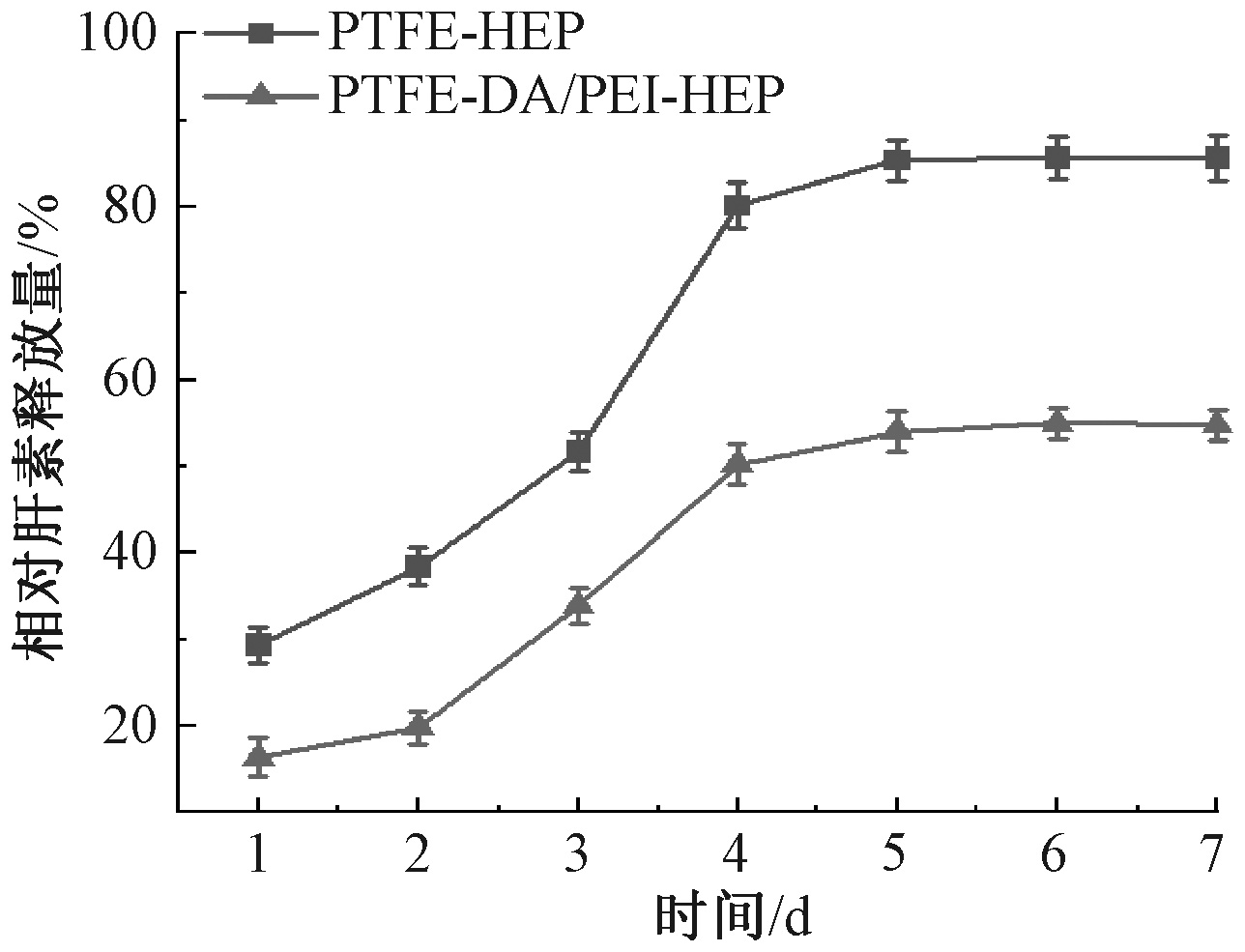
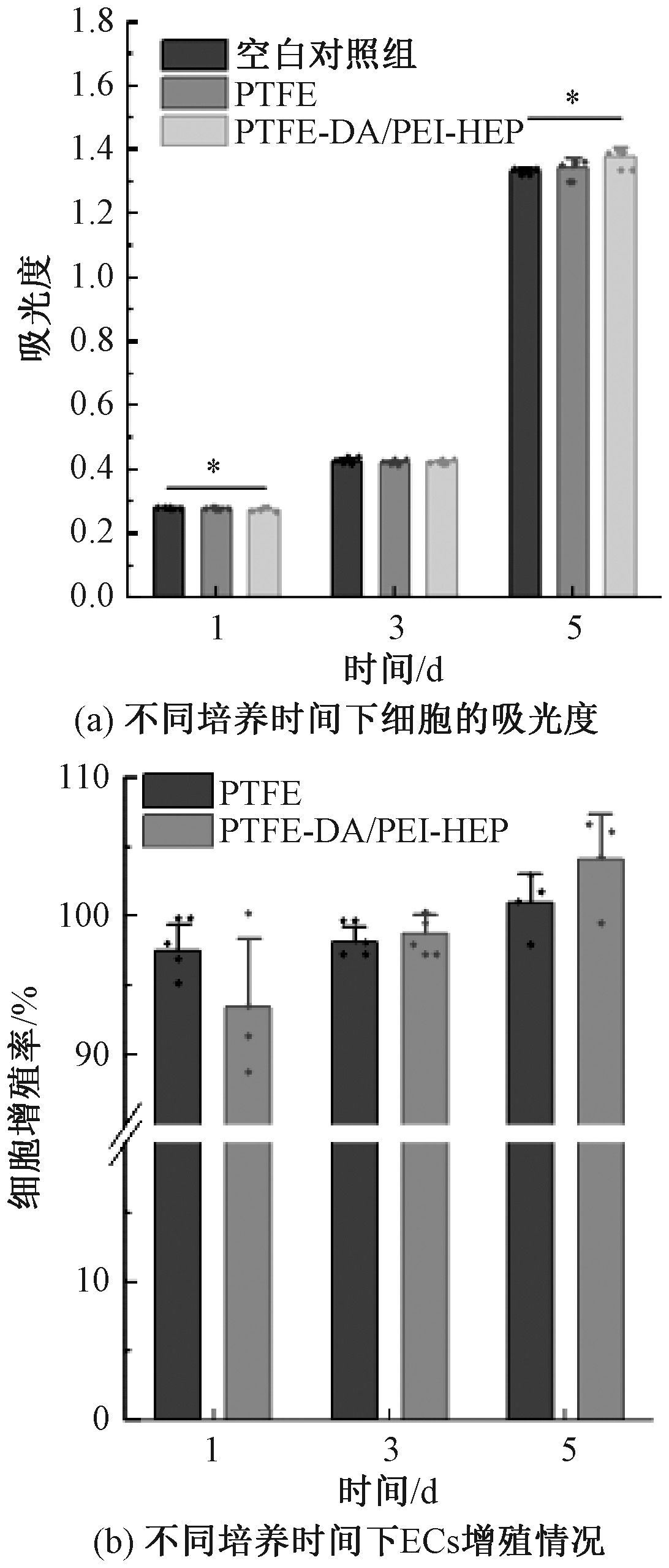
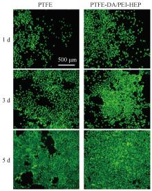
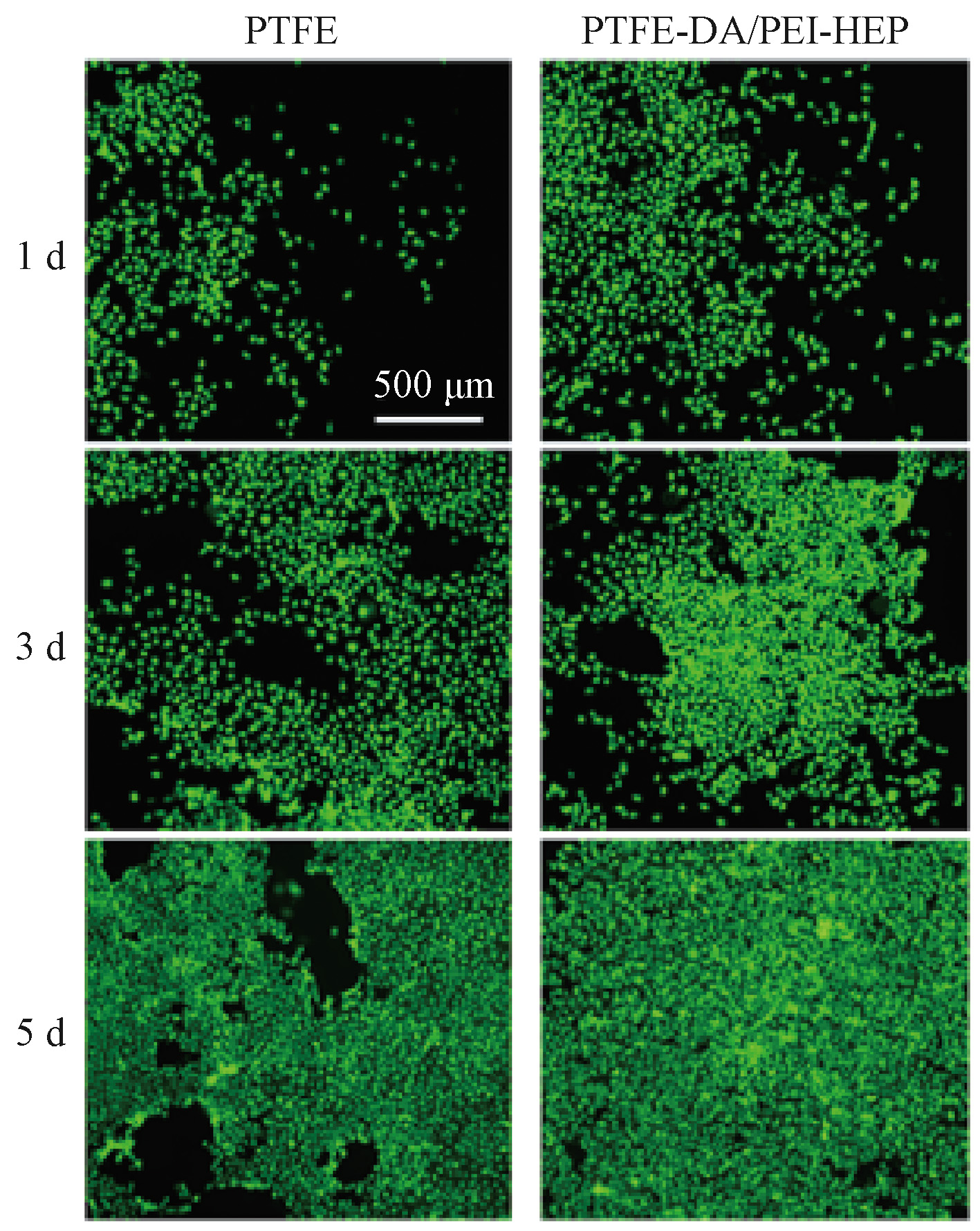
Results Airflow assisting significantly suppressed the whipping instability of the electrospinning jet, resulting in more uniform and finer fibers compared to the conventional method. The airflow-assisted process achieved a unit time production of 0.012 8 g/min, a 4.13 time increase over the 0.003 1 g/min rate of conventional electrospinning. This denser packing improved mechanical performance, with the tensile strength of the PTFE stent membrane increasing significantly to 13.38 MPa. Surface analysis confirmed the successful deposition of the functional coating, with the water contact angle dropping drastically to near 0°, indicating a transition from superhydrophobicity to superhydrophilicity. The DA/PEI intermediate layer is proved highly effective for drug loading; the functionalized PTFE-DA/PEI-HEP surface achieved a heparin density of 32.914 μg/cm2, a 4.7 time increase compared to direct adsorption (P<0.01), and demonstrated a sustained release profile over 7 d. Regarding hemocompatibility, the modified membranes exhibited an extremely low hemolysis ratio of 0.72%, far below the 5% international standard. The BBCI value was improved by 24.97% compared to unmodified PTFE, reaching 74.23%. Furthermore, SEM revealed that platelet adhesion density decreased by 44.19% (to 480 platelets/mm2), with minimal platelet activation observed. Cytocompatibility assays demonstrated that the coating created a favorable microenvironment for endothelial cells; by 5 d, the relative proliferation rate of endothelial cells on the modified surface reached 104.13%, representing a 3.1% enhancement over the unmodified control, with fluorescence imaging confirming a dense, healthy cell monolayer.

Conclusion This study establishes a scalable airflow-assisted electrospinning protocol that overcomes the production efficiency bottleneck of PTFE nanofiber membranes while significantly reducing costs compared to thermal stretching methods. The combination of plasma treatment and DA/PEI-mediated heparin grafting transforms the bio-inert PTFE surface into a bioactive interface. The resulting vascular stent membranes possess excellent mechanical strength, superior hemocompatibility, and the ability to promote endothelialization. These findings suggest that the developed PTFE-DA/PEI-HEP membranes offer a robust solution for replacing ePTFE stent membranes, which is promising for future clinical applications.

Key words: airflow-assisted electrospinning, polytetrafluoroethylene vascular stent membrane, functional modification, anticoagulation, endothelialization promotion

CLC Number: 

  • TS151

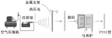
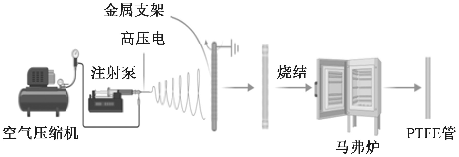
Fig.1

Schematic diagram of preparation process of PTFE electrospun membrane by airflow-assisted electrospinning"

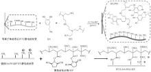
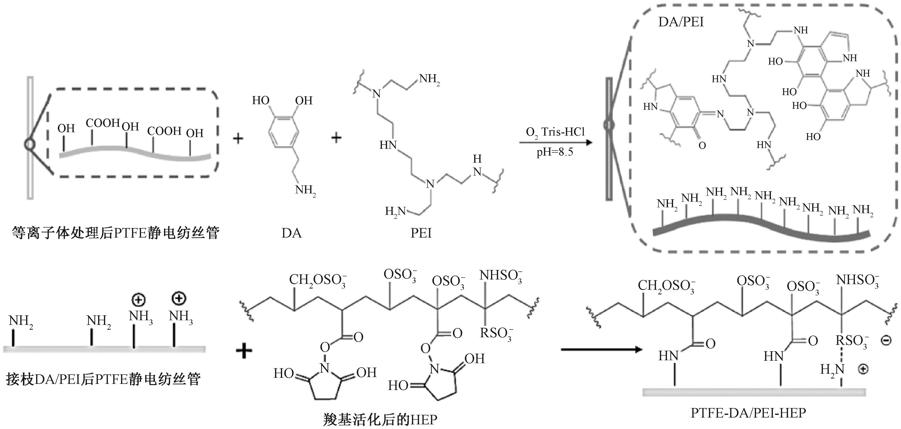
Fig.2

Reaction mechanism of DA/PEI-HEP coating"

Fig.3

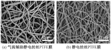
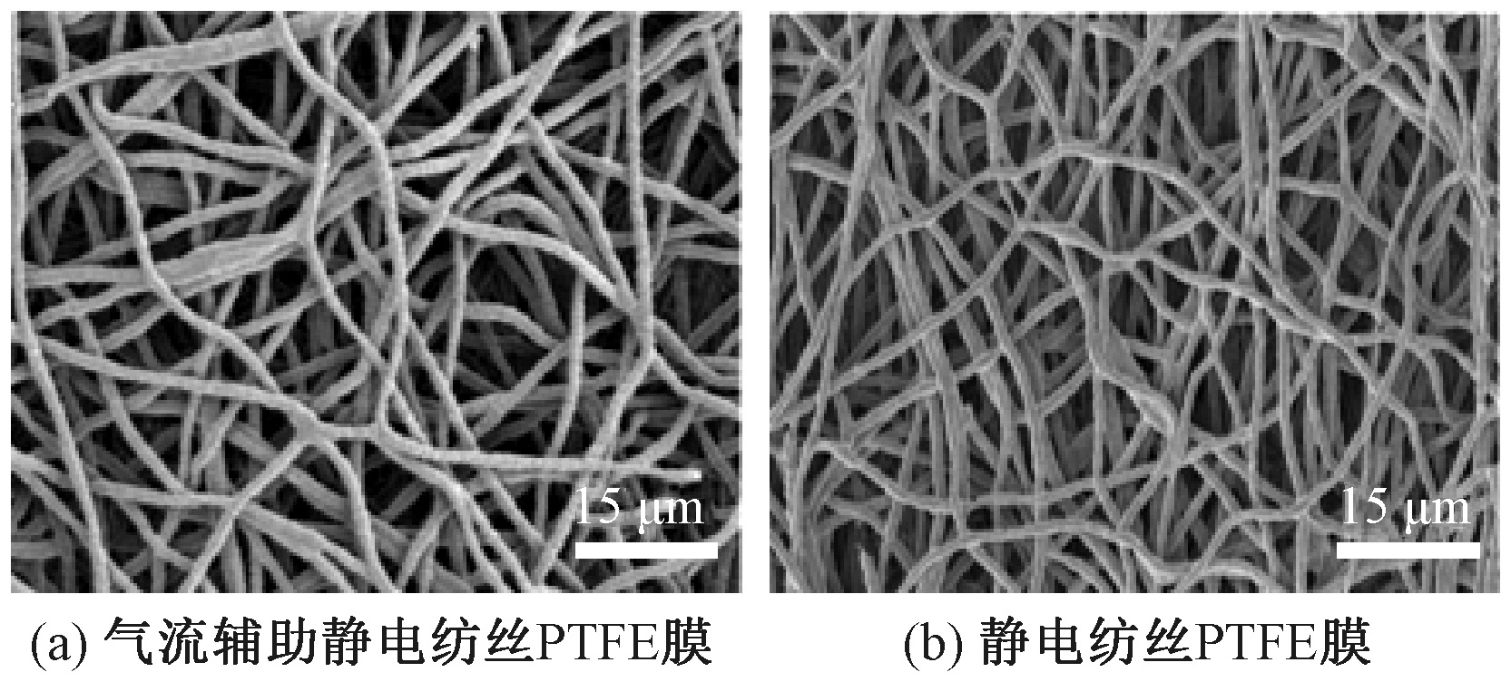
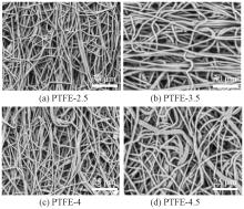
Microstructures of PTFE membranes prepared by different methods.(a)Airflow-assisted electrospun PTFE membrane;(b)Electrospun PTFE membrane"

Fig.4

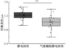
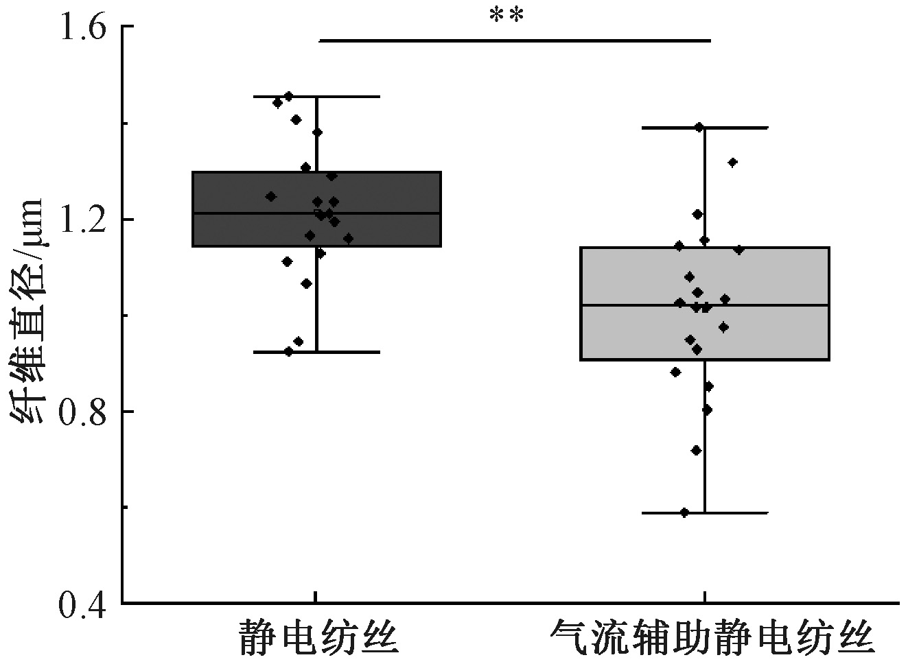
Diameters of PTFE fibers prepared by different methods"

Fig.5

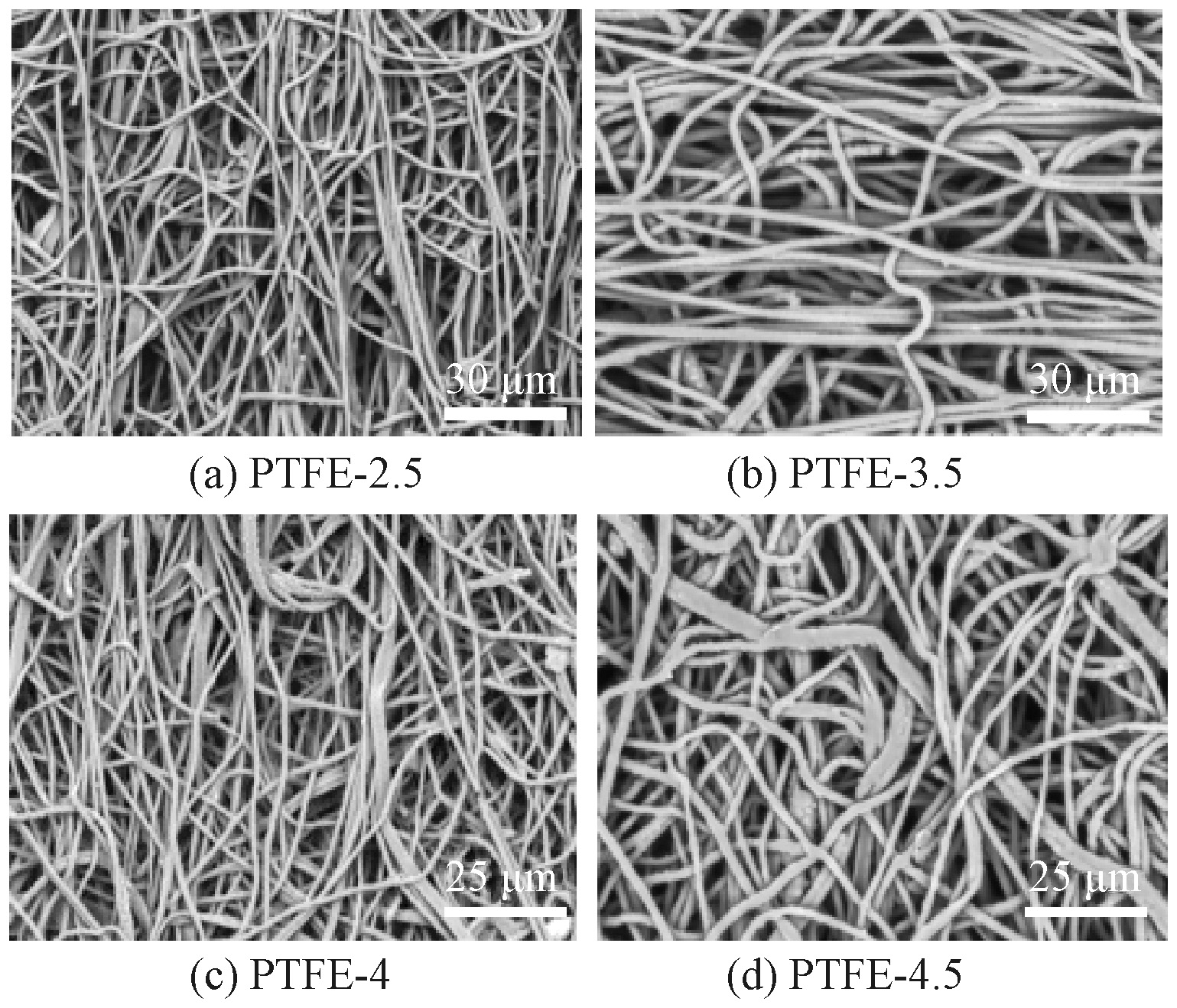
Microstructures of airflow-assisted electrospun PTFE membranes prepared at different extrusion rates"

Fig.6

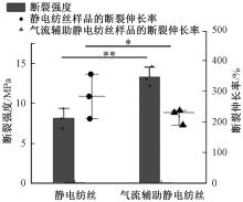
Mechanical performance of different samples"

Fig.7

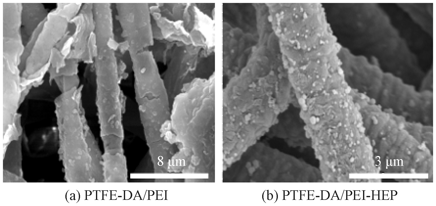
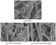
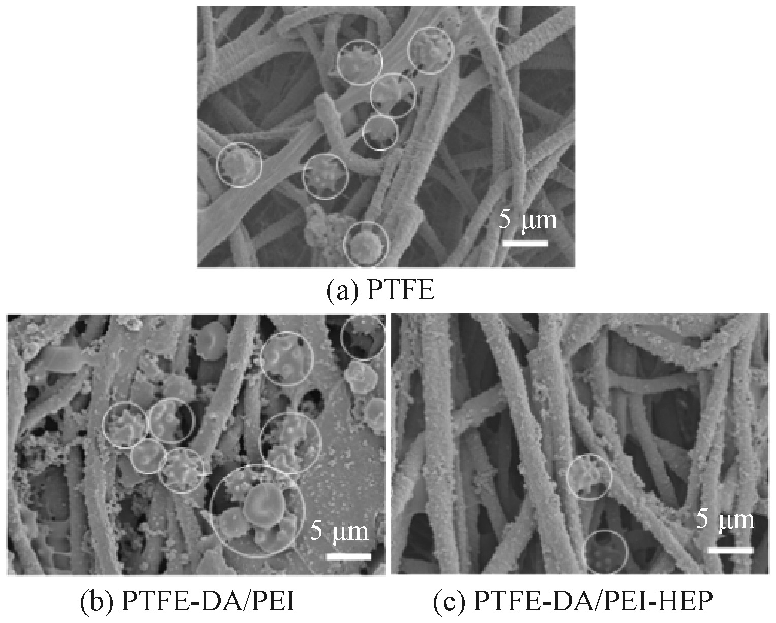
Microstructures of PTFE membranes before and after heparin modification"

Fig.8

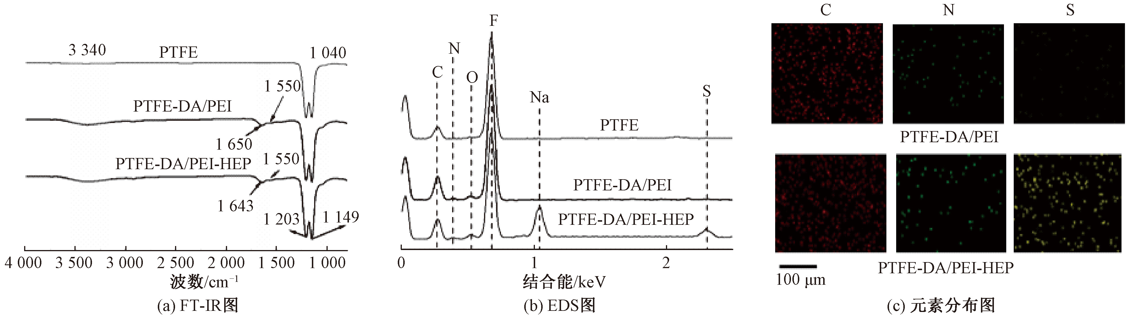
FT-IR spectra (a), EDS spectra (b), and element distribution images (c) of PTFE stent membrane before and after grafting DA/PEI and HEP"

Fig.9

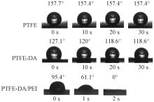
Contact angles photos of different coatings"

Fig.10

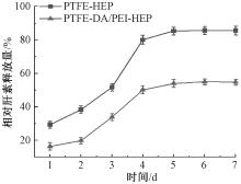
Heparin release curves from surfaces of different samples"

Fig.11

Platelet adhesion status on different samples"

Fig.12

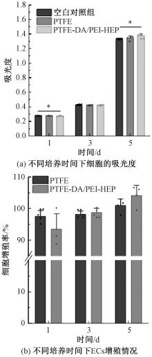
Absorbance of different samples and proliferation of endothelial cells.(a)Optical density values over culture time;(b)Proliferation status of ECs over culture time"

Fig.13

Fluorescence images of endothelial cells cultured on different materials for 1, 3, and 5 d"

[1] SHAMAKI G R, MARKSON F, SOJI-AYOADE D, et al. Peripheral artery disease: a comprehensive updated review[J]. Current Problems in Cardiology, 2022, 47(11): 101082.
doi: 10.1016/j.cpcardiol.2021.101082
[2] BRAUER M, ROTH G A, ARAVKIN A Y, et al. Global burden and strength of evidence for 88 risk factors in 204 countries and 811 subnational locations, 1990-2021: a systematic analysis for the Global Burden of Disease Study 2021[J]. The Lancet, 2024, 403(10440): 2162-2203.
doi: 10.1016/S0140-6736(24)00933-4
[3] PALANIAPPAN L P, ALLEN N B, ALMARZOOQ Z I, et al. 2026 heart disease and stroke statistics: a report of US and global data from the American heart association[J]. Circulation, 2026, 153(9): e275-e906.
[4] THUKKANI A K, KINLAY S. Endovascular intervention for peripheral artery disease[J]. Circulation Research, 2015, 116(9): 1599-1613.
doi: 10.1161/CIRCRESAHA.116.303503 pmid: 25908731
[5] WATSON N W, MOSARLA R C, SECEMSKY E A. Endovascular interventions for peripheral artery disease: a contemporary review[J]. Current Cardiology Reports, 2023, 25(11): 1611-1622.
doi: 10.1007/s11886-023-01973-9 pmid: 37804391
[6] ZHU P, DONG S T, SUN P, et al. Expanded polytetrafluoroethylene (ePTFE)-covered stents versus bare stents for transjugular intrahepatic portosystemic shunt in people with liver cirrhosis[J]. Cochrane Database of Systematic Reviews, 2023, 2023(8): 123-158.
[7] CASSADY A I, HIDZIR N M, GRØNDAHL L. Enhancing expanded poly(tetrafluoroethylene) (ePTFE) for biomaterials applications[J]. Journal of Applied Polymer Science, 2014, 131(15): 40533.
[8] ZHANG L L, HAM S W, WEAVER F A, et al. Performance of gore viabahn VBX compared with atrium iCast as bridging stents during fenestrated endovascular aortic repairs[J]. Journal of Vascular Surgery, 2019, 69(6): e215-e216.
[9] SOUKAS P, BECKER M, STARK K, et al. Three-year results of the GORE VIABAHN endoprosthesis in the superficial femoral artery for in-stent restenosis[J]. Journal of the Society for Cardiovascular Angiography & Interventions, 2023, 2(6): 101183.
[10] CHEN E, LI Z T, TURNG L S. Effects of expansion rate and sintering time on the morphology and mechanical properties of double-expanded polytetrafluoroethylene tubes[J]. Polymer Engineering & Science, 2025, 65(3): 1483-1496.
doi: 10.1002/pen.v65.3
[11] WANG G, FENG Y S, GAO C Y, et al. Biaxial stretching of polytetrafluoroethylene in industrial scale to fabricate medical ePTFE membrane with node-fibril microstructure[J]. Regenerative Biomaterials, 2023, 10: rbad056.
[12] MIURA H, NISHIBE T, YASUDA K, et al. The influence of node-fibril morphology on healing of high-porosity expanded polytetrafluoroethylene grafts[J]. European Surgical Research Europaische Chirurgische Forschung Recherches Chirurgicales Europeennes, 2002, 34(3): 224-231.
[13] GUO Q, HUANG Y, XU M D, et al. PTFE porous membrane technology: a comprehensive review[J]. Journal of Membrane Science, 2022, 664: 121115.
doi: 10.1016/j.memsci.2022.121115
[14] SCHMIDT II G A, LIN Y J, XU Y Y, et al. Viscosity characterization and flow simulation and visualization of polytetrafluoroethylene paste extrusion using a green and biofriendly lubricant[J]. Polymer Engineering & Science, 2021, 61(4): 1050-1065.
doi: 10.1002/pen.v61.4
[15] TOMKOVIC T, HATZIKIRIAKOS S G. Rheology and processing of polytetrafluoroethylene (PTFE) paste[J]. The Canadian Journal of Chemical Engineering, 2020, 98(9): 1852-1865.
doi: 10.1002/cjce.v98.9
[16] WANG H J, XU R, SHE S Y, et al. PTFE stent membrane based on the electrospinning technique and its potential for replacing ePTFE[J]. ACS Applied Bio Materials, 2024, 7(12): 8608-8620.
doi: 10.1021/acsabm.4c01392 pmid: 39601771
[17] HASAN A, MEMIC A, ANNABI N, et al. Electrospun scaffolds for tissue engineering of vascular grafts[J]. Acta Biomaterialia, 2014, 10(1): 11-25.
doi: 10.1016/j.actbio.2013.08.022 pmid: 23973391
[18] ZHANG Y, LIN L N, NIU M Y, et al. Artificial human blood vessels for tissue engineering[J]. ACS Materials Letters, 2025, 7(4): 1626-1645.
doi: 10.1021/acsmaterialslett.5c00038
[19] JIANG S Y, ZHANG A Y, AKHAVAN B, et al. Biofunctionalization of electrospun silk scaffolds with perlecan for vascular tissue engineering[J]. Biomaterials Science, 2025, 13(13): 3598-3616.
doi: 10.1039/D5BM00364D
[20] CASSANO R, PERRI P, ESPOSITO A, et al. Expanded polytetrafluoroethylene membranes for vascular stent coating: manufacturing, biomedical and surgical applications, innovations and case reports[J]. Membranes, 2023, 13(2): 240.
doi: 10.3390/membranes13020240
[21] LUO Y, GONG X S, XU Z W, et al. PTFE electrospun stent graft: preparation, properties and its industrialization prospect[J]. Chemical Research in Chinese Universities, 2021, 37(3): 589-597.
doi: 10.1007/s40242-021-1177-4
[22] 徐溶. PTFE电纺血管支架覆膜的制备及性能优化[D]. 苏州: 苏州大学, 2023:22-26.
XU Rong. Preparation and performance optimization of PTFE electrospun vascular stent covering[D]. Suzhou: Soochow University, 2023:22-26.
[23] XIONG J, HUO P F, KO F K. Fabrication of ultrafine fibrous polytetrafluoroethylene porous membranes by electrospinning[J]. Journal of Materials Research, 2009, 24(9): 2755-2761.
doi: 10.1557/jmr.2009.0347
[1] LIU Qingqing, MAO Xiaohui, YAO Yan, SUN Ying, CHEN Yu, ZHANG Xiaozhe, ZHU Liping, WANG Xuefen. Recent advances in fibrous separation membranes for functional modification and applications [J]. Journal of Textile Research, 2025, 46(12): 39-48.
[2] REN Tianxiang, CHEN Jiangbing, FANG Taorong, ZHAN Yingtao, HONG Yujie, XU Zhiqiang, XU Yudong, ZHAN Haihua. Research progress in preparation, modification and application of polyphenylene sulfide film [J]. Journal of Textile Research, 2025, 46(08): 245-253.
[3] JIN Rushi, CHEN Wanming, LIU Guojin, LIU Chenghai, QI Dongming, ZHAI Shimin. Application progress in biochars in printing and dyeing wastewater treatment [J]. Journal of Textile Research, 2025, 46(04): 235-243.
Viewed
Full text


Abstract

Cited

  Shared   
  Discussed   
No Suggested Reading articles found!