


近年来,随着脑神经科学、运动科学及心脏健康等医学诊疗方面的发展,生理电信号监测已成为当前该领域的重要研究热点。同时,在材料科学和制备技术的发展背景下,用于采集信号的神经电极材料也得到了多方面发展,通过生物体内部或外部的神经电极,可以记录跨越多个时间和空间尺度的定量数据,包括脑电图(EEG)、心电图(ECG)、肌电图(EMG)、局部场电位(LFPs)以及单个神经元的峰值活动(AP)等[3-4]。为获取高信噪比、高时空分辨率的电信号,优化神经电极的结构与功能至关重要,而导电纤维因其独特的小型化和轻量化、结构可设计及导电性等优点,成为神经电极和智能器件开发的理想选择,能够在更少干扰自然生理活动的情况下,实现高精度的神经电信号记录,推动了神经科学以及生物医学等领域的发展[2,5-6]。
本文聚焦于导电纤维的理化性质,从电极分类、原料选择、结构设计以及应用探索方面入手,系统综述了导电纤维在生理电信号监测中的研究进展并展望了该领域当前所面临的挑战以及未来发展方向。为高精度信号采集用导电纤维的结构、功能设计及其在医疗领域应用方面提供一定的理论参考。
1 导电纤维的分类及其制备
图1
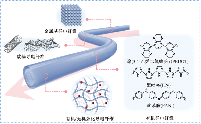
1.1 无机导电纤维
碳纳米管作为神经电极具有诸多优势,包括优异的导电性能以及长期稳定性,且制备工艺成熟,可通过湿法纺丝、化学气相沉积等方法进行规模化制备[18]。Lu等[19]使用直径为5~20 μm的碳纳米管(CNT)纤维制备与核磁共振成像兼容的柔软神经电极。可以精确定位特定脑区,记录高质量生理电信号。与坚硬的金属电极相比,能够连续监测大鼠的单个神经元信号长达4~5个月,且无需重新定位电极,同时大大降低了脑部炎症反应。Qian等[20]将刚性二维缺陷石墨烯纳米片集成在柔性CNT纤维上,设计了一种自适应、可拉伸和生物相容的碳烯纤维神经电极,可定制成各种复杂形状,也可在很宽的范围(0.5~600 kPa)内定制模量,类组织模量的设计减少了对生物体的影响,有利于生理电信号的长期监测。
1.2 有机导电纤维
此外,基于离子导电的有机导电纤维凭借其独特的导电机制,在柔韧性、环境适应性等方面表现出优势。Zhao等[24]通过调节质子供体和受体的序列来调节分子的静电相互作用,制备了一种力学性能优异的黏性水凝胶纤维,优良的离子电导率使其可用于肌电信号和心电信号监测,为生物电极、脑机接口和可穿戴电子设备等领域提供新的设计思路。
1.3 有机/无机杂化导电纤维
在杂化纤维中引入水凝胶基质可以显著提升纤维的多功能性和适应性,是有机/无机杂化导电纤维未来研究应用的主要趋势。Peng等[30]开发了一种微纤维状神经探针,核层为碳纳米管纤维,鞘层为海藻酸钙凝胶。植入生物体内后,鞘层凝胶吸水,该神经探针的弹性模量可从10 GPa降至10 kPa,接近脑组织的弹性模量。同时,水凝胶基质内可以加入各种纳米级材料,在保持力学适配的前提下扩展其功能。例如Rao等[31]在聚乙烯醇(PVA)水凝胶交联过程中引入导电CNT。通过酸化促进聚合物链相互作用以及力学拉伸促进CNT 在聚合物基质中的分散,确保与 PVA链的缠结从而增强了导电性,可以对自发的与光刺激下的生理电信号进行稳定记录。
2 导电纤维用于生理电信号的采集
图2
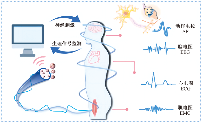
图2
导电纤维对不同生理电信号监测
Fig.2
Monitoring of different electrophysiological signals by conductive fibers
2.1 非侵入式生理电信号采集
非侵入式神经电极具备较低的风险性,无创的生理电信号记录形式更易被接受以及普及应用。但是三维结构的皮肤组织增加了电信号采集的难度,高接触阻抗导致记录信号的振幅和信噪比较低,难以精确定位深层的电生理活动,影响生理信号记录的准确性和一致性。而导电纤维微型的结构导致较低的空间分辨率,可通过适当的结构设计和材料选择来改善。
Gao等[33]制备了一种具有高柔韧性的导电针形碳纤维干电极,针尖进一步加工成碳纤维刷毛,并在表面进行金电镀处理,从而减少皮肤与干电极之间的接触阻抗,其平均值为133 kΩ。其能够在对皮肤影响较小的情况下对毛发区域的脑电图进行记录与分析。干电极易于集成到外部设备中,但缺乏固定在皮肤上的黏附性能。Chang等[34]将丙烯酸-丙烯酰胺有机水凝胶均匀涂覆在干燥纤维电极的表面,获得具有良好皮肤顺应性、界面黏附性和生物相容性的导电复合纤维,减轻了干燥纤维电极的运动伪影,可应用于日常穿戴心电图设备。Chang等[35]基于热塑性弹性体苯乙烯-乙烯-丁烯-苯乙烯制备了一种导电弹性纤维电极,该电极具有低阻抗(大于4×10⁴ Ω)和优异的拉伸性,可以通过编织技术开发可穿戴心电系统,用于长期ECG监测。
2.2 侵入式生理电信号采集
侵入式神经电极可以按照应用需求,通过有创的方式植入特定的生物体器官或组织内部,从而提取到生物体内部的生理电信号,并实现稳定的信号记录。相比于非侵入式电极具有更高的空间和时间分辨率,在医疗诊断和治疗、脑机接口和康复医学等领域具有重要应用价值[36]。根据植入深度可进一步细分为半侵入式和全侵入式电极。
2.2.1 半侵入式电极
半侵入式电极可以共形附着在器官的复杂三维表面,如大脑表面(即硬脑膜)或颅骨内。导电纤维由于其独特的结构更易贴合,当器官在生理活动中变形时,仍能保证信号传输的稳定。Chen等[37]设计了一种具有快速响应能力的纤维有机电化学晶体管,通过涂覆PEDOT:PSS形成垂直通道,具有较高的力学性能、电稳定性以及良好的生物相容性。皮下植入大鼠后可以捕获振幅为12 mA、心室搏动间隔约为0.2 s的心电图谱,在连续监测长达7 d后,振幅没有显著变化。证明该纤维电极具有长期电生理诊断的能力。Hu等[38]采用溶剂置换和多级氢键增强的策略,连续制备了一种导电离子凝胶纤维,以PVA为固态弹性相,纤维素纳米纤维为增强相,深层共晶溶剂为液体离子导电相,用作监测表面肌电图信号,此外将纤维电极包裹在坐骨神经周围,可以清晰记录复合神经动作电位,且增加刺激电流强度后检测的电位峰值更加明显。
2.2.2 全侵入式电极
全侵入式电极通常需要植入真皮层下,可放置于特定的神经元或神经组织上,极大程度地增加了与组织的接触面积,减少了噪音干扰,提高了信号记录质量。Wang等[39]制备了一种多通道植入式氧化铱神经探针,较高的电荷存储容量(39.6 mC/cm2)使其具备优越的电生理记录和神经刺激性能,实现了对同一神经元放电长达4周的持续跟踪,并在电刺激期间成功捕获了瞬时群体尖峰。
实现多通道对同一神经元群体的稳定记录,对于理解神经元的动态功能和脑机接口等实际应用具有重要意义。然而免疫系统以及生物体的运动特性使刚性植入器件易造成组织损伤,并影响电极材料长期的信号记录。因此为满足体内长期服役的需求以及信号质量的稳定性,微量化及柔性化设计的导电纤维得到广泛应用,通过不断优化电极设计和植入技术,可进一步提高信号传输能力和保障患者的安全性。Won等[29]制备了一种由金纳米颗粒掺杂的聚氨酯纤维神经探针,具有优异的电学性能和接近脑组织模量的力学性能。因此成功应用于记录麻醉状态下小鼠海马区的神经信号,获得了高信噪比(3.1和4.2 dB)的单个神经元峰值。植入生物体内4个月后仍可以对自发和诱发的生理电信号进行稳定记录。Wang等[17]选择石墨烯纤维作为柔性基底,低阻抗和多孔结构提供了强的电荷注入能力,附加薄铂层可以有效地沿着微电极传输收集到的信号。植入大鼠大脑皮层的微电极可以检测到信噪比为9.2 dB的单个神经元活动。
此外,在生物体剧烈活动下会导致强烈的大脑变形,使得神经元的信号提取和记录仍是一大挑战,因此需要满足低模量、低界面阻抗和高电导率的条件,以实现稳定的脑机接口连接和高质量的神经信号监测。例如,Tang等[40]制备了一种液体网络结构的PEDOT:PSS导电凝胶纤维,能够稳定追踪中型动物深脑部的单个神经元活动信号。将带有16个通道的神经纤维束植入猫的视觉皮层后,该纤维束记录了猫在自由移动和跑步状态时的生理电信号,并稳定跟踪记录单个神经元活动的峰值,可作为复杂条件下的神经生物学研究的可靠工具,进一步拓展了侵入式神经电极的应用领域。
3 结束语
尽管导电纤维在生理电信号采集方面相较于传统金属基或硅基电极具有一系列优势,包括增强信号采集连续稳定性、易于功能结构集成等,但仍有部分关键问题亟需解决,例如导电性与力学性能之间的平衡、长期稳定及耐用性等,以确保理论研究与实际应用的匹配。
随着材料科学和纳米技术的进步,导电纤维逐渐由刚性材料向柔性材料、单功能监测向多功能集成过渡。为满足导电纤维在可穿戴器件、生物体内长期服役等场景需求,需要进一步优化其力学性能和生物相容性,实现更加微型化和智能化的设计,从而减少生理电信号监测过程造成的组织创伤和免疫反应。此外,多功能集成是导电纤维用于临床试验的重要需求,结合无线通信和大数据分析等技术,生理信号监测用导电纤维将推动可穿戴技术和植入式医疗设备的发展进步,在远程医疗、个性化健康管理以及神经科学研究中发挥更大作用。
参考文献
Recent advances in flexible noninvasive electrodes for surface electromyography acquisition
[J].
Flexible and implantable microelectrodes for chronically stable neural inter-faces
[J].
Mechanically tissue-like and highly conductive au nanoparticles embedded elastomeric fiber electrodes of brain-machine interfaces for chronic in vivo brain neural record-ing
[J].
Advanced electrode technologies for noninvasive brain-computer interfaces
[J].
DOI:10.1021/acsnano.3c06781
PMID:38064282
[本文引用: 2]

Brain-computer interfaces (BCIs) have garnered significant attention in recent years due to their potential applications in medical, assistive, and communication technologies. Building on this, noninvasive BCIs stand out as they provide a safe and user-friendly method for interacting with the human brain. In this work, we provide a comprehensive overview of the latest developments and advancements in material, design, and application of noninvasive BCIs electrode technology. We also explore the challenges and limitations currently faced by noninvasive BCI electrode technology and sketch out the technological roadmap from three dimensions: Materials and Design; Performances; Mode and Function. We aim to unite research efforts within the field of noninvasive BCI electrode technology, focusing on the consolidation of shared goals and fostering integrated development strategies among a diverse array of multidisciplinary researchers.
Electrospun fiber-based flexible electronics: fiber fabrication, device platform, functionality integration and applications
[J].
Flexible electrodes for in vivo and in vitro electrophysiological signal recording
[J].
Next-generation probes, particles, and proteins for neural interfacing
[J].
Material considerations for peripheral nerve interfacing
[J].
Hydrogel fiber-based biointerfacing
[J].
Scalable layered heterogeneous hydrogel fibers with strain-induced crystallization for tough, resilient, and highly conductive soft bioelectronics
[J].
Conductive fibers for biomedical applications
[J].
DOI:10.1016/j.bioactmat.2022.10.014
PMID:36311045
[本文引用: 2]
Bioelectricity has been stated as a key factor in regulating cell activity and tissue function in electroactive tissues. Thus, various biomedical electronic constructs have been developed to interfere with cell behaviors to promote tissue regeneration, or to interface with cells or tissue/organ surfaces to acquire physiological status electrical signals. Benefiting from the outstanding advantages of flexibility, structural diversity, customizable mechanical properties, and tunable distribution of conductive components, conductive fibers are able to avoid the damage-inducing mechanical mismatch between the construct and the biological environment, in return to ensure stable functioning of such constructs during physiological deformation. Herein, this review starts by presenting current fabrication technologies of conductive fibers including wet spinning, microfluidic spinning, electrospinning and 3D printing as well as surface modification on fibers and fiber assemblies. To provide an update on the biomedical applications of conductive fibers and fiber assemblies, we further elaborate conductive fibrous constructs utilized in tissue engineering and regeneration, implantable healthcare bioelectronics, and wearable healthcare bioelectronics. To conclude, current challenges and future perspectives of biomedical electronic constructs built by conductive fibers are discussed.© 2022 The Authors.
A robust core-shell nanofabric with personal protection, health monitoring and physical comfort for smart sportswear
[J].
Wearable fiber-based visual strain sensors with high sensitivity and excellent cyclic stability for health monitoring and thermal management
[J].
Nano-yarn carbon nanotube fiber based enzymatic glucose biosensor
[J].
Transparent and flexible low noise graphene electrodes for simultaneous electrophysiology and neuroimaging
[J].
U1 snRNP regulates cancer cell migration and invasion in vitro
[J].
High-performance graphene-fiber-based neural recording microelectrodes
[J].
Neural stimulation and recording with bidirectional, soft carbon nanotube fiber microelectrodes
[J].
DOI:10.1021/acsnano.5b01060
PMID:25803728
[本文引用: 1]
The development of microelectrodes capable of safely stimulating and recording neural activity is a critical step in the design of many prosthetic devices, brain-machine interfaces, and therapies for neurologic or nervous-system-mediated disorders. Metal electrodes are inadequate prospects for the miniaturization needed to attain neuronal-scale stimulation and recording because of their poor electrochemical properties, high stiffness, and propensity to fail due to bending fatigue. Here we demonstrate neural recording and stimulation using carbon nanotube (CNT) fiber electrodes. In vitro characterization shows that the tissue contact impedance of CNT fibers is remarkably lower than that of state-of-the-art metal electrodes, making them suitable for recording single-neuron activity without additional surface treatments. In vivo chronic studies in parkinsonian rodents show that CNT fiber microelectrodes stimulate neurons as effectively as metal electrodes with 10 times larger surface area, while eliciting a significantly reduced inflammatory response. The same CNT fiber microelectrodes can record neural activity for weeks, paving the way for the development of novel multifunctional and dynamic neural interfaces with long-term stability.
Soft and mRI compatible neural electrodes from carbon nanotube fibers
[J].
DOI:10.1021/acs.nanolett.8b04456
PMID:30798604
[本文引用: 1]
Soft and magnetic resonance imaging (MRI) compatible neural electrodes enable stable chronic electrophysiological measurements and anatomical or functional MRI studies of the entire brain without electrode interference with MRI images. These properties are important for many studies, ranging from a fundamental neurophysiological study of functional MRI signals to a chronic neuromodulatory effect investigation of therapeutic deep brain stimulation. Here we develop soft and MRI compatible neural electrodes using carbon nanotube (CNT) fibers with a diameter from 20 μm down to 5 μm. The CNT fiber electrodes demonstrate excellent interfacial electrochemical properties and greatly reduced MRI artifacts than PtIr electrodes under a 7.0 T MRI scanner. With a shuttle-assisted implantation strategy, we show that the soft CNT fiber electrodes can precisely target specific brain regions and record high-quality single-unit neural signals. Significantly, they are capable of continuously detecting and isolating single neuronal units from rats for up to 4-5 months without electrode repositioning, with greatly reduced brain inflammatory responses as compared to their stiff metal counterparts. In addition, we show that due to their high tensile strength, the CNT fiber electrodes can be retracted controllably postinsertion, which provides an effective and convenient way to do multidepth recording or potentially selecting cells with particular response properties. The chronic recording stability and MRI compatibility, together with their small size, provide the CNT fiber electrodes unique research capabilities for both basic and applied neuroscience studies.
Modulus-tailorable, stretchable, and biocompatible carbonene fiber for adaptive neural electrode
[J].
Pure PEDOT:PSS hydrogels
[J].
Support-free PEDOT:PSS fibers as multifunctional microelectrodes for in vivo neural recording and modulation
[J].
Well-modulated interfacial ion transport enables D-sorbitol/PEDOT:PSS fibers to sense brain electrophysiological signals in vivo
[J].
Neuron-inspired sticky artificial spider silk for signal transmission
[J].
Graphene-polymer nanofiber membrane for ultrafast photonics
[J].
Wearable fiber-shaped energy conversion and storage devices based on aligned carbon nanotubes
[J].
Conductive nanocomposites based on polystyrene microspheres and silver nanowires by latex blending
[J].
Flexible and stretchable nanowire-coated fibers for optoelectronic probing of spinal cord circuits
[J].
Mechanically tissue-like and highly conductive AU nanoparticles embedded elastomeric fiber electrodes of brain-machine interfaces for chronic in vivo brain neural recording
[J].
A fiber-shaped neural probe with alterable elastic moduli for direct implantation and stable electronic-brain inter-faces
[J].
Control of polymers' amorphous-crystalline transition enables miniaturization and multifunctional integration for hydrogel bioelectronics
[J].
Advances in conductive hydrogels for neural recording and stimul-ation
[J].
Soft pin-shaped dry electrode with bristles for EEG signal measurements
[J].
A high-performance composite fiber with an organohydrogel sheath for electrocardiogram monitoring
[J].
Conductive elastic composite electrode and its application in electrocardiogram monitoring clothing
[J].
Multifunctional neural probes enable bidirectional electrical, optical, and chemical recording and stimulation in vivo
[J].
Fast-response fiber organic electrochemical transistor with vertical channel design for electrophysiological monitoring
[J].
Mechanically and conductively robust eutectogel fiber produced by continuous wet spinning enables epidermal and implantable electrophysiological monitoring
[J].
Multifunctional IrOx neural probe for in situ dynamic brain hypoxia evalu-ation
[J].
A soft-fiber bioelectronic device with axon-like architecture enables reliable neural recording in vivo under vigorous activi-ties
[J].

 京公网安备11010502044800号