



心血管疾病(CVD)及其并发症是全球人类主要死因之一[1-2]。病情严重时往往需要通过手术治疗,但术后短期内仍易引发血管硬化、狭窄甚至闭塞,进而导致手术失败[3-
左旋聚乳酸(PLLA)是一类具备优良生物相容性与生物降解性的有机压电材料,可用于体内植入式自供电传感器的研发[8]。但相较于铁电PVDF基材料来说,PLLA的压电输出性能较弱,导致其在压力与应变监测中的精度及灵敏度偏低,因此提升PLLA的压电输出性能已成为近年研究热点。静电纺丝是制备PLLA纳米纤维的常用技术。当外部电场施加到PLLA上时,其带电官能团受到电场力作用,诱导PLLA聚合物链沿电场线方向伸长和排列,调控纤维结构和性能。Tai等[9]制备了直径为30~500 nm的取向PLLA纳米纤维,研究发现,纤维直径减小可强化压电偶极子的取向,进而提升材料的电压输出性能;其中30 nm样品的电压输出约为500 nm样品的8倍。刘古月等[10]探究纺丝电压对PLLA静电纺丝过程及纤维性能的影响发现:低电压下电场力不足,难以克服纺丝液的表面张力,导致纺丝液无法有效牵伸;高电压则会使射流速度过快,同样不利于纺丝液的稳定牵伸与分裂;18 kV为最优纺丝电压,可获得形貌优良且压电输出最高的纤维膜。Cuong等[11]证实,在PLLA的玻璃化转变温度以上进行热退火处理,能有效改善纤维膜的形貌规整性并提高其结晶度,进而提升其力学及压电性能。
尽管PLLA压电性能优化研究已取得诸多进展,但对压电性能的调控机制仍不明确。目前普遍认为PLLA的压电性能来源于其主链酯基中的C=O偶极子,其中氧原子带负电,碳原子带正电,二者形成了偶极矩。PLLA具有α、α'、β、γ 4种不同的晶体结构[12-13]。其中α和α'晶体形式是热力学稳定构象,β相需经力学拉伸形成。研究指出,α和α'-PLLA中C=O偶极子沿主链随机取向,极化互相抵消,导致压电性能较弱[9,14-15]。β-PLLA中偶极子呈有序排列,表现出高且稳定的压电性能,因此认为需将10/3螺旋结构的α相转变为3/1螺旋结构的β相,才能提升PLLA的压电性能[16-17]。但也有研究认为α-PLLA具有良好的压电输出性能,α'晶型因结构问题,其纤维内互连性易被非晶区破坏,形成阻碍电子迁移、降低电导率的电子陷阱,而α晶型的晶体互连且紧密堆积,能显著减少非晶区的破坏作用,因此认为促进α晶型形成并提高结晶度利于优化压电性能[18-
基于此,本研究通过改变PLLA静电纺丝过程中纺丝液的质量分数、溶剂配比、纺丝电压以及热处理温度,系统探究提高其压电性能的方法;结合纳米纤维膜的微观形貌、结晶结构、化学特性与压电输出性能,明确静电纺丝参数对PLLA压电性能的调控规律。此外,进一步结合封装技术构建PLLA柔性压电传感器,并在体外脉动模型上开展血管脉动应变的实时、连续化监测研究。
1 实验部分
1.1 实验材料
左旋聚乳酸(PLLA)颗粒(重均分子量为850 000),购自济南岱罡生物工程有限公司;N,N-二甲基甲酰胺(DMF)、二氯甲烷(DCM),购自国药集团化学试剂有限公司;铜电极,购自上海浩铁电子科技有限公司;模拟血管,购自杭州远点商贸有限公司。
1.2 纳米纤维膜的制备
首先,称取适量PLLA粉末溶解于DCM溶剂中,在30 ℃下磁力搅拌20 min。之后,将适量DMF溶剂加入该混合溶液中,在30 ℃下磁力搅拌 1 h,配制成纺丝液。随后,将纺丝液装入10 mL K50针管注射器(天津科迈生物科技有限公司)中进行静电纺丝。工艺参数设定如下:纺丝液推进速度4 mL/h,接收距离15 cm,滚筒转速140 r/min,环境相对湿度20%~30%,纺丝温度25~35 ℃。纺丝结束后,将所得纤维膜置于30 ℃烘箱中干燥3 d,备用。
探究纺丝液质量分数影响时,PLLA质量分数分别设置为2%、4%和6%;探究溶剂配比影响时,将DCM和DMF体积比分别设置为6∶4、7∶3、8∶2、9∶1和10∶0;探究纺丝电压影响时,将电压分别设置为15、18.5、22 kV。探究热处理温度影响时,将纤维膜分别放入30、100、150 ℃烘箱中干燥8 h。
1.3 压电传感器的制备
为测试纳米纤维膜的压电输出性能,将制备的纳米纤维膜切割成2.5 cm×2.5 cm尺寸,在其上下表面分别附着直径为2 cm的铜电极,从铜电极表面分别引出宽度为0.5 cm的铜条作为输出导线。最后,将整个压电传感器用聚酰亚胺薄膜封装。
1.4 测试与表征
1.4.1 纳米纤维膜形貌与结构表征
采用FLEX 1000型场发射扫描电子显微镜(日本株式会社日立制作所)观察纳米纤维膜的形貌。
采用Bruker D8型X射线衍射仪(德国布鲁克公司)表征纳米纤维膜的结晶结构,扫描速度为5(°)/min,扫描范围(2θ)为2°~90°,电压为40 kV,电流为50 mA。
采用Nicolet 6700型傅里叶红外显微成像光谱仪(美国赛默飞公司)表征纤维膜的化学结构,扫描范围为4 000~400 cm-1,分辨率为4 cm-1。
采用STA8000型差示扫描量热仪(美国珀金埃尔默公司)分析纤维膜的结晶度,在N2气氛中加热,以10 ℃/min从0 ℃加热到210 ℃,根据DSC曲线计算结晶度,公式如下:
式中:Xc为结晶度,%;
1.4.2 纳米纤维膜压电输出性能测试
采用Linmot-PS01-37×120-C型直线电机(德国LINMOT公司)作为施力系统,采用Keithley-6517B型静电计系统(美国吉时利公司)测量纳米纤维膜在1 Hz频率下的开路电压、短路电流和输出电荷。
1.4.3 纳米纤维膜体外血管模拟测试
将压电传感器固定在模拟血管上,用硅胶管连接模拟血管与蠕动泵,形成闭环系统,模拟血流环境。通过DDBT-201智能型蠕动泵(上海之信仪器有限公司)模拟血管收缩膨胀行为。采用静电计系统测量纳米纤维膜在不同频率和不同血管直径下的压电输出性能。
2 结果与讨论
2.1 纺丝参数对纤维膜形貌和结构的影响
2.1.1 PLLA质量分数的影响
为探究PLLA质量分数对静电纺纤维膜微观形貌及纤维直径的影响,设置 PLLA 质量分数为 2%、4% 和 6%,并固定DCM与 DMF体积比为6∶4、纺丝电压 15 kV、热处理温度 30 ℃,结果如图1、2 所示。由图2可知:随PLLA质量分数增加,纤维直径显著提升。PLLA质量分数为2%、4%、6%制备的纳米纤维膜的纤维平均直径d分别为(0.325±0.071)、(0.537±0.103)、(1.513±0.355)μm。当PLLA质量分数为2%时,纤维虽然直径最细,但其易形成串珠结构。这一现象的成因与纺丝液黏度变化直接相关:随PLLA质量分数增加,纺丝液黏度相应升高,聚合物链的缠结程度随之增大;反之,低质量分数下纺丝液黏度偏低,分子链缠结度不足,在静电纺丝过程中受到的静电拉伸力分布不均,导致分子链取向协同性较差,最终促使串珠结构的形成[9]。当PLLA质量分数为4%时,所制备的纤维具有更规整的形貌及较低的直径。
图1
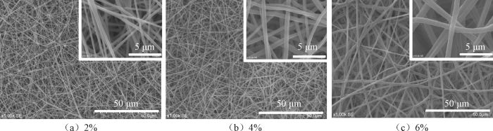
图1
不同PLLA质量分数下纳米纤维膜的扫描电镜照片
Fig.1
SEM images of nanofiber membranes with different PLLA mass fractions
图2
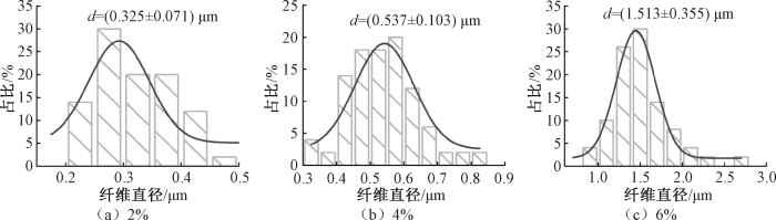
图2
不同PLLA质量分数下纳米纤维膜的直径分布图
Fig.2
Diameter distribution diagrams of nanofiber membranes with different PLLA mass fractions
图3
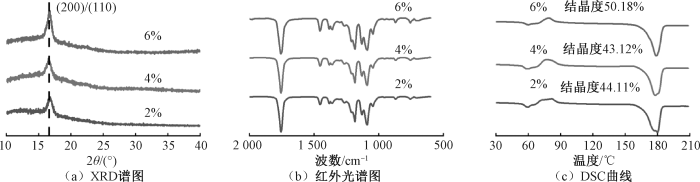
图3
不同PLLA质量分数下纳米纤维膜的结晶结构与化学结构
Fig.3
Crystalline structures and chemical structure of nanofiber membranes with different PLLA mass fractions. (a) XRD pattern; (b) FT-IR spectra; (c) DSC curves
图3(c)示出不同质量分数下纳米纤维膜的DSC曲线。由图可知,2%、4%、6%PLLA质量分数下纤维膜的结晶度分别为44.11%、43.12%、50.18%,这与XRD分析结果相吻合,证实高PLLA质量分数下更易制备出结晶度较高的纳米纤维膜。值得注意的是,2%PLLA质量分数样品的结晶度未出现明显下降,推测其原因是串珠结构的形成导致纤维膜中纤维直径分布不均,进而影响了纤维膜的整体结晶特性。
2.1.2 溶剂配比的影响
图4

图4
不同溶剂配比下纳米纤维膜的扫描电镜照片
Fig.4
SEM images of nanofiber membranes at different solvent ratios
图5
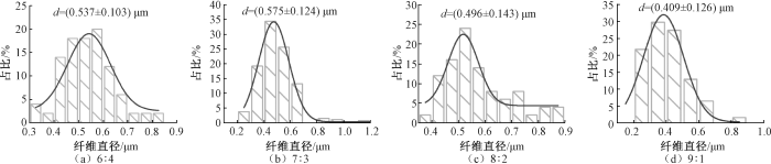
图5
不同溶剂配比下纳米纤维膜的直径分布图
Fig.5
Diameter distribution diagrams of nanofiber membranes with different solvent ratios
由图4(e)可见,10∶0溶剂配比下纤维以串珠形态存在,无法准确测定其直径分布,且纤维膜强度过低,难以满足后续结晶结构与化学特性分析的需求,因此后续表征仅针对其它4组溶剂配比展开。由图5可知:不同DCM与DMF体积比下,纤维平均直径d分别为(0.537 ± 0.103)μm(6∶4)、(0.575 ± 0.124)μm(7∶3)、(0.496 ± 0.143)μm(8∶2)、(0.409 ± 0.126)μm(9∶1)。尽管9∶1体积比下纤维直径最细,但易形成串珠结构。这是由于DCM是PLLA的良溶剂,当PLLA仅溶解于DCM时,溶液黏度较低,且在纺丝过程中DCM会快速挥发,最终导致纤维呈现串珠且多孔的形貌;而DMF的加入则可有效抑制DCM的快速挥发[26],因此随着DMF占比的提高,纤维直径呈现出相应的增大趋势。
图6
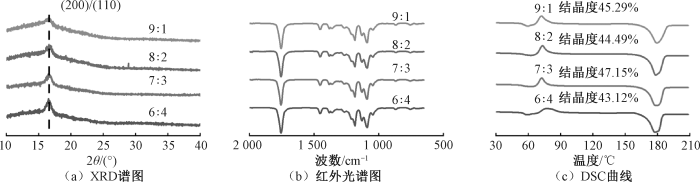
图6
不同溶剂配比下纳米纤维膜的结晶结构与化学结构
Fig.6
Crystalline structures and chemical structure of nanofiber membranes at different solvent ratios. (a) XRD pattern; (b) FT-IR spectra; (c) DSC curves
图6(c)示出不同溶剂配比下纳米纤维膜的DSC曲线。由图可知,6∶4、7∶3、8∶2、9∶1这4种溶剂体积比下,纤维膜的结晶度分别为43.12%、47.15%、44.49%、45.29%。结合上述数据可推断,改变溶剂体积比对PLLA纳米纤维膜的结晶度影响较小。
2.1.3 纺丝电压的影响
图7
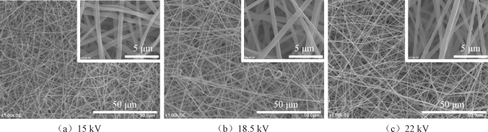
图7
不同纺丝电压下纳米纤维膜的扫描电镜照片
Fig.7
SEM images of nanofiber membranes under different electrospinning voltages
图8
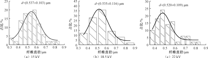
图8
不同纺丝电压下纳米纤维膜的直径分布图
Fig.8
Diameter distribution diagrams of nanofiber membranes under different electrospinning voltages
图9
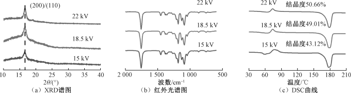
图9
不同纺丝电压下纳米纤维膜的结晶结构与化学结构
Fig.9
Crystalline structures and chemical structure of nanofiber membranes under different electrospinning voltages. (a) XRD pattern; (b) FT-IR spectra; (c) DSC curves
2.1.4 热处理温度的影响
图10
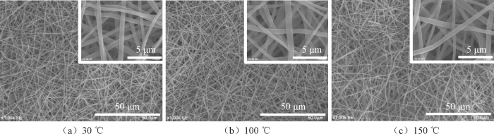
图10
不同热处理温度下纳米纤维膜的扫描电镜照片
Fig.10
SEM images of nanofiber membranes at different heat treatment temperatures
图11
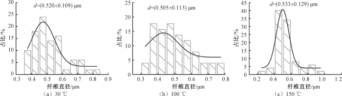
图11
不同热处理温度下纳米纤维膜的直径分布图
Fig.11
Diameter distribution diagrams of nanofiber membranes at different heat treatment temperatures
图12
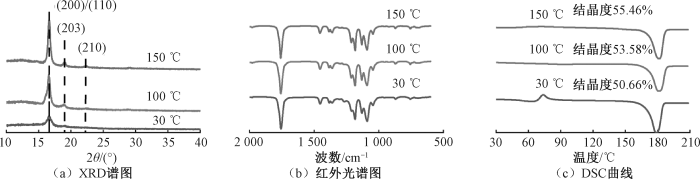
图12
不同热处理温度下纳米纤维膜的结晶结构与化学结构
Fig.12
Crystalline structures and chemical structure of nanofiber membranes at different heat treatment temperatures. (a) XRD pattern; (b) FT-IR spectra; (c) DSC curves
图12(b)示出不同热处理温度下纳米纤维膜的红外光谱图,在所有光谱中均未观察到对应PLLA β相的特征谱带。
图12(c)示出不同热处理温度下纳米纤维膜的DSC曲线。由图可知,当热处理温度为30、100、150 ℃时,纤维膜的结晶度分别为50.66%、53.58%、55.46%。该结果与XRD分析结果相一致,证实提高热处理温度可在一定程度上提升PLLA纳米纤维膜的结晶度。
2.2 纺丝参数对纤维膜压电性能的影响
固定施加压力87.7 N、频率1 Hz、负载电阻1×10⁹ Ω,测试各纺丝参数下纤维膜的压电输出性能,结果如图13所示。
图13
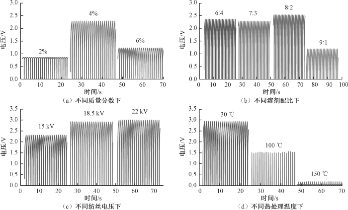
图13
不同纺丝条件下纳米纤维膜的输出电压
Fig.13
Output voltages of nanofiber membranes under different spinning conditions. (a) Under different mass fraction; (b) Under different solvent ratio; (c) Under different spinning voltage; (d) Under different heat treatment temperature
基于以上结果可知,纤维直径、纤维形貌和结晶度均显著影响PLLA纳米纤维膜的压电输出性能,具体表现如下:1)纤维直径变细源于聚合物链受到良好的拉伸作用,这一过程可促使偶极子高度取向排列,进而有利于获得高压电性能;2)压电PLLA纳米纤维膜的主要晶型为α相,该晶型晶体互连且紧密堆积,因此提高其结晶度有利于分子链规整排列,使偶极矩方向趋于一致,从而优化压电性能;而当α'晶型出现时,提高其结晶度不利于压电性能的提高;3)PLLA的压电性能并非单纯随结晶度的提高而增强,较细直径、光滑表面形貌和特定晶相这些因素对压电性能的影响程度高于结晶度的单一作用。
2.3 压电传感器的输出性能
图14示出以最优纺丝参数(PLLA质量分数4%、DCM与DMF体积比为6∶4、纺丝电压22 kV、热处理温度30 ℃)制备的PLLA压电传感器的输出性能。如图14(a)~(c)所示,该传感器的平均压电输出为2.933 V,平均短路电流为766.26 nA,平均输出电荷为1.22 nC,据此计算其电荷密度为1.95 μC/m2。从图14(d)可看出,压电传感器在连续击打2 500次后,压电输出仍保持稳定。由图14(e)可见,该传感器在8.3~186.4 kPa范围内可保持优异的线性度,灵敏度为0.014 V/kPa。在压电传感器的实际应用中,输出功率取决于负载本身的电阻,因此,对PLLA压电传感器在1×104~1×1010 Ω负载范围内的功率密度变化进行表征,结果如图14(f)所示。可知,当负载电阻为1×10⁹ Ω时,传感器达到最大功率密度,为4.23 mW/m2。
图14
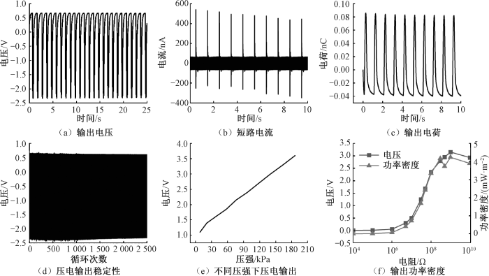
图14
PLLA压电传感器的输出性能
Fig.14
Output performance of PLLA piezoelectric sensor. (a) Output valtage; (b) Short-circuit current; (c) Output charge; (d) Stability of piezoelectric output; (e) Piezoelectric output under different pressures; (f) Output power density
2.4 压电传感器的体外模拟血管监测应用
图15
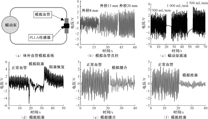
图15
PLLA压电传感器在不同血管模拟条件下获得的电压
Fig.15
Voltage obtained by PLLA piezoelectric sensor under different vascular simulation conditions. (a) In vitro blood vessel simulation system;(b) Simulated blood vessel diameter;(c) Peristaltic pump flow rate; (d) Simulated vessel occlusion;(e) Simulated suture;(f) Simulated thrombosis
图15(b)示出压电传感器在不同模拟血管直径下的监测结果。不同模拟血管直径对应的平均输出电压分别为2.14 V(外径8 mm)、3.34 V(外径13 mm)、4.39 V(外径20 mm)。分析可知,血管直径越粗,在相同血压下产生的形变越大,对应的传感器输出电压也越高。图15(c)示出压电传感器在不同脉动频率下的监测结果。通过改变蠕动泵的流速可模拟血管的搏动频率,不同流速下的平均输出电压分别为3.34 V(500 mL/min)、3.53 V(1 000 mL/min)、3.51 V(1 500 mL/min)。图15(d)示出压电传感器对血管阻塞的监测结果。采用镊子夹紧血管以模拟血管阻塞情况,结果显示,当血管受到阻塞时,传感器输出电压显著降低且呈现出不稳定波形;当阻塞解除、血管恢复正常后,输出电压也恢复至正常水平。图15(e)示出压电传感器对缝合血管的监测结果。将模拟血管剪开后用生物胶水重新黏合,模拟血管缝合场景,此时平均输出电压由正常血管的3.34 V降低至缝合后的2.25 V。这是由于重新黏合后血管形变能力下降,进而导致传感器输出电压减小。图15(f)示出压电传感器对血管栓塞的监测结果。在模拟血管内放置1颗与其直径相当的硬质石块模拟栓塞,此时平均输出电压由正常血管的3.34 V降低至栓塞后的0.65 V。这是由于栓塞后血管形变减小,导致输出电压降低。上述结果表明,该传感器可实现对模拟血管直径、脉动频率、血管狭窄、缝合血管、血栓阻塞的监测,在血管术后监测领域具有良好的应用潜力。
3 结论
本研究通过调控静电纺丝参数,系统探究了左旋聚乳酸(PLLA)纳米纤维膜的微观形貌、结晶结构、化学特性与压电输出性能之间的关系,结果表明:仅形成α晶型的PLLA纳米纤维膜表现出优异的压电性能;纤维直径、纤维形貌和结晶度均对纤维膜的压电输出强度有显著影响,其中纤维直径越细且无串珠结构的PLLA压电输出性能最优;在纤维形貌一定时,PLLA的压电输出随α相结晶度的提高而增大;但当纤维经热处理形成α'晶型时,其结晶度的提高反而会抑制压电性能;以最优纺丝参数(PLLA质量分数4%,DCM与DMF体积比为6∶4,纺丝电压22 kV,30 ℃干燥)制备的压电传感器,其输出电压达2.933 V、短路电流达766.26 nA、电荷密度达1.95 μC/m2、功率密度达4.23 mW/m2。该传感器在体外模拟血管监测中,表现出高输出电压和明显的波形差异,可实现对模拟血管直径、脉动频率、血管狭窄、缝合血管、血栓阻塞的监测,有望作为可植入柔性传感器用于血管疾病术后实时连续监测。
参考文献
Cell therapy for the treatment of heart disease: renovation work on the broken heart is still in progress
[J].DOI:10.1016/j.freeradbiomed.2020.12.444 URL [本文引用: 1]
Forecasting the future of cardiovascular disease in the United States: a policy statement from the American Heart Association
[J].
DOI:10.1161/CIR.0b013e31820a55f5
PMID:21262990
[本文引用: 1]

Cardiovascular disease (CVD) is the leading cause of death in the United States and is responsible for 17% of national health expenditures. As the population ages, these costs are expected to increase substantially.To prepare for future cardiovascular care needs, the American Heart Association developed methodology to project future costs of care for hypertension, coronary heart disease, heart failure, stroke, and all other CVD from 2010 to 2030. This methodology avoided double counting of costs for patients with multiple cardiovascular conditions. By 2030, 40.5% of the US population is projected to have some form of CVD. Between 2010 and 2030, real (2008$) total direct medical costs of CVD are projected to triple, from $273 billion to $818 billion. Real indirect costs (due to lost productivity) for all CVD are estimated to increase from $172 billion in 2010 to $276 billion in 2030, an increase of 61%.These findings indicate CVD prevalence and costs are projected to increase substantially. Effective prevention strategies are needed if we are to limit the growing burden of CVD.
体外循环心内直视术后急性心包堵塞17例报告
[J].
Report of 17 cases of acute pericardial obstruction after open heart surgery under cardiopulmonary bypass
[J].
用于心血管介入治疗的柔性MEMS电化学阻抗传感器
[J].
Flexible MEMS sensors based on electrochemical impedance method for percutaneous coronary interventions
[J].
Nanomaterials based flexible devices for monitoring and treatment of cardiovascular diseases (CVDs)
[J].DOI:10.1007/s12274-022-4551-8 [本文引用: 1]
Unconstrained piezoelectric vascular electronics for wireless monitoring of hemodynamics and cardiovascular health
[J].DOI:10.1002/smll.v20.3 URL [本文引用: 1]
A review of recent advances of piezoelectric poly-L-lactic acid for biomedical applications
[J].DOI:10.1016/j.ijbiomac.2024.133748 URL [本文引用: 1]
Modulation of piezoelectric properties in electrospun PLLA nanofibers for application-specific self-powered stem cell culture platforms
[J].DOI:10.1016/j.nanoen.2021.106444 URL [本文引用: 5]
甲壳素/聚乳酸压电复合膜的制备及性能研究
[J].
Research for preparation and properties of chitin/polylactic acid piezoelectriccomposite membrane
[J].
On the nanoscale mapping of the mechanical and piezoelectric properties of poly (L-lactic acid) electrospun nano-fibers
[J].
Fractionated crystallization kinetics and polymorphic homocrystalline structure of poly(L-lactic acid)/poly(D-lactic acid) blends: effect of blend ratio
[J].DOI:10.1007/s10118-022-2658-8 [本文引用: 1]
Poly(lactic acid) crystallization
[J].DOI:10.1016/j.progpolymsci.2012.07.005 URL [本文引用: 1]
Phase transition mechanism of poly(L-lactic acid) among the α, δ, and β forms on the basis of the reinvestigated crystal structure of the β form
[J].DOI:10.1021/acs.macromol.7b00272 URL [本文引用: 1]
Helix distortion and crystal structure of the α-form of poly(L-lactide)
[J].DOI:10.1021/ma0348674 URL [本文引用: 1]
Protective effect of resveratrol against cisplatin-induced ototoxicity in HEI-OC1 auditory cells
[J].
DOI:10.1016/j.ijporl.2014.11.008
PMID:25434479
[本文引用: 1]
Cisplatin is an effective chemotherapeutic drug, but it generates reactive oxygen species (ROS) that induce severe adverse effects such as ototoxicity. Resveratrol reportedly prevents oxidative stress-induced cell death. Thus, we hypothesized that the anti-oxidative effect of resveratrol could protect against cisplatin-induced ototoxicity. The present study examined the protective effect of resveratrol against cisplatin-induced ototoxicity in HEI-OC1 auditory cells.HEI-OC1 cells were pretreated with resveratrol at 1μM for 24h and then exposed to 15μM cisplatin for 48h. Resulting cytotoxicity was measured by the MTT method, and intracellular ROS was measured using flow cytometry. Protective effect of resveratrol was compared with other anti-oxidants.Pretreatment with resveratrol 1μM protected HEI-OC1 auditory cells against cisplatin-induced cytotoxicity and significantly reduced a cisplatin-induced increase in ROS. Resveratrol provided significant protection against 15μM cisplatin applied for 48h (50.8% cell viability in the cisplatin group vs. 57.6% in the cisplatin-plus-resveratrol group), and there was a 9% decrease in cisplatin-induced ROS associated with resveratrol.This is the study investigating the protective effects of resveratrol against cisplatin-induced ototoxicity in an auditory cell line. Resveratrol significantly reduced a cisplatin-induced increase in ROS and thereby inhibited cisplatin-induced cytotoxicity.Copyright © 2014 Elsevier Ireland Ltd. All rights reserved.
Dominant β-form of poly(L-lactic acid) obtained directly from melt under shear and pressure fields
[J].DOI:10.1021/acs.macromol.6b00595 URL [本文引用: 1]
Regulated crystallization and piezoelectric properties of bio-based poly(L-lactic acid)/diatomite composite fibers by electrospinning
[J].DOI:10.1007/s42114-024-01034-x [本文引用: 1]
Tailoring the morphology and crystallinity of poly(L-lactide acid) electrospun membranes
[J].DOI:10.1088/1468-6996/12/1/015001 URL
Poly-L-lactic acid/graphene electrospun composite nanofibers for wearable sensors
[J].DOI:10.1002/ente.v8.5 URL [本文引用: 1]
Key factors affecting the piezoelectric response of poly-L-lactic acid electrospun fibers
[J].DOI:10.1016/j.polymer.2025.128286 URL [本文引用: 1]
Enthalpy of melting of α'- and α-crystals of poly(L-lactic acid)
[J].DOI:10.1016/j.eurpolymj.2015.07.024 URL [本文引用: 2]
Comparison study of hydrolytic degradation behaviors between α'-and α-poly(L-lactide)
[J].DOI:10.1016/j.polymdegradstab.2017.12.014 URL [本文引用: 2]
Tunable piezoelectric PLLA nanofiber membranes for enhanced mandibular repair with optimal self-powering stimulation
[J].
Study of field-induced chain conformation transformation in poly(L-lactic acid) based piezoelectric film by infrared spectroscopy
[J].
DOI:10.1063/1.4965716
URL
[本文引用: 1]
In this investigation, the chain conformation transformation of the piezoelectric polymer of a poly(L-Lactic Acid) (PLLA) film was analyzed under an electric field for the first time using infrared spectroscopy. It is revealed that the piezoelectric shear mode coefficient d14 (∼10 pC/N) of a stretched α form PLLA film mainly comes from the rotation of C O dipoles inside the polymer main chain. The reorientation of the dipoles causes the deformation of the crystal structure, which corresponds to a shear mode strain macroscopically in the PLLA film along a 45° direction to the polymer length. The back-bone of the molecular chain keeps its own conformation of a 103 helix under an external field up to 100 MV/m.
Polymorphous crystallization and multiple melting behavior of poly(l-lactide): molecular weight dependence
[J].DOI:10.1021/ma071258d URL [本文引用: 1]
聚乳酸基压电材料的研究和应用
[J].
Research and application of polylactic acid-based piezoelectric materials
[J].
Decoupling the effects of crystallinity and orientation on the shear piezoelectricity of polylactic acid
[J].DOI:10.1002/polb.v49.21 URL [本文引用: 1]

 京公网安备11010502044800号


