纺织学报 ›› 2026, Vol. 47 ›› Issue (03): 118-128.doi: 10.13475/j.fzxb.20250902201
郭一铭1,2, 喻爽1,2, 赵帆1,2( ), 王富军1,2
GUO Yiming1,2, YU Shuang1,2, ZHAO Fan1,2(), WANG Fujun1,2
摘要:
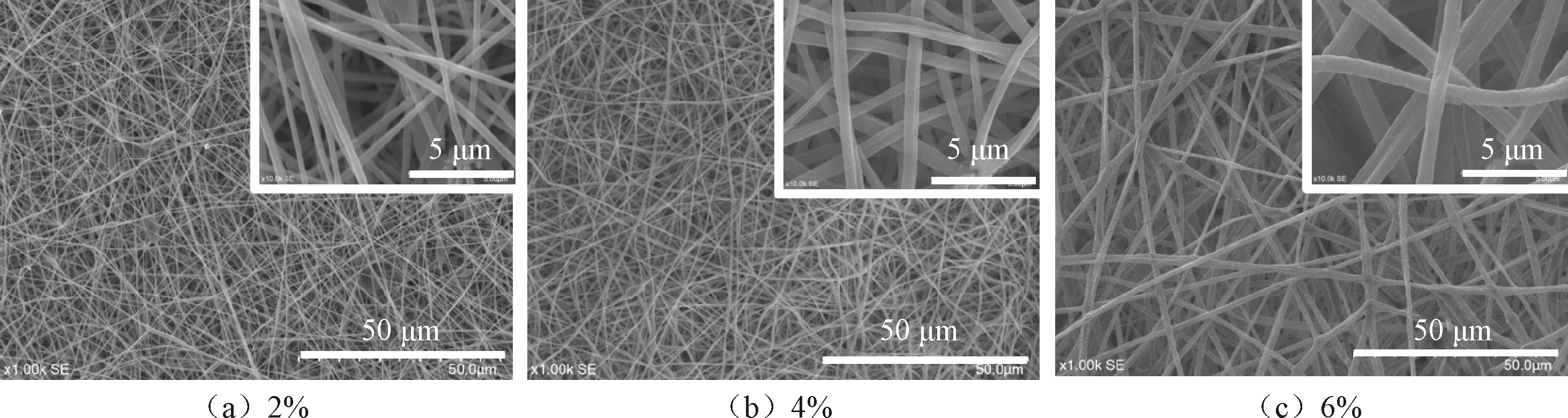
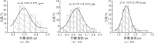
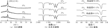
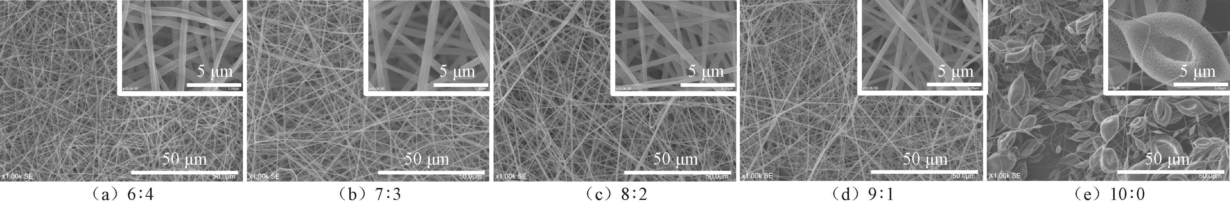
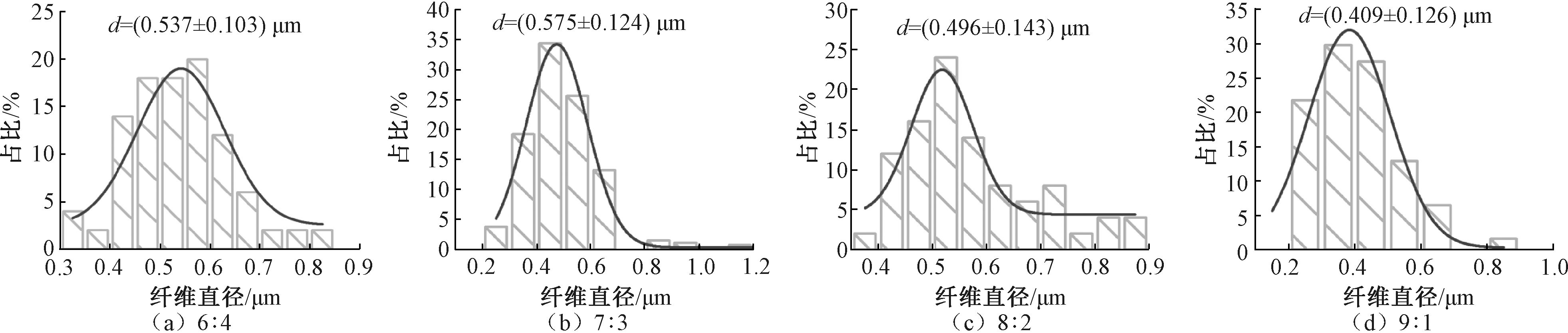
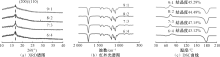
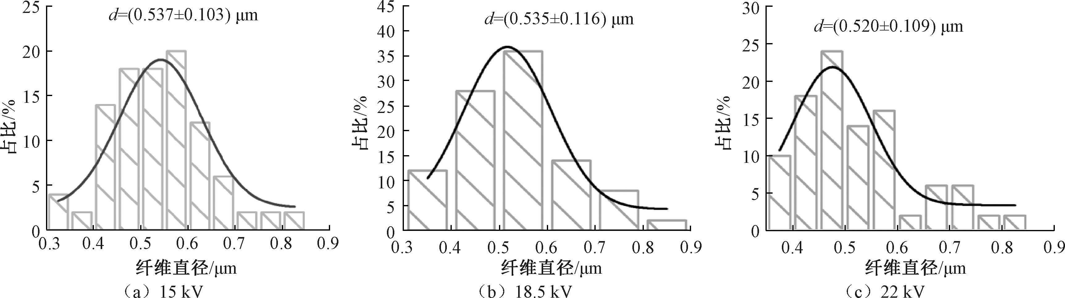
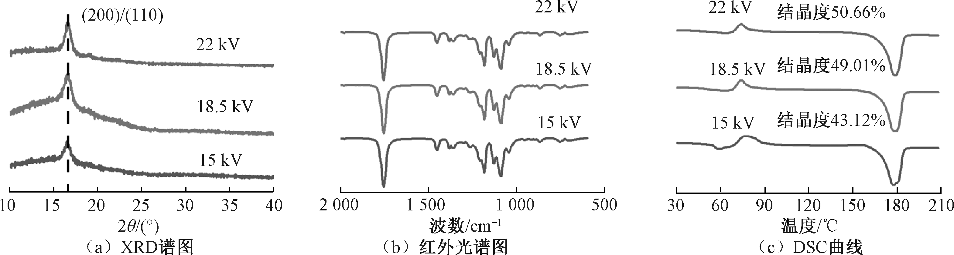
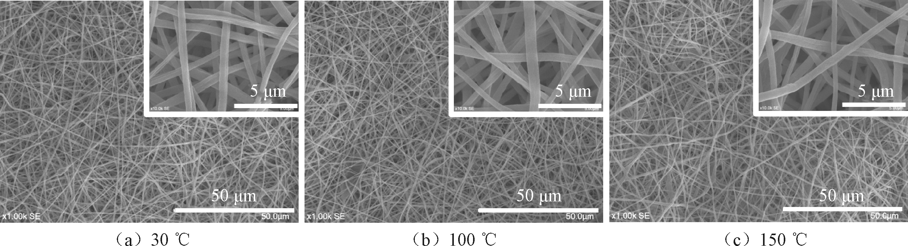
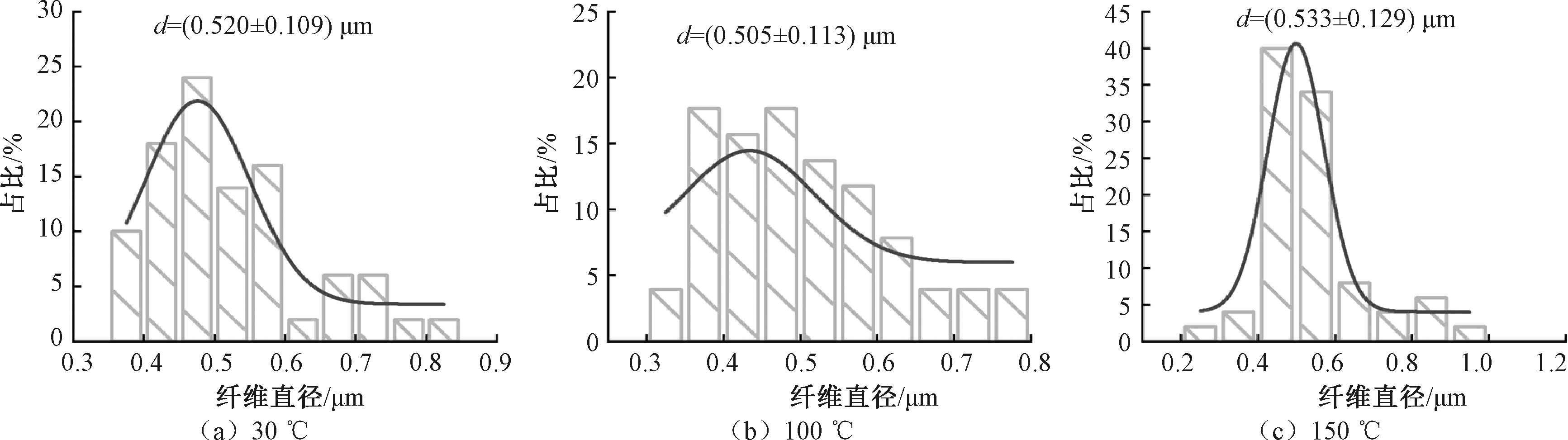
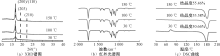
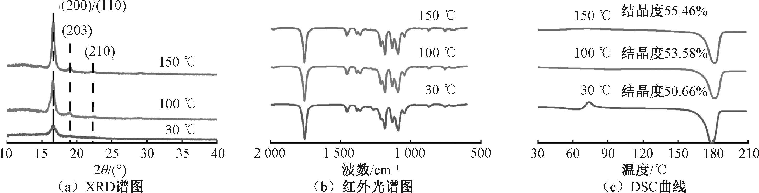
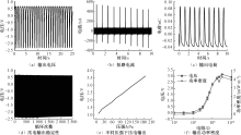
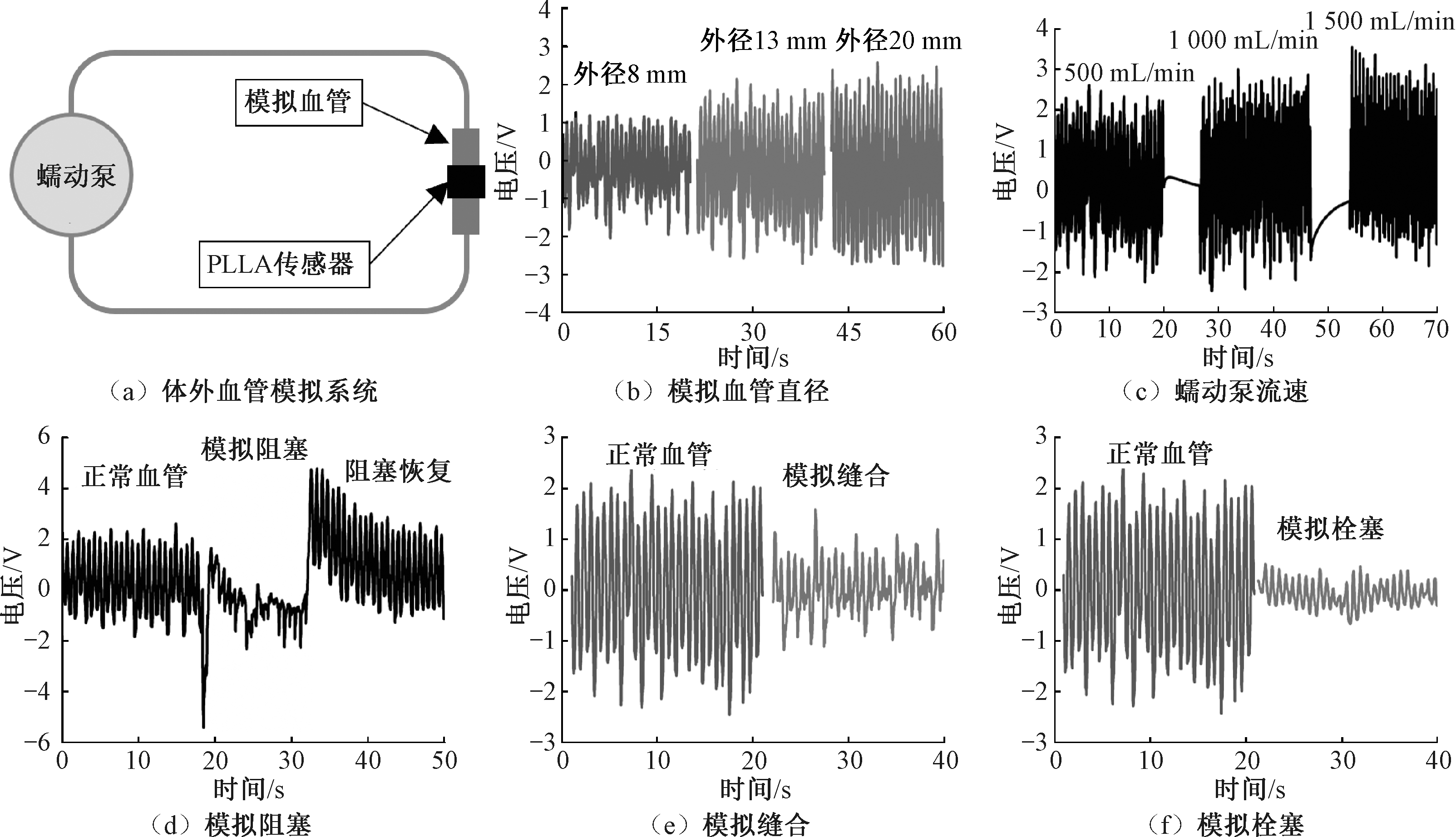
为解决现有血管疾病术后监测方法如血管造影、核磁共振等需要大型设备且繁琐操作、缺乏持续监测方法的问题,采用左旋聚乳酸(PLLA)开发了可持续监测血管疾病修复状态的柔性植入式传感器。针对PLLA压电输出性能较低且调控机制尚不明确的问题,通过改变静电纺丝参数和热处理参数系统探究了纤维形貌和分子结构对PLLA纳米纤维膜压电性的影响规律。结果表明:纤维形貌和结晶度都会影响PLLA纳米纤维膜的压电输出性能;纤维直径越细且无串珠结构的PLLA压电输出性能最好;在纤维形貌良好时,PLLA的输出压电随α相晶型结晶度的提高而增大;纤维膜经热处理后出现α'晶型,提高了其结晶度,但导致压电性能降低;最优参数下PLLA纳米纤维膜的输出电压为2.933 V(87.7 N,1 Hz),电流为766.26 nA,电荷密度为1.95 μC/m2,最大输出功率密度为4.23 mW/m2,且在8.3~186.4 kPa范围内保持优异的线性度。体外血管模拟结果证实,该柔性传感器可有效感知环状脉动应变。
中图分类号:
| [1] |
MADEDDU P. Cell therapy for the treatment of heart disease: renovation work on the broken heart is still in progress[J]. Free Radical Biology and Medicine, 2021, 164(20): 206-222.
doi: 10.1016/j.freeradbiomed.2020.12.444 |
| [2] |
HEIDENREICH P A, TROGDON J G, KHAVJOU O A, et al. Forecasting the future of cardiovascular disease in the United States: a policy statement from the American Heart Association[J]. Circulation, 2011, 123(8): 933-944.
doi: 10.1161/CIR.0b013e31820a55f5 pmid: 21262990 |
| [3] | 王清宇. 血管搭桥术后静脉血管动脉化动物模型建立及形成进程研究[D]. 贵阳: 贵州大学, 2010:1-10. |
| WANG Qingyu. Study on animal model establishment and time course of arterializations of implanted vein after vascular bypass surgery[D]. Guiyang: Guizhou University, 2010:1-10. | |
| [4] | 张亚军, 张霖, 火旭东, 等. 体外循环心内直视术后急性心包堵塞17例报告[J]. 实用临床医药杂志, 2003, 7(2): 156. |
| ZHANG Yajun, ZHANG Lin, HUO Xudong, et al. Report of 17 cases of acute pericardial obstruction after open heart surgery under cardiopulmonary bypass[J]. Journal of Jiangsu Clinical Medicine, 2003, 7(2): 156. | |
| [5] | 唐龙军, 杨斌, 陈翔, 等. 用于心血管介入治疗的柔性MEMS电化学阻抗传感器[J]. 中国科技论文, 2017, 12(8): 957-961. |
| TANG Longjun, YANG Bin, CHEN Xiang, et al. Flexible MEMS sensors based on electrochemical impedance method for percutaneous coronary interventions[J]. China Sciencepaper, 2017, 12(8): 957-961. | |
| [6] |
XIANG Z H, HAN M D, ZHANG H X. Nanomaterials based flexible devices for monitoring and treatment of cardiovascular diseases (CVDs)[J]. Nano Research, 2023, 16(3): 3939-3955.
doi: 10.1007/s12274-022-4551-8 |
| [7] |
TANG C Y, LIU Z R, HU Q H, et al. Unconstrained piezoelectric vascular electronics for wireless monitoring of hemodynamics and cardiovascular health[J]. Small, 2024, 20(3): 2304752.
doi: 10.1002/smll.v20.3 |
| [8] |
ZHANG S W, ZHANG H S, SUN J T, et al. A review of recent advances of piezoelectric poly-L-lactic acid for biomedical applications[J]. International Journal of Biological Macromolecules, 2024, 276(1): 133748.
doi: 10.1016/j.ijbiomac.2024.133748 |
| [9] |
TAI Y Y, YANG S, YU S, et al. Modulation of piezoelectric properties in electrospun PLLA nanofibers for application-specific self-powered stem cell culture platforms[J]. Nano Energy, 2021, 89: 106444.
doi: 10.1016/j.nanoen.2021.106444 |
| [10] | 刘古月, 张博韬, 谷潇夏, 等. 甲壳素/聚乳酸压电复合膜的制备及性能研究[J]. 北京服装学院学报(自然科学版), 2024, 44(1): 10-18. |
| LIU Guyue, ZHANG Botao, GU Xiaoxia, et al. Research for preparation and properties of chitin/polylactic acid piezoelectriccomposite membrane[J]. Journal of Beijing Institute of Fashion Technology (Natural Science Edition), 2024, 44(1): 10-18. | |
| [11] | CUONG N T, BARRAU S, DUFAY M, et al. On the nanoscale mapping of the mechanical and piezoelectric properties of poly (L-lactic acid) electrospun nano-fibers[J]. Applied Sciences, 2020, 10(2), 652. |
| [12] |
XIANG W K, XIE Q, XU S S, et al. Fractionated crystallization kinetics and polymorphic homocrystalline structure of poly(L-lactic acid)/poly(D-lactic acid) blends: effect of blend ratio[J]. Chinese Journal of Polymer Science, 2022, 40(6): 567-575.
doi: 10.1007/s10118-022-2658-8 |
| [13] |
SAEIDLOU S, HUNEAULT M A, LI H B, et al. Poly(lactic acid) crystallization[J]. Progress in Polymer Science, 2012, 37(12): 1657-1677.
doi: 10.1016/j.progpolymsci.2012.07.005 |
| [14] |
WANG H, ZHANG J M, TASHIRO K. Phase transition mechanism of poly(L-lactic acid) among the α, δ, and β forms on the basis of the reinvestigated crystal structure of the β form[J]. Macromolecules, 2017, 50(8): 3285-3300.
doi: 10.1021/acs.macromol.7b00272 |
| [15] |
SASAKI S, ASAKURA T. Helix distortion and crystal structure of the α-form of poly(L-lactide)[J]. Macromolecules, 2003, 36(22): 8385-8390.
doi: 10.1021/ma0348674 |
| [16] |
LEE S H, KIM H S, AN Y S, et al. Protective effect of resveratrol against cisplatin-induced ototoxicity in HEI-OC1 auditory cells[J]. International Journal of Pediatric Otorhinolaryngology, 2015, 79(1): 58-62.
doi: 10.1016/j.ijporl.2014.11.008 pmid: 25434479 |
| [17] |
RU J F, YANG S G, ZHOU D, et al. Dominant β-form of poly(L-lactic acid) obtained directly from melt under shear and pressure fields[J]. Macromolecules, 2016, 49(10): 3826-3837.
doi: 10.1021/acs.macromol.6b00595 |
| [18] |
ZHAO J, LI T, SUN H Y, et al. Regulated crystallization and piezoelectric properties of bio-based poly(L-lactic acid)/diatomite composite fibers by electrospinning[J]. Advanced Composites and Hybrid Materials, 2024, 7(6): 218.
doi: 10.1007/s42114-024-01034-x |
| [19] |
RIBEIRO C, SENCADAS V, COSTA C M, et al. Tailoring the morphology and crystallinity of poly(L-lactide acid) electrospun membranes[J]. Science and Technology of Advanced Materials, 2011, 12(1): 015001.
doi: 10.1088/1468-6996/12/1/015001 |
| [20] |
LI X, CHEN S, ZHANG X Y, et al. Poly-L-lactic acid/graphene electrospun composite nanofibers for wearable sensors[J]. Energy Technology, 2020, 8(5): 1901252.
doi: 10.1002/ente.v8.5 |
| [21] |
RENTERO C, AMORÍN H, JIMÉNEZ R, et al. Key factors affecting the piezoelectric response of poly-L-lactic acid electrospun fibers[J]. Polymer, 2025, 325(22): 128286.
doi: 10.1016/j.polymer.2025.128286 |
| [22] |
RIGHETTI M C, GAZZANO M, DI LORENZO M L, et al. Enthalpy of melting of α'- and α-crystals of poly(L-lactic acid)[J]. European Polymer Journal, 2015, 70: 215-220.
doi: 10.1016/j.eurpolymj.2015.07.024 |
| [23] |
ZHANG N, YU X, DUAN J, et al. Comparison study of hydrolytic degradation behaviors between α'-and α-poly(L-lactide)[J]. Polymer Degradation and Stability, 2018, 148: 1-9.
doi: 10.1016/j.polymdegradstab.2017.12.014 |
| [24] | CHEN S, WANG X Q, ZHANG D, et al. Tunable piezoelectric PLLA nanofiber membranes for enhanced mandibular repair with optimal self-powering stimulation[J]. Regenerative Biomaterials, 2024, 12: rbae150. |
| [25] |
DU X Y, ZHAO C L, ZHANG J X, et al. Study of field-induced chain conformation transformation in poly(L-lactic acid) based piezoelectric film by infrared spectroscopy[J]. Journal of Applied Physics, 2016, 120(16): 164101.
doi: 10.1063/1.4965716 |
| [26] | 张海杨. 可生物降解的高压电性镁基PLLA复合导管在神经再生中的应用研究[D]. 南京: 南京理工大学, 2021:1-10. |
| ZHANG Haiyang. The application of biodegradable high piezoelectricl magnesium-based PLLA composite conduit in nerve regeneration[D]. Nanjing: Nanjing University of Science and Technology, 2021:1-10. | |
| [27] |
PAN P J, KAI W H, ZHU B, et al. Polymorphous crystallization and multiple melting behavior of poly(l-lactide): molecular weight dependence[J]. Macromolecules, 2007, 40(19): 6898-6905.
doi: 10.1021/ma071258d |
| [28] | 段瑞侠, 陈金周, 刘文涛, 等. 聚乳酸基压电材料的研究和应用[J]. 材料导报, 2022, 36(10): 215-222. |
| DUAN Ruixia, CHEN Jinzhou, LIU Wentao, et al. Research and application of polylactic acid-based piezoelectric materials[J]. Materials Reports, 2022, 36(10): 215-222. | |
| [29] |
LOVELL C S, FITZ-GERALD J M, PARK C. Decoupling the effects of crystallinity and orientation on the shear piezoelectricity of polylactic acid[J]. Journal of Polymer Science Part B: Polymer Physics, 2011, 49(21): 1555-1562.
doi: 10.1002/polb.v49.21 |
| [1] | 冯晓莉, 宫钧耀, 夏良君, 徐卫林. 磁电式柔性传感器研究进展[J]. 纺织学报, 2026, 47(03): 107-117. |
| [2] | 孙小芸, 岳程飞, 张如全. 基于激光诱导石墨烯的柔性温度传感器制备及其性能[J]. 纺织学报, 2026, 47(03): 129-138. |
| [3] | 陈泳良, 杨潇, 王朝荣, 黄俊鸿, 李彦, 王璐. 静电纺丝-恒应力退火协同构建的湿态稳定型聚乳酸/I型胶原肩袖补片[J]. 纺织学报, 2026, 47(03): 60-69. |
| [4] | 李好义, 田鑫哲, 张毅, 牟文英, 张超, 赵千龙, 杨卫民. 导电各向异性复合心脏补片的熔体静电纺丝/直写构建及体外评价[J]. 纺织学报, 2026, 47(03): 70-76. |
| [5] | 刘金枝, 赵回汇, 吴焕友, 张建明, 高晶. 壳聚糖/聚己内酯取向纳米纤维膜的结构调控与物理引导作用[J]. 纺织学报, 2026, 47(03): 9-17. |
| [6] | 张曼琦, 孙艳丽, 张晓茹, 李博, 刘哲. 共轭静电纺双模态调温织物的制备及其性能[J]. 纺织学报, 2026, 47(02): 153-161. |
| [7] | 孔珂欣, 张怡帆, 卢哲, 王哲. 载钴钌原子无机微纳米纤维的制备及其电催化水分解性能[J]. 纺织学报, 2026, 47(02): 26-36. |
| [8] | 王世杰, 孙辉, 于斌. 聚乙烯醇/牡丹皮提取物复合纳米静电纺丝膜的制备及其抗菌性能[J]. 纺织学报, 2026, 47(02): 56-64. |
| [9] | 孔艳辉, 张琳萍, 毛志平, 徐红. 甲基丙烯酰化明胶纤维膜的制备及其止血性能[J]. 纺织学报, 2026, 47(01): 1-10. |
| [10] | 刘一鸣, 李琳, 杜鲜晶, 刘攀, 殷霞, 田明伟. 内螺旋结构弹性导电纱线的制备及其应变不敏感性能的调控[J]. 纺织学报, 2026, 47(01): 115-122. |
| [11] | 邵剑波, 岳欣琰, 陈雨, 韩潇, 洪剑寒. 全针织结构多模态柔性电容传感器的构筑及其传感性能[J]. 纺织学报, 2026, 47(01): 123-131. |
| [12] | 赵婧雯, 袁香楠, 高晶, 王璐. 聚丙烯腈-普鲁士蓝/月桂酸/环丙沙星光热响应性抗菌敷料的制备及其性能[J]. 纺织学报, 2026, 47(01): 20-28. |
| [13] | 王世豪, 徐晓禹, 郑挺, 王金星, 姚德刚, 王俊, 叶翔宇, 田慧, 李婷, 朱斐超. 碳纤维非织造材料的研究应用及展望[J]. 纺织学报, 2026, 47(01): 240-249. |
| [14] | 胡崴琳, 白洁, 刘丹, 白濛, 李娟, 李启正. 基于机器学习模型的电子纺织品研究进展[J]. 纺织学报, 2026, 47(01): 268-276. |
| [15] | 罗家俊, 何耀权, 赵振鸿, 黎锦稻, 赵景, 黄钢, 王先锋. 苯乙烯-乙烯-丁烯-苯乙烯/氟化聚酰亚胺防水透湿纤维膜的制备及其性能[J]. 纺织学报, 2026, 47(01): 38-45. |
|
||
 京公网安备11010502044800号