



皮肤是人体重要的屏障,可护卫身体免受环境危害和微生物威胁,同时调节生理稳态[1],但皮肤也易受到烧伤、手术、外伤等伤害,形成急性或慢性伤口[2]。其中慢性伤口有很高的细菌感染风险,会使伤口恶化并阻碍伤口愈合过程[3]。伤口微环境的pH值是反映其愈合状态的重要指标:健康皮肤表面呈弱酸性(pH=5.5),有利于抑制微生物生长;当感染发生时,因细菌代谢与组织液成分改变,伤口pH值常升高至7.2的碱性范围[4],因此,实时、直观监测伤口pH值变化,对早期识别感染、及时干预具有重要意义[5]。水凝胶因与细胞外基质结构相似,且具备维持创面湿润微环境、吸收伤口渗出液、促进氧交换、防止继发性损伤及生物降解性等特性[6],成为一种性能优异的功能性敷料。在水凝胶中负载pH响应变色材料,通过水凝胶颜色的变化直观反映伤口pH值,进而反映伤口状态,是监测伤口最简单经济的方式之一。
目前,用于伤口pH值监测的指示剂主要为溴百里酚蓝(BTB)和姜黄素等合成或天然小分子。BTB作为合成染料,其潜在的细胞毒性与生物降解性欠佳的问题引发了对长期敷用安全性的担忧,同时其单一的pH值指示功能也无法满足实际的需求[7];姜黄素虽为天然产物,但其水溶性差,且在实际伤口敷料应用中变色对比度不够显著,限制了其监测可靠性[8];花青素作为水凝胶敷料的pH值指示剂,不仅生物相容性优、变色效果显著,而且能赋予水凝胶优秀的抗氧化性能[9]。纳米纤维素来源广泛,具有高强度、高比表面积和表面性能可调控等优势,是制备水凝胶敷料的理想材料[10]。羧甲基壳聚糖(CMCS)继承了壳聚糖优异的抗菌性、生物相容性与可生物降解性,并因其羧甲基基团而增强了水溶性,常被作为抗菌水凝胶的主要材料[11]。
1 实验部分
1.1 实验材料
纤维素纳米纤维溶液(CNF,质量分数1.0%),愉悦家纺有限公司;羧甲基壳聚糖(CMCS,取代度≥90%)、蓝莓花青素(BA,纯度25%),上海阿拉丁生化科技股份有限公司;高碘酸钠(NaIO4,分析纯)、乙二醇(分析纯),国药集团化学试剂有限公司;金黄色葡萄球菌(ATCC25922)、大肠埃希菌(DH10B),上海鲁威微生物科技有限公司;人包皮成纤维细胞(HFF),中国科学院典型培养库;CCK-8试剂盒,碧云天生物技术有限公司;磷酸盐缓冲液(PBS)、1,1-二苯基-2-三硝基苯肼(DPPH),福州飞净生物科技有限公司。
1.2 试样的制备
1.2.1 双醛基纳米纤维素溶液的制备
纳米纤维素醛基化改性参考文献[14]的方法。取200 mL质量分数为1%的CNF悬浮液,经超声波分散1 h后,与溶解了7.96 g NaIO4的80 mL水溶液混合。用PBS调节pH值至4.5,于45 ℃避光搅拌反应4 h。随后加入等量乙二醇猝灭反应30 min,最后将产物在去离子水中透析3 d(每8 h换水1次)。将透析后的双醛基纳米纤维素(DACNF),使用SCIENTZ-18N冷冻干燥机(宁波新芝生物科技有限公司)进行冷冻干燥,得到DACNF粉末,取1 g双醛基纳米纤维素加入到99 mL去离子水中,使用JY92-ⅡDN超声波细胞破碎机(宁波新芝生物科技有限公司)分散,制得DACNF分散液。
1.2.2 DACNF-BA混合溶液及CMCS溶液的制备
将BA添加到DACNF溶液中,用细胞破碎机分散后配制成质量分数分别为0%、0.2%、0.25%、0.3%、0.35%的DACNF-BA混合溶液。称取4 g CMCS,加入96 mL去离子水,50 ℃下搅拌3 h,制得CMCS溶液。
1.2.3 水凝胶的制备
将不同BA质量分数(0%、0.1%、0.125%、0.15%、0.175%)的DACNF-BA混合溶液与CMCS溶液等体积混合,搅拌均匀,离心20 min后,倒入培养皿中放置24 h,形成稳定水凝胶,根据BA含量将样品依次记作1#、2#、3#、4#、5#。
1.3 测试与表征
1.3.1 化学结构表征
使用Antaris II傅里叶变换近红外光谱仪(赛默飞世尔科技公司)对CNF、DACNF、CMCS以及冷冻干燥后的水凝胶样品进行表征分析,扫描范围为4 000~500 cm-1。
1.3.2 结晶结构表征
使用Bruker D8 X射线衍射仪(德国布鲁克AXS有限公司)分析CNF、DACNF、CMCS以及冷冻干燥后的水凝胶样品的结晶结构,扫描速率为1(°)/min,扫描范围(2θ)为5°~60°,电压为20 kV,电流为20 mA。结晶度(C)的计算公式为
式中:I200为结晶区衍射峰的强度,2θ约22.6°时衍射角的最大强度;Iam为无定形区衍射峰的强度,2θ约18.7°时衍射峰的强度。
1.3.3 微观形貌观察
将水凝胶样品冷冻干燥、镀金后,利用FLEX1000扫描电子显微镜(日本日立高新技术集团有限公司)观察其截面微观形貌和结构。
1.3.4 冻干水凝胶吸水性能测试
首先称取冻干样品质量(记为W0),然后将其分别浸入pH值为7.2和5.5的0.01 mol/L PBS中。最后在预定时间取出样品,用滤纸吸干表面缓冲液后再次称量(记为Wt)。每个样品测试5次,结果取平均值。吸水率(W)的计算公式为
1.3.5 水蒸气透过率测试
根据YY/T 0471.2—2004《接触性创面敷料试验方法 第2部分:透气膜敷料水蒸气透过率》,对水凝胶的水蒸气透过率进行测试,每个样品测试5次,结果取平均值。将厚度1 mm的水凝胶制为直径2 cm的圆形试样,测试容器中加入足量的水,使液面与放置后的样品间的距离为(5±1) mm。称量并记录样品、容器和水的质量,放入37 ℃的THZ-100恒温振荡箱(上海一恒科学仪器有限公司)中,样品正放,24 h后再次称量并记录样品、容器和水的质量。水蒸气透过率(V)的计算公式为
式中:W1为容器中水的初始质量,g;W2为24 h后容器中水的质量,g;S为有效实验面积,m2;T为实验时间,本次测试为24 h。
1.3.6 抗氧化性能测试
采用DPPH自由基清除法评估水凝胶的抗氧化性能。具体步骤为:将40 mg冻干水凝胶置于pH值为7.2的40 mL PBS中。取1 mL PBS浸泡液加入DPPH-乙醇溶液中,室温下避光反应30 min后,使用UV2600紫外-可见光谱仪(岛津仪器(苏州)有限公司)测定波长为517 nm处的紫外吸光度,每个样品测试5次,结果取平均值。对照组是PBS和DPPH-乙醇溶液的混合溶液。DPPH自由基清除率(S)的计算公式为
式中:AS为样品吸光度;AC为对照组吸光度。
1.3.7 抗菌性能测试
采用金黄色葡萄球菌(革兰阳性菌)和大肠埃希菌(革兰阴性菌)为模型,表征水凝胶的抗菌活性。具体步骤为:将20 mg冻干水凝胶样品经紫外线灭菌1 h后,与1 mL浓度为106 CFU/mL的菌液混合。将混合物于37 ℃静置培养12 h后,用无菌PBS对菌液进行连续10倍稀释。取100 μL特定稀释度的菌液涂布于琼脂平板,再次于37 ℃静置培养18 h,最后记录菌落数。每个样品测试5次,结果取平均值。抑菌率(A)的计算公式为
式中:QS样品组菌落数;QC对照组菌落数。
1.3.8 pH响应变色指示性能测试
将1 mm厚的水凝胶片状样品裁成圆形,分别加入pH值为5.5(模拟人体健康皮肤)和pH值为7.2(模拟慢性创面)的PBS溶液中,在温度为37 ℃,转速为120 r/min恒温培养箱中浸泡0、20、40、60、90 min后,观察水凝胶颜色变化,并用Datacolor 850测色配色仪(德塔颜色商贸(上海)有限公司)记录水凝胶红绿值(a)、黄蓝值(b)和总色差(ΔE)。
1.3.9 细胞毒性测试
为评估水凝胶的生物相容性,采用人包皮成纤维细胞(HFF)作为体外模型。将样品经紫外线灭菌处理后置于温度为37 ℃、体积分数为5%的CO2湿润培养环境中预孵育3 h。随后将100 μL HFF细胞悬液接种至48孔板,在相同条件下培养约3 h,使细胞充分贴壁。参考GB/T 16886.5—2017《医疗器械生物学评价 第5部分:体外细胞毒性试验》,不同水凝胶按照0.2 g/mL在37 ℃完全培养基中浸提12 h制备浸提液。贴壁完成后,每孔更换为200 μL新鲜培养基,然后加入100 μL水凝胶浸提液继续培养。之后更换为含有CCK-8试剂的100 μL新鲜培养基,于37 ℃下反应4 h。反应结束后,测定450 nm波长处各孔吸光度值,以评价细胞增殖能力。同时使用活/死细胞染色试剂盒对细胞生存情况进行检测,并在培养1、3、7 d后通过显微镜观察并记录细胞形态变化。细胞活力(VC)的计算公式为
式中:Ac为配有细胞、CCK-8溶液和样品的孔的吸光度;Ab为配有培养基、CCK-8溶液但没有细胞的孔的吸光度;Ad为配有细胞、CCK-8溶液但没有样品的孔的吸光度。
2 结果与讨论
2.1 化学结构分析
纳米纤维素的醛基化是合成水凝胶的必要条件,因此通过红外光谱对CNF、DACNF、CMCS、BA和水凝胶的化学结构进行分析,结果如图1所示。
图1
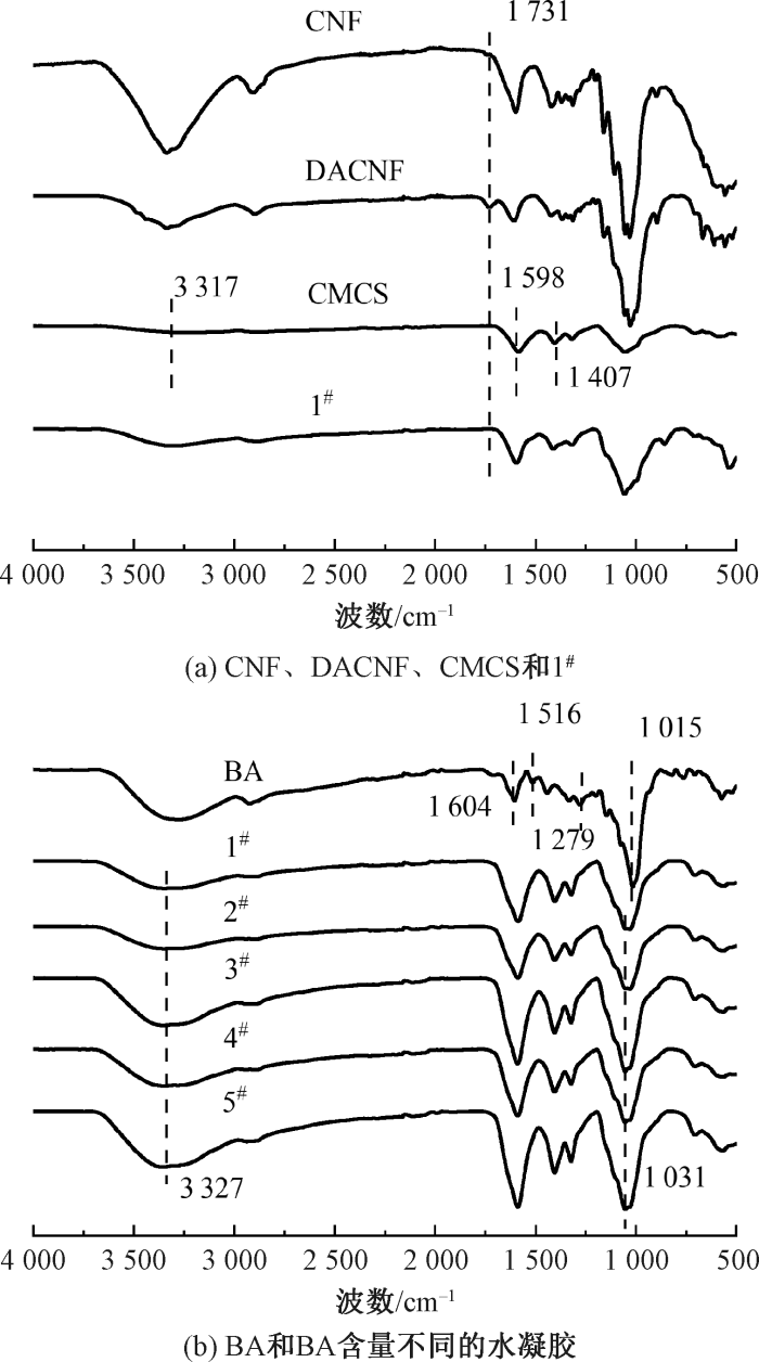
图1
不同试样的红外光谱图
Fig.1
FT-IR spectra of different samples. (a) CNF,DACNF, CMCS and 1#;(b) BA and hydrogels with different BA contents
由图1(a)可知,在1 731 cm-1处出现显著的C=O伸缩振动峰,证实CNF已被氧化为DACNF。CMCS谱图上3 317 cm-1处的宽峰归属于—OH/—NH2伸缩振动,1 598、1 407 cm-1处的吸收峰则分别对应 COO-的不对称和对称伸缩振动。这些官能团,特别是氨基,为DACNF的醛基通过席夫碱反应与之形成共价交联提供了反应位点。相较于DACNF,水凝胶1#的红外谱图中1 731 cm-1处的醛基吸收峰已消失,可推断DACNF的醛基与CMCS的氨基已通过席夫碱键合,反应成功[15]。在图1(b)所示蓝莓花青素(属黄酮类)的红外谱图中,1 604、1 516 cm-1处的特征峰归属于芳香环C=C的伸缩振动[16];1 279 cm-1处的吸收峰归属于黄酮吡喃环的伸缩振动;1 015 cm-1处的峰则归属于芳香环的C—H弯曲振动[17]。根据图示分析,在水凝胶中添加BA后,3 327 cm-1处的拉伸振动峰发生位移且增强,同时1 031 cm-1处的吸收峰也出现了位移和增强。这表明BA的加入引起新的氢键形成,并导致氢键结构的变化[18]。
2.2 结晶结构分析
为研究化学改性和凝胶化过程对纳米纤维素和水凝胶晶体结构的影响,对材料的晶体结构进行测试分析,结果如图2所示。可见,CNF的特征衍射峰(2θ为16.5°、22.7°处)在被高碘酸钠氧化为DACNF后位置不变,表明晶体结构未发生改变,但其结晶度由77.1%显著降低至56.9%,说明氧化作用部分破坏了规整的结晶区,并与吡喃葡萄糖环的断裂重组有关。同时,半结晶的CMCS在2θ为11.0°和20.4°处显示出其特征衍射峰。在1#水凝胶的XRD谱图中,上述所有晶体衍射峰均消失,并在2θ为20°附近出现宽化弥散峰,这证实了席夫碱反应彻底破坏了DACNF与CMCS的晶体结构,形成非晶态的交联网络。
图2
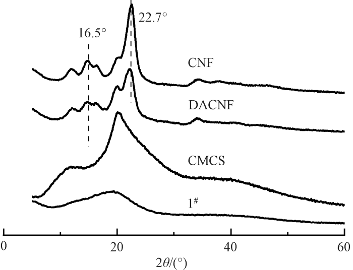
图2
CNF、DACNF、CMCS、1#的X射线衍射谱图
Fig.2
X-ray diffraction patterns of CNF,DACNF,CMCS and 1#
2.3 微观形貌分析
图3
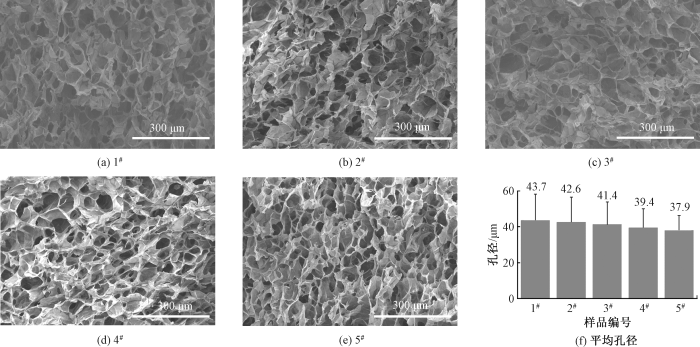
图3
不同水凝胶的SEM照片和孔径
Fig.3
SEM images and pore sizes of different hydrogels. (a)1#; (b)2#; (c)3#; (d)4#; (e)5#; (f) Average pore size of five hydrogel samples
2.4 吸水性能分析
吸收伤口渗出液是伤口敷料的重要功能。对水凝胶在pH值为7.2和5.5下的吸水率进行测试,结果如图4所示。所有的水凝胶都能吸水溶胀,这是由于DACNF和CMCS分子骨架上有大量的亲水性基团。样品3#在pH值为5.5和7.2的PBS中,分别在5 h和8 h时到达最高吸水率1 296.65%和1 523.26%,平衡吸水率均比未添加BA的高,这是由于BA的引入带来大量的亲水基团。水凝胶在pH=7.2比在pH值为5.5时具有更高的吸水率,这是因为在碱性条件下,TEMPO氧化CNF与CMCS分子链上的羧基解离为—COO-,产生的静电排斥力使聚合物链伸展,便于水分子渗入三维网络;而在pH值为5.5的酸性条件下,羧基质子化(—COOH)并与氨基产生静电吸引,同时在网络中形成强氢键,导致聚合物链收缩[9],从而使溶胀率的降低。
图4
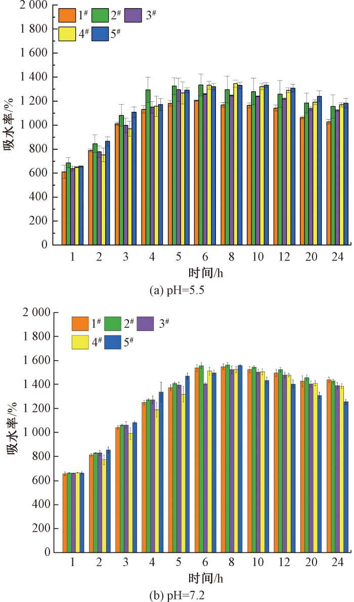
图4
冻干水凝胶在不同pH值时的吸水率
Fig.4
Water absorption rates of freeze-dried hydrogels at different pH values
2.5 水蒸气透过率分析
适当的水蒸气透过率可以保持伤口环境的湿润,促进细胞增殖和迁移,从而加速伤口愈合。表1示出不同BA含量水凝胶的水蒸气透过率和DPPH自由基清除率。BA含量为0的1#水凝胶,其水蒸气透过率为2 705.31 g/(m2·24 h),添加BA后,水蒸气透过率持续降低,BA含量到达0.125%(3#)时,水蒸气透过率降到2 611.43 g/(m2·24 h)。这是因为添加BA后,BA与DACNF和CMCS之间可通过氢键作用增强交联,水凝胶的孔隙变小,形成更为致密的三维网络结构,使得水蒸气渗透难度增大,从而降低了水凝胶的水蒸气透过率。一般伤口敷料的水蒸气透过率在2 000~2 500 g/(m2·24 h)之间[19],本文材料的水蒸气透过率略大,故更加适合处理渗出液量大的伤口。
表1 不同水凝胶的水蒸气透过率和DPPH自由基清除率
Tab.1
| 样品编号 | 水蒸气透过率/ (g·(m2·24 h)-1) | DPPH自由基 清除率/% |
|---|---|---|
| 1# | 2 705.31±57.55 | 17.98±1.75 |
| 2# | 2 643.41±7.24 | 66.90±3.70 |
| 3# | 2 611.43±5.84 | 75.83±0.83 |
| 4# | 2 577.38±45.41 | 80.60±2.61 |
| 5# | 2 523.14±18.62 | 94.64±1.22 |
2.6 抗氧化性能分析
创面微环境中活性氧(ROS)的过度积累会造成氧化应激损伤,阻碍愈合进程,因此,需对水凝胶进行抗氧化能力评价,考察其能否有效清除过量ROS,缓解氧化应激,从而为创面修复提供有利微环境。用DPPH自由基清除法测试水凝胶的体外抗氧化活性,结果如表1所示。可知,不含花青素的1#水凝胶的DPPH自由基清除率只有17.98%,这是因为羧甲基壳聚糖中的氨基和羧基可作为电子供体中和自由基的活性,但这种自由基清除能力有限。3#水凝胶的DPPH自由基清除率达到75.83%,这是因为加入BA后,花青素通过酚羟基的供氢和电子转移能力达到中和DPPH自由基的效果。随着BA含量的增加,BA酚羟基数量增加,水凝胶对DPPH自由基清除率从66.90%(2#)增加到94.64%(5#),证明添加BA的水凝胶具有优异的抗氧化性能。
2.7 抗菌性能分析
表2 不同水凝胶的抑菌率
Tab.2
| 样品编号 | 抑菌率/% | |
|---|---|---|
| 对金黄色葡萄球菌 | 对大肠埃希菌 | |
| 1# | 98.38±0.81 | 98.61±1.33 |
| 2# | 97.60±0.43 | 97.26±1.11 |
| 3# | 97.35±1.01 | 97.21±0.98 |
| 4# | 97.61±1.13 | 97.32±0.36 |
| 5# | 99.97±0.77 | 98.49±0.53 |
图5
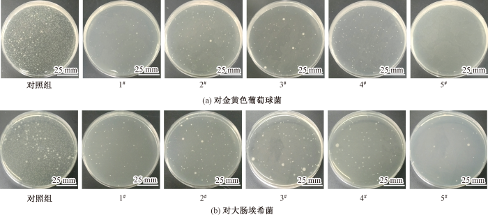
图5
水凝胶的抗菌性能
Fig.5
Antibacterial properties of different hydrogels against Staphylococcus aureus (a) and Escherichia coli (b)
2.8 细胞毒性分析
具有优良生物相容性的水凝胶可维持细胞活力并促进细胞增殖。图6示出用CCK-8检测法测试的不同水凝胶浸提液培养1、3、7 d的细胞活力。
图6
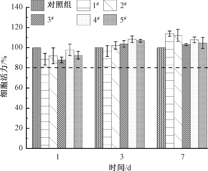
图6
在不同时间下水凝胶浸提液的细胞活力
Fig.6
Cell viability of different hydrogels at various time periods
图7
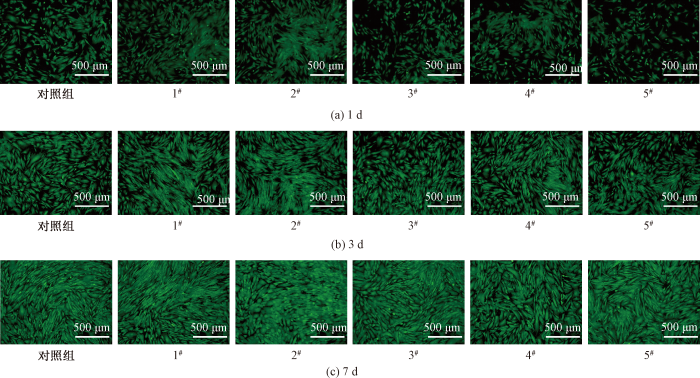
图7
水凝胶在不同时间下活/死细胞染色观察结果
Fig.7
Observation results of live/dead cell staining of different hydrogels at various time periods
2.9 pH值变色指示性能分析
花青素是一种对pH值非常敏感的材料,将花青素载入水凝胶中,水凝胶即可在不同pH值的PBS中呈现变色效果(见图8)。如图所示,经20 min后,水凝胶在pH值为5.5和7.2时,分别变为红色和蓝紫色,具有显著的pH响应变色指示效果。
图8
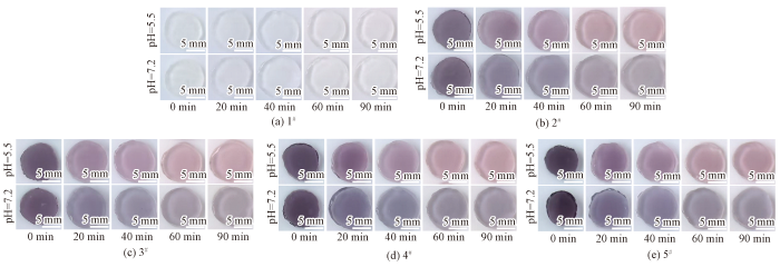
图8
不同水凝胶在不同pH值下经不同时间处理后的实物图
Fig.8
Images of different hydrogels at various pH values after treatment for different time periods
如图9(a)所示,在pH值为5.5时,20 min后3#水凝胶的a值从1.10变为4.34,水凝胶红色饱和度增加,随着时间的延长,a值增加,水凝胶颜色逐渐变红,且经过20 min后,ΔE值从0增加到8.02(>5.00),是人眼可清晰识别出的颜色差异[21]。这是因为部分花青素结合质子,其分子结构变成花青素阳离子,呈现出红色。在图9(b)中,在pH值为7.2环境下,经过20 min后,水凝胶的b值从-6.36增加到-1.70,水凝胶的蓝色饱和度降低。这是由于水凝胶制备过程中溶液pH值也是7.2,颜色不变化,然而溶胀度的增加导致水凝胶颜色变淡,b值增加。与此同时,3#水凝胶的ΔE增加到6.28,这种颜色变化仍可被人眼捕捉(见图9(c))。综上,水凝胶的这种变色特性能够比较快速、直观地反映创面pH值变化,辅助临床快速诊断。
图9
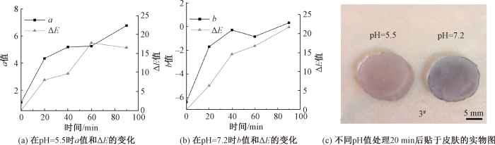
图9
3#水凝胶的变色图
Fig.9
pH color change chart of 3# hydrogel. (a) Changes in a value and ΔE over time at pH=5.5; (b) Changes in b value and ΔE over time at pH=7.2; (c) Physical images on human skill after treatment for 20 min at different pH value
3 结论
本研究成功制备了一种基于DACNF与CMCS交联网络,并负载BA的pH响应水凝胶敷料。BA含量为0.125%的水凝胶展示出优异的抗氧化和伤口pH值变色指示性能。在抗菌测试中,BA含量为0.125%的水凝胶对金黄色葡萄球菌和大肠埃希菌的抑菌率分别达97.35%和97.21%。BA通过氢键与DACNF和CMCS连接,表现出75.83%的DPPH自由基清除率。此外,敷料在模拟健康皮肤的酸性环境(pH=5.5)下呈现红色,而在模拟感染伤口的偏碱性环境(pH=7.2)下则转变为蓝紫色;二者颜色对比鲜明,易于被人眼识别,从而建立起pH值与伤口感染状态之间直观、可靠的视觉关联。细胞毒性测试结果表明,水凝胶浸提液在1~7 d内的细胞活力均超过80%,表明其没有细胞毒性。综上所述,水凝胶敷料不仅通过抗氧化与抗菌功能促进伤口愈合,更借助明确的pH值与颜色响应关系,实现了对伤口感染的实时、无创监测,展现出作为智能诊断一体化敷料的广阔应用前景。
参考文献
Functional hydrogels as wound dressing to enhance wound healing
[J].
DOI:10.1021/acsnano.1c04206
PMID:34374515
[本文引用: 1]

Hydrogels, due to their excellent biochemical and mechnical property, have shown attractive advantages in the field of wound dressings. However, a comprehensive review of the functional hydrogel as a wound dressing is still lacking. This work first summarizes the skin wound healing process and relates evaluation parameters and then reviews the advanced functions of hydrogel dressings such as antimicrobial property, adhesion and hemostasis, anti-inflammatory and anti-oxidation, substance delivery, self-healing, stimulus response, conductivity, and the recently emerged wound monitoring feature, and the strategies adopted to achieve these functions are all classified and discussed. Furthermore, applications of hydrogel wound dressing for the treatment of different types of wounds such as incisional wound and the excisional wound are summarized. Chronic wounds are also mentioned, and the focus of attention on infected wounds, burn wounds, and diabetic wounds is discussed. Finally, the future directions of hydrogel wound dressings for wound healing are further proposed.
Wound repair and regeneration: mechanisms, signaling, and translation
[J].
An antioxidant hydrogel dressing with wound pH indication function prepared based on silanized bacterial nanocellulose crosslinked with beet red pigment extract
[J].DOI:10.1016/j.ijbiomac.2024.131824 URL [本文引用: 1]
Nanocellulose composite wound dressings for real-time pH wound monitoring
[J].DOI:10.1016/j.mtbio.2023.100574 URL [本文引用: 1]
Wound healing: aiming for perfect skin regeneration
[J].
DOI:10.1126/science.276.5309.75
URL
[本文引用: 1]
The healing of an adult skin wound is a complex process requiring the collaborative efforts of many different tissues and cell lineages. The behavior of each of the contributing cell types during the phases of proliferation, migration, matrix synthesis, and contraction, as well as the growth factor and matrix signals present at a wound site, are now roughly understood. Details of how these signals control wound cell activities are beginning to emerge, and studies of healing in embryos have begun to show how the normal adult repair process might be readjusted to make it less like patching up and more like regeneration.
Antibacterial anti-oxidant electroactive injectable hydrogel as self-healing wound dressing with hemostasis and adhesiveness for cutaneous wound healing
[J].
DOI:S0142-9612(17)30019-4
PMID:28107663
[本文引用: 1]
Injectable self-healing hydrogel dressing with multifunctional properties including anti-infection, anti-oxidative and conductivity promoting wound healing process will be highly desired in wound healing application and its design is still a challenge. We developed a series of injectable conductive self-healed hydrogels based on quaternized chitosan-g-polyaniline (QCSP) and benzaldehyde group functionalized poly(ethylene glycol)-co-poly(glycerol sebacate) (PEGS-FA) as antibacterial, anti-oxidant and electroactive dressing for cutaneous wound healing. These hydrogels presented good self-healing, electroactivity, free radical scavenging capacity, antibacterial activity, adhesiveness, conductivity, swelling ratio, and biocompatibility. Interestingly, the hydrogel with an optimal crosslinker concentration of 1.5 wt% PEGS-FA showed excellent in vivo blood clotting capacity, and it significantly enhanced in vivo wound healing process in a full-thickness skin defect model than quaternized chitosan/PEGS-FA hydrogel and commercial dressing (Tegaderm™ film) by upregulating the gene expression of growth factors including VEGF, EGF and TGF-β and then promoting granulation tissue thickness and collagen deposition. Taken together, the antibacterial electroactive injectable hydrogel dressing prolonged the lifespan of dressing relying on self-healing ability and significantly promoted the in vivo wound healing process attributed to its multifunctional properties, meaning that they are excellent candidates for full-thickness skin wound healing.Copyright © 2017 Elsevier Ltd. All rights reserved.
Nanofiber-reinforced self-healing polysaccharide-based hydrogel dressings for pH discoloration monitoring and treatment of infected wounds
[J].DOI:10.1016/j.carbpol.2024.122209 URL [本文引用: 1]
一种通过变色效应指示伤口感染情况的纳米银/双改性壳聚糖抗菌水凝胶敷料及其制备方法: 113975458B
[P]. 2022-07-26.
A nano-silver/double-modified chitosan antibacterial gel dressing for indicating wound infection through color change effect and its preparation method, 11397545813
[P].2022-07-26.
Self-healing carboxymethyl chitosan hydrogel with anthocyanin for monitoring the spoilage of flesh foods
[J].DOI:10.1016/j.foodhyd.2025.111270 URL [本文引用: 2]
纤维素基止血材料的研究进展
[J].
Research progress in cellulose-based hemostatic materials
[J].
Synthesis and characterization of antibacterial carboxymethyl Chitosan/ZnO nanocomposite hydrogels
[J].
DOI:10.1016/j.ijbiomac.2016.03.044
PMID:27017980
[本文引用: 1]
The antibacterial carboxymethyl chitosan/ZnO nanocomposite hydrogels were successfully prepared via in situ formation of ZnO nanorods in the crosslinked carboxymethyl chitosan (CMCh) matrix, by treating the CMCh hydrogel matrix with zinc nitrate solution followed by the oxidation of zinc ions with alkaline solution. The resulting CMCh/ZnO hydrogels were characterized by using FTIR spectroscopy, X-ray diffractormetry and scanning electron microscopy (SEM). SEM micrographs revealed the formation of ZnO nanorods in the hydrogel matrix with the size ranging from 190nm to 600nm. The swelling behavior of the prepared nanocomposite hydrogels was also investigated in different pH solutions. The CMCh/ZnO nanocomposite hydrogel showed rather higher swelling behavior in different pH solutions in comparison with neat CMCh hydrogel. Furthermore, the antibacterial activity of CMCh/ZnO hydrogel was studied against Escherichia coli and Staphylococcus aureus by CFU assay. The results demonstrated an excellent antibacterial activity of the nanocomposite hydrogel. Therefore, the developed CMCh/ZnO nanocomposite hydrogel can be used effectively in biomedical field. Copyright © 2016 Elsevier B.V. All rights reserved.
Wound dressing double-crosslinked quick self-healing hydrogel based on carboxymethyl chitosan and modified nanocellulose
[J].DOI:10.3390/polym15163389 URL [本文引用: 1]
Sodium alginate/chitosan-based intelligent bilayer film with antimicrobial activity for pork preservation and freshness monitoring
[J].DOI:10.1016/j.foodcont.2023.109615 URL [本文引用: 1]
Cellulose nanofiber-based hydrogels with high mechanical strength
[J].DOI:10.1007/s10570-012-9784-3 URL [本文引用: 1]
纳米纤维素基高吸水抗菌抗氧化双相凝胶的制备与性能
[J].
Preparation and properties of nanocellulose-based superabsorbent antibacterial and antioxidant biphasic gels
[J].
Active chitosan/PVA films with anthocyanins from Brassica Oleraceae (red cabbage) as time-temperature Indicators for application in intelligent food packaging
[J].DOI:10.1016/j.foodhyd.2014.05.014 URL [本文引用: 1]
淀粉-羧甲基纤维素钠-花青素指示膜的制备及在鱼肉鲜度指示中的应用
[J].
DOI:10.7506/spkx1002-6630-20190715-202
[本文引用: 1]
以马铃薯淀粉、羧甲基纤维素钠为成膜基材,紫薯花青素作为指示剂制备指示膜。研究马铃薯淀粉和羧甲基纤维素钠的比例、花青素质量分数、甘油质量分数对指示膜性能抗拉伸强度、断裂伸长率、水溶性、颜色(a值)的影响,将指示膜性能综合分作为响应值,采用响应面法优化制备工艺参数,并对最优指示膜进行表征。结果表明,当马铃薯淀粉和羧甲基纤维素钠质量比6∶4、花青素质量分数0.5%、甘油质量分数0.6%时,指示膜的综合分为63.8,此时对应的抗拉伸强度为(21.48±0.12)MPa,断裂伸长率为(28.88±0.08)%,水溶性为(19.24±0.21)%,a值为4.95±0.02。通过傅里叶变换红外光谱分析、X射线衍射和扫描电镜分析各组分之间相容性好,截面平整光滑。将指示膜用于监测草鱼新鲜度,鱼肉的挥发性盐基氮值、pH值以及菌落总数分别达到21.55 mg/100 g、7.58、7.3(lg(CFU/g))时,指示膜由红色变为蓝紫色。研究结果可为指示膜用于食品货架期指示应用提供参考。
Preparation of indicator films based on sodium carboxymethyl cellulose/starch and purple sweet potato anthocyanins for monitoring fish freshness
[J].
DOI:10.7506/spkx1002-6630-20190715-202
[本文引用: 1]
Indicator films were prepared through using starch (S) and sodium carboxymethyl cellulose (CMC-Na) as the film forming material and purple sweet potato anthocyanins (PA) as the indicator. The effects of S/CMC-Na ratio, anthocyanin concentration and glycerol concentration on the tensile strength (TS), elongation at break (EAB), water solubility and color (a value) of films were investigated. Using the comprehensive score of indicator film performance was as the response value, the process parameters were optimized by the response surface methodology, and the optimal indicator film was characterized. The results demonstrated that the optimum formulation of indicator films was determined as follows: starch/CMC-Na ratio 6:4, anthocyanin concentration 0.5%, and glycerol concentration 0.6%. The overall score of the indicator film prepared using the optimized formulation were the highest, up to 63.8, and it showed tensile strength of (21.48 ± 0.12) MPa, elongation at break of (28.88 ± 0.08)%, water solubility of (19.24 ± 0.21)%, and a value of 4.95 ± 0.02. The results of Fourier-transform infrared (FT-IR) spectroscopy, X-ray-differentiation (XRD) and scanning electron microscopy (SEM) indicated that the cross section of the SCA film was smooth and showed a good compatibility among the ingredients. Furthermore, the indicator film was used to monitor the freshness of grass carp. When the total volatile basic nitrogen (TVB-N) value, pH and total viable count reached 21.55 mg/100 g, 7.58 and 7.3 (lg(CFU/g)), respectively, the indicator film changed from red to blue-violet. These research results provide a basis for the application of indicator films for food shelf life evaluation.
Preparation of an indicator film based on pectin, sodium alginate, and xanthan gum containing blueberry anthocyanin extract and its application in blueberry freshness monitoring
[J].DOI:10.1016/j.heliyon.2023.e14421 URL [本文引用: 1]
Evaluation of an in situ forming hydrogel wound dressing based on oxidized alginate and gelatin
[J].
DOI:10.1016/j.biomaterials.2005.04.012
PMID:15919113
[本文引用: 1]
Wound dressings that can be formed in situ offer several advantages over the use of preformed dressings such as conformability without wrinkling or fluting in the wound bed, ease of application and improved patient compliance and comfort. Here we describe such an in situ forming hydrogel wound dressing from gelatin, oxidized alginate and borax. Periodate oxidized alginate rapidly cross-links proteins such as gelatin in the presence of borax to give in situ forming hydrogels that are both non-toxic and biodegradable. The composite matrix has the haemostatic effect of gelatin, the wound healing-promoting feature of alginate and the antiseptic property of borax to make it a potential wound dressing material. The hydrogel was found to have a fluid uptake of 90% of its weight which would prevent the wound bed from accumulation of exudates. The water vapour transmission rate (WVTR) of the hydrogel was found to be 2686+/-124 g/m2/day indicating that the hydrogel can maintain a moist environment over wound bed in moderate to heavily exuding wound which would enhance epithelial cell migration during the healing process. The wound healing efficacy of hydrogel was evaluated in experimental full thickness wounds using a rat model which demonstrated that within 2 weeks, the wound covered with gel was completely filled with new epithelium without any significant adverse reactions. These in situ forming hydrogels fulfil many critical elements desirable in a wound dressing material.
伤口渗出液对抗菌敷料效果影响的研究
[J].
Influence of wound exudate on the efficacy of antimicrobial dressings
[J].
Color discrimination threshold of human vision for textiles under different illumination conditions
[J].
DOI:10.1177/00405175221148257
URL
[本文引用: 1]
\n In this work, the varying color discrimination thresholds of human vision for textiles with a wide range of physical color attributes under different illumination conditions were examined. It was found that, regardless of the illumination condition and the color attributes of the textiles, people cannot generally perceive the color differences of textiles smaller than 4.25 Δ E*\n ab\n. Meanwhile, the color discrimination threshold was found to be significantly affected by the illuminance of illuminants and all the physical color attributes of lightness, chroma, and hue of the textiles observed. People were generally better able to distinguish color differences in bright environments with the illuminance of 2000 lux than in darker ones with 100 lux. In addition, color differences between dark and less-saturated textiles were found to be generally easier to be perceived than those between light and saturated textiles under various illuminants. In addition, regardless of the illuminant, color differences between neutral colored textiles were easier to be perceived than those between chromatic colored textiles with the mean color discrimination threshold of 2.85 and 4.42 Δ E*\n ab\n, respectively. In particular, among chromatic colored textiles, color differences between yellow textiles were generally the hardest to be perceived with the mean threshold of 5.58 Δ E*\n ab\n.\n

 京公网安备11010502044800号


