



相较而言,将稳定性更高、易于规模化生产的纳米酶负载于伤口敷料发挥ROS抗菌效力极具应用前景。静电纺纳米纤维敷料凭借高比表面积、孔隙率大、易于表面功能化等优点,被认为是负载纳米酶的良好载体[8],但纳米酶负载的关键难点在于如何将这类功能材料以分散状态均匀组装到敷料基材中,充分暴露其活性位点并保留ROS生成能力。目前常用的负载方法包括表面涂敷、原位合成、共混静电纺丝等[9-10]。表面负载相比于直接共混或包埋更能有效保持纳米酶活性,并且纳米酶在纤维上的均匀分布有利于提高多酶级联催化反应的反应效率[11];然而,由于纳米纤维膜固有的致密堆叠结构,传统的纤维表面负载方法难以实现纳米酶在三维空间内的均匀锚定[12],且常规的表面静电吸附稳定性差,纳米酶易从纤维上脱落,导致级联催化反应效率降低[13],因此,有必要开发一种简便有效的组装途径,能够将纳米酶稳定且均匀地固定在纳米纤维载体上,并保持其高效催化活性,从而充分发挥ROS抗菌作用。
针对上述问题,本研究基于慢性DFUs创面高葡萄糖含量的特征,设计了一种双层结构复合敷料。下层为疏水聚丙烯(PP)非织造布层,上层为纳米酶负载层,有望为DFUs的治疗提供一种先进的材料设计方案。
1 实验部分
1.1 材料与仪器
材料:聚丙烯(PP)非织造布(面密度为40 g/m2),信达无纺布有限公司;葡萄糖、2-氨基对苯二甲酸(NH2-BDC)、聚偏氟乙烯(PVDF)、3,3,5,5-四甲基联苯胺(TMB)、甲基红指示剂,上海阿拉丁生化科技股份有限公司;单宁酸基黏合剂(TBA),实验室自制[14];金纳米颗粒(Au NPs,15 nm),博梵生命科技有限公司;六水合三氯化铁(FeCl3·6H2O)、甲醇、乙醇、N,N-二甲基甲酰胺(DMF)、冰醋酸、醋酸钠、丙酮、氢氧化钠(NaOH),国药集团化学试剂有限公司;棕色墨水,上海精细文化用品有限公司;N-(2-羟乙基)-N-2-乙烷磺酸(HEPES),梯希爱化成工业发展有限公司;LB固体培养基、琼脂粉,北京索莱宝科技有限公司;金黄色葡萄球菌(S. aureus)、大肠埃希菌(E. coli),中国工业微生物菌种保藏管理中心;人包皮成纤维细胞(HFF),中国科学院细胞库;细胞计数试剂盒(CCK-8),上海翊圣生物科技有限公司。
仪器:SU8010场发射扫描电子显微镜,日本日立株式会社;METASH-X6紫外可见分光光度计,上海元析仪器有限公司;高速剪切机,孚莱(上海)流体设备有限公司;瓦格纳尔W590电动喷枪,德国瓦格纳尔公司;DZF-6050TG真空干燥箱,上海精宏实验设备有限公司;PS-40AD超声波清洗机,深科洁环境科技有限公司;H17.5R台式高速冷冻离心机,上海卢湘仪离心机仪器有限公司;MQL-61HR摇床,上海仪天科学仪器有限公司;Spectrum Two傅里叶变换红外光谱仪,美国珀金埃尔默股份有限公司;OCA15EC接触角测量仪,德国Data Physics公司;YG(B)026G纺织品多功能强力仪,浙江大荣纺织仪器有限公司。
1.2 复合抗菌敷料的制备
1.2.1 Fe-MIL-88NH2的合成
称取0.126 g NH2-BDC和0.187 g FeCl3·6H2O加入7.5 mL超干DMF溶剂中,超声波处理15 min至溶解,120 ℃油浴加热反应4 h。在反应开始15 min时加入197 μL浓度为3.45 mmol/L冰醋酸。待冷却至室温后,以10 000 r/min转速离心15 min后收集棕色沉淀,用DMF离心洗涤3次,60 ℃下真空干燥12 h,得到棕色固体粉末,避光保存。
1.2.2 PVDF纳米纤维膜的制备
以丙酮和DMF(体积比为7∶3)为溶剂,取1.2 g PVDF配制质量分数为12%的PVDF溶液,60 ℃下加热12 h。将溶液加入10 mL注射器中,纺丝参数如下:负电压2.0 kV、正电压19.0 kV、推注速度2 mL/h、接收距离15 cm、滚筒转速200 r/min,环境温度(30 ± 5) ℃,相对湿度(60 ± 5)%,针头直径0.8 mm,最终在铝箔上形成PVDF纳米纤维膜,取下铝箔,将纤维膜于60 ℃真空干燥12 h,待用。
1.2.3 复合敷料的制备
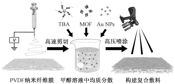
将PVDF纤维膜裁成碎片,称取6 g PVDF碎片加入310 mL甲醇溶液,用高速剪切机在8 000 r/min下分散均匀后,加入50 mL TBA-甲醇溶液(10 mg/mL),20 mL Fe-MIL-88NH2分散液(8 mg/mL)和20 mL Au NPs(0.3 mg/mL),得到PVDF短纤维分散液。裁剪50 cm × 50 cm的PP非织造布并平铺,使用高压喷枪将短纤维均匀喷涂在非织造布表面,50 ℃下真空干燥12 h,即得双层结构复合敷料(记为PVDF/PP),制备过程如图1所示。
图1
1.3 性能测试与表征
1.3.1 微观形貌观察
采用扫描电子显微镜对样品的微观表面形貌进行观察,以表征其表面特征与微观结构;借助图像分析软件对样品中的Fe-MIL-88NH2颗粒及纤维的直径进行精准测量,为样品的微观结构分析提供可靠的量化依据。
1.3.2 理化性能表征
使用TMB显色实验表征不同pH值下Fe-MIL-88NH2的类过氧化物酶催化活性。Fe-MIL-88NH2类过氧化物模拟酶能够催化H2O2产生·OH,产生的·OH可将无色的TMB氧化为蓝色的oxTMB。采用醋酸-醋酸钠配制不同pH值(3、4、5、6、7)缓冲液,加入0.8 mL TMB-乙醇溶液(0.2 mmol/L)、0.8 mL H2O2-水溶液(1 mmol/L)和0.8 mL Fe-MIL-88NH2(1 mg/mL),充分混合后在室温环境下避光静置20 min,移入比色皿中,用紫外可见分光光度计测量产物在652 nm处的吸光度。如果吸收峰明显升高,说明该pH条件下有更多的oxTMB生成,证明模拟酶催化活性高。每组平行测量3次。
采用傅里叶变换红外光谱仪对PVDF、甲醇活化后的PVDF及PVDF/PP进行红外光谱(FT-IR)扫描分析,以表征敷料的表面化学组成。
将PP非织造布、PVDF、甲醇活化后的PVDF及PVDF/PP固定于接触角测量仪的台面上,采用液滴法测试其水接触角。
1.3.3 力学性能测试
参照ASTM D5035—2011(R2019)《织物断裂强力和伸长率的标准试验方法(条样法)》对PP和PVDF/PP分别从生产线运行方向(MD方向)和垂直于生产线方向(CD方向)进行单轴拉伸测试。将样品裁剪成50 mm×5 mm的条状试样,设置隔距为20 mm,以10 mm/min的速度拉伸,直至样品断裂。绘制应力-应变曲线,得出试样的断裂强度及断裂应变。
1.3.4 单向导湿性能测试
将50 μL的棕色墨水滴在敷料的上、下表面,观察墨水在60 s内的扩散面积随时间的变化,并记录墨滴在纤维膜上、下两面的传递过程,以分析复合纤维膜的单向导湿性能。
1.3.5 级联催化性能测试
pH值调节能力测试:取2 cm × 2 cm大小的含Au NPs和不含Au NPs的PVDF/PP敷料浸泡到含有3 mL葡萄糖(50 mg/mL)的HEPES缓冲液中,并加入30 μL的0.001%甲基红指示剂,在37 ℃下孵育,每间隔30 min拍照记录1次,对比每组颜色变化。
取2 cm×2 cm复合敷料加入含有3 mL葡萄糖(50 mg/mL)的HEPES缓冲液(pH = 7.4)中,37 ℃下孵育60 min,之后加入1 mL TMB-乙醇溶液(0.4 mmol/L),再反应60 min,之后观察颜色变化。
1.3.6 抗菌性能测试
选择S. aureus和E. coli为实验模型,未放置敷料的组别为空白样。将空白样和复合敷料裁成直径为2 cm的圆片,将样品灭菌后放入菌液浓度为1 × 10⁷ CFU/mL的5 mL LB培养基中,在37 ℃、摇床120 r/min条件下培养24 h。培养后将样品在PBS溶液中超声波洗脱,将洗脱液按10倍浓度梯度稀释3次,在琼脂培养板的4个区域依次点板,培养12 h,记录菌落形成单位数(CFU)。每组样品重复测试3次。
1.3.7 细胞毒性测试
采用CCK-8法测试复合敷料的细胞毒性。以1 × 10⁴个/孔密度将人包皮成纤维细胞(HFF)接种在24孔板中,4 h贴壁后,加入灭菌后的PP、PVDF重构层和PVDF/PP复合敷料,设置空白对照样,每个样品设置3个平行样。培养24 h后将CCK-8染液在黑暗条件下加入孔板中孵育2 h,测试其在450 nm处的吸光度,实验组的吸光度与空白对照组的吸光度比值即为细胞存活率。
2 结果与讨论
2.1 Fe-MIL-88NH2形貌与最优pH值分析
图2
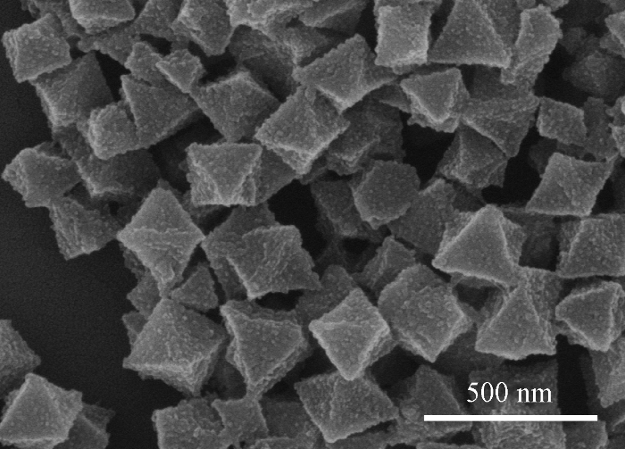
图3示出不同pH值下Fe-MIL-88NH2溶液的吸光度(652 nm)。可知,随着pH值增大,溶液在652 nm处的吸光度呈现先增后降趋势,在pH值为4时吸光度最大,说明该条件下MOF的催化活性最高,表现出最高的类过氧化物酶活性,而随着pH值继续增大,催化活性逐渐降低,且在碱性条件下几乎无法表现类酶催化活性。
图3
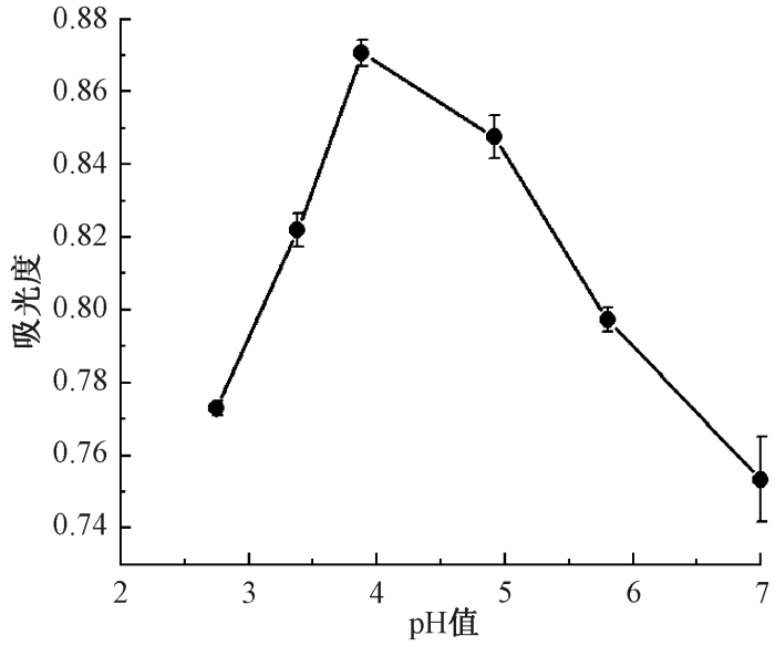
图3
不同pH值下Fe-MIL-88NH2溶液的吸光度
Fig.3
Absorbance of Fe-MIL-88NH2 solution at different pH values
2.2 微观形貌结构分析
图4(a)示出复合敷料下层PP非织造布和上层重构PVDF纤维层的表面SEM照片。可以看出2种纤维均匀分布。其中:PP层纤维直径较粗,平均值为(15.68±0.26) μm,纤维交织形成相互连通的多孔结构;重构PVDF层纤维直径较细,平均值为(515±19.8) nm,未见明显串珠缺陷。PVDF纤维上均匀负载有MOF颗粒,未出现明显团聚现象,并且MOF保持了完整的正八面体晶型结构,说明MOF在甲醇体系以及高速匀浆分散条件下能够稳定存在。Au NPs由于尺寸较小,难以被SEM观察到,后续通过验证其葡萄糖氧化酶活性证明其负载。从图4(b)示出的截面SEM照片可发现,重构后的PVDF纤维层在PP基底上均匀堆叠,形成了蓬松多孔的网络结构。这种多孔网络有利于吸收和传导伤口渗出液,有望提升敷料的导湿性能。
图4
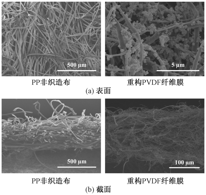
图4
复合敷料下层PP非织造布及上层重构PVDF纤维表面和截面的SEM照片
Fig.4
Surface (a)and cross-section (b) SEM images of lower PP nonwoven fabric and upper reconstituted PVDF fiber membrane of composite dressing
2.3 表面理化性能分析
图5示出复合敷料亲水层制备前后的红外光谱。由图可见,PVDF/PP纤维膜在保留PVDF特征吸收峰的同时,在1 576 cm-1处出现羟基与铁离子螯合形成的伸缩振动峰,570 cm-1处出现Fe—O键的伸缩振动峰[16],证明Fe-MIL-88NH2纳米酶已成功负载到PVDF短纤维上。重构PVDF短纤维表面涂敷了TBA作为黏合层,在2 979 cm-1处出现饱和脂肪族C—H伸缩振动峰[17],表明TBA已成功附着于纤维表面。此外,由于Fe-MIL-88NH2与PVDF分子间的相互作用,在1 500~1 000 cm-1区间的吸收峰强度显著增加。上述结果表明,通过短纤维重构的方法,已在PVDF纤维表面同时负载了TBA涂层和Fe-MIL-88NH2纳米酶。
图5
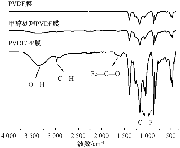
图5
PVDF膜、甲醇处理PVDF膜及PVDF/PP短纤维膜的红外光谱
Fig.5
FT-IR spectra of PVDF, methanol-treated PVDF, and PVDF/PP short-fiber membrane
表1示出复合敷料亲水层制备前后材料的水接触角变化。下层PP非织造布呈现出强疏水性,而经甲醇处理后的PVDF纤维膜接触角从(132.13±1.63)°降至(75.80±2.24)°,亲水性显著提升。主要是因为甲醇会引起PVDF链段的溶胀与重新取向,促使非极性的α相部分转变为极性更高的β相,从而增加表面偶极矩与表面能[18];同时,甲醇处理可改变纤维表面的微观形貌与粗糙度,并使亲水性较强的—CH2—基团在表面暴露的比例增加[19],这些结构与化学状态的变化协同降低了水在膜表面的接触角,使固有亲水性得到持久提升。在PP基底上重构的PVDF亲水纤维层同时保持了良好的亲水性,其静态接触角明显小于PP层,实现润湿性差异构筑。
表1 复合敷料制备过程中各层材料的水接触角
Tab.1
| 试样名称 | 接触角/(°) |
|---|---|
| PP非织造布 | 137.94±1.09a |
| PVDF膜 | 132.13±1.63a |
| 甲醇处理PVDF膜 | 75.80±2.24b |
| PVDF/PP复合敷料 | 75.51±6.65b |
注:采用均值±标准差表示各组与空白对照组的差异对比,上标字母不同时,表示组间存在显著性差异(P<0.05);相同时,表示组间无显著性差异。
2.4 单向导湿性能分析
图6
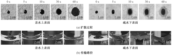
图6
墨滴在敷料上下表面的单向导湿性能
Fig.6
Unidirectional wetting properties of ink droplets on upper and lower surfaces of dressings. (a)Diffusion process;(b)Transport path
图6(b)示出敷料上下表面的液体传输路径。通过连续多次滴加墨水发现:当墨水持续滴加在下层疏水侧时,液体能够单向透过疏水层进入外层亲水层;当墨水滴加在上层亲水侧时,墨水会被亲水层迅速吸收铺展,且不会反渗回疏水层。上述结果说明,该内疏水外亲水的双层复合敷料依靠内外层纤维之间形成的润湿性梯度形成了有效的单向液体驱动作用,只允许液体从疏水层泵送至亲水层而不会发生反向渗透。此外,重构得到的蓬松多孔亲水纤维层具有更大的储液空间,进一步增强了敷料的单向导湿效果。
2.5 力学性能分析
图7示出PP非织造布和PVDF/PP复合敷料的应力-应变曲线。由此得出,PP非织造布在MD方向和CD方向的断裂强度分别为(9.1±0.3)、(4.3±0.2)MPa,断裂伸长率分别为(71±12)%和(137±12)%。负载重构PVDF纤维层后,敷料在MD和CD方向的断裂强度分别提高至(11.4±0.3)和(7.3±0.4)MPa,断裂伸长率分别为(53±8)%和(142±20)%。可见,复合敷料的力学性能相比单层PP有所提升,其断裂强度和伸长率均接近人体皮肤的力学参数,能够与伤口完整贴合并起到保护和支撑作用。
图7
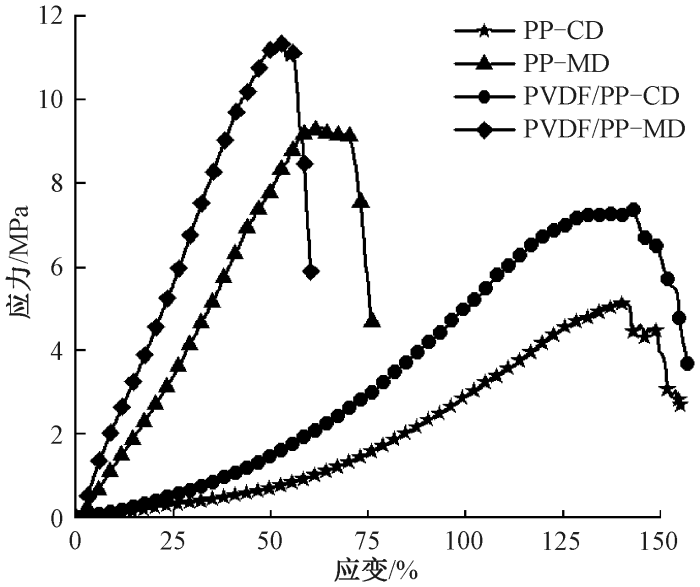
图7
PP非织造布与PVDF/PP复合敷料MD和CD方向的应力-应变曲线
Fig.7
Stress-strain curves of PP nonworen and PVDF/PP composite dressing in MD and CD directions
2.6 级联催化性能分析
Au NPs和Fe-MIL-88NH2纳米酶的催化活性是复合敷料实现抗菌功能的关键。通过甲基红和TMB显色实验验证了PVDF/PP复合敷料中Au NPs和Fe-MIL-88NH2的类酶活性及协同催化产生活性物质的能力。具有类葡萄糖氧化酶活性的Au NPs可催化葡萄糖生成葡萄糖酸和过氧化氢[20],其中葡萄糖酸使体系pH值降低,过氧化氢则作为Fe-MIL-88NH2催化反应的底物,为后续反应创造条件。加入甲基红指示剂可以直观呈现反应体系在不同pH值下的颜色变化(见表2):当pH>6.2时溶液呈黄色,pH=4.4~6.2时呈橙色,pH<4.4时呈红色[21]。说明复合敷料中的Au NPs保持了模拟葡萄糖氧化酶的活性,能够有效催化葡萄糖氧化生成葡萄糖酸,显著降低体系pH值。
表2 复合敷料催化葡萄糖反应体系的溶液pH值随时间变化
Tab.2
| 时间/min | 颜色变化 | pH值区间 |
|---|---|---|
| 0 | 黄色 | >6.2 |
| 30 | 橙色 | 4.4~6.2 |
| 60 | 浅红 | <4.4 |
| 90 | 红色 | <4.4 |
| 120 | 深红 | <4.4 |
图8
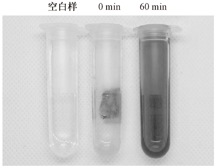
图8
复合敷料在级联催化反应中TMB显色结果
Fig.8
TMB colorimetric reaction results for composite dressing
2.7 抗菌性能分析
为验证级联催化反应产生的·OH自由基能有效杀菌,通过平板计数法比较了空白对照组和复合敷料组对S. aureus和E. coli的抑菌效果,结果如图9所示。由图可见,空白组2种细菌均形成大量菌落,复合敷料组菌落数大幅减少,仅有零星菌落形成。通过计算得到对S. aureus和E. coli的抑菌率均超过97%。由此可见,所构建的利用DFUs创面高糖环境触发酶促级联催化反应的敷料能够有效杀灭细菌,表现出优异的广谱抗菌性能。
图9

图9
复合敷料的抗菌性能
Fig.9
Antibacterial properties of composite dressings. (a) Control samples; (b) Composite dressings
2.8 细胞毒性分析
将HFF与复合敷料共培养24 h后,通过CCK-8法检测细胞存活率,以评估敷料的细胞相容性。复合敷料的细胞毒性如表3所示。虽然实验组细胞存活率略低于空白对照组(P<0.05),但PP非织造布、PVDF重构层、PVDF/PP复合敷料之间的细胞存活率不存在显著性差异(P>0.05),证明在PP非织造布基底上增加的纳米酶、短纤维等材料并未引入额外的细胞毒性,且细胞存活率均保持在84%以上,说明该复合敷料对HFF细胞无明显毒性,具有良好的细胞相容性。
表3 复合敷料的细胞毒性
Tab.3
| 试样名称 | 细胞存活率/% |
|---|---|
| 空白对照样 | 99.81±1.87a |
| PP非织造布 | 84.88±6.07b |
| PVDF重构层 | 87.41±8.94b |
| PVDF/PP复合敷料 | 83.91±0.67b |
注:采用均值±标准差表示各组与空白对照组的差异对比,上标字母不同时,表示组间存在显著性差异(P<0.05);相同时,表示组间无显著性差异。
3 结论
本文制备了一种面向糖尿病足溃疡(DFUs)治疗的双层纳米纤维复合敷料:上层为亲水的纳米酶复合聚偏氟乙烯(PVDF)纤维,下层为疏水的聚丙烯(PP)非织造布基底。所构建的纳米酶级联复合敷料可借助DFUs创面中高浓度葡萄糖触发金纳米颗粒(Au NPs)和Fe-MIL-88NH2的级联催化反应,局部产生·OH自由基杀菌,可使体外抗菌率在97%以上。敷料内外层的润湿性梯度赋予其单向导湿性能,可将伤口渗出液迅速抽离至外层并避免回渗,有效保持创面干爽洁净。复合敷料还具备接近人体皮肤的力学强度,且对细胞无明显毒性,表现出良好的细胞相容性。综上,这种同时具备主动抗菌和定向导湿功能的纳米纤维复合敷料满足对抗菌性、排液性和细胞相容性的多重要求,为DFUs的治疗提供了一种新颖有效的材料设计策略,在增强感染控制和促进创面修复方面具有重要的应用潜力。
参考文献
Advances in the development of medical dressings for the treatment of diabetic foot wounds
[J].DOI:10.1016/j.cej.2024.155575 URL [本文引用: 1]
A bio-inspired Janus patch for treating abdominal wall defects
[J].DOI:10.1002/adfm.v34.41 URL [本文引用: 1]
Regulating wound moisture for accelerated healing: a strategy for the continuous drainage of wound exudates by mimicking plant transpiration
[J].DOI:10.1016/j.cej.2021.131964 URL [本文引用: 1]
Multimodal enzyme-carrying suprastructures for rapid and sensitive biocatalytic cascade reactions
[J].DOI:10.1002/advs.v9.10 URL [本文引用: 1]
Antibacterial effects of low-temperature plasma generated by atmospheric-pressure plasma jet are mediated by reactive oxygen species
[J].DOI:10.1038/s41598-020-59652-6 [本文引用: 2]
Nanocatalytic tumor therapy by biomimetic dual inorganic nanozyme-catalyzed cascade reaction
[J].DOI:10.1002/advs.v6.3 URL [本文引用: 1]
Mitochondrial-mimicking nanozyme-catalyzed cascade reactions for aging attenuation
[J].DOI:10.1016/j.nantod.2023.101757 URL [本文引用: 1]
Functionalized silk fibroin nanofibers as drug carriers: advantages and challenges
[J].
DOI:S0168-3659(20)30112-7
PMID:32061791
[本文引用: 1]

Electrospinning is well thought of as the most potent nanofiber producing technique, which is applicable in biomedical fields. The generation of electrospun nanofibers as drug carriers has widely been shown much interest over the past years. Electrospun nanofibers meet various advantages as drug delivery platforms including high surface area, acceptable mechanical properties based on the choice of the polymer, high processability, and the possibilities for surface modifications. Silk fibroin protein has gained a great attention as a drug delivery carrier due to ease of purification, sterilization, processability without using chemical crosslinkers, good biocompatibility, tailorable biodegradability, low immunogenicity, and high capacity to stabilize the loaded drugs. These characteristics along with advantageous benefits of electrospinning provide opportunities for producing suitable nanofibers based on silk fibroin for drug delivery purposes. It is also possible to incorporate various functional moieties to the electrospun silk fibroin nanofibers to enhance its biological activities. This review covers the progress in electrospinning of silk fibroin as a drug carrier in recent years.Copyright © 2020 Elsevier B.V. All rights reserved.
Janus LAAM-loaded electrospun fibrous buccal films for treating opioid use disorder
[J].DOI:10.1016/j.biomaterials.2024.123041 URL [本文引用: 1]
Electrospinning in situ synthesis of graphene-doped porous copper indium disulfide/carbon composite nanofibers for highly efficient counter electrode in dye-sensitized solar cells
[J].DOI:10.1016/j.electacta.2016.08.101 URL [本文引用: 1]
Elongated magnetic nanorobots with multi-enzymatic cascades for active in vivo tumor targeting and enhanced chemodynamic therapy
[J].
DOI:10.1021/acsnano.5c01566
PMID:40223775
[本文引用: 1]
Targeted delivery of therapeutic agents to malignant tissues is crucial for enhancing clinical outcomes and reducing side effects. Magnetic nanorobots (MNRs) present a promising strategy for controlled delivery, leveraging external magnetic fields to achieve precise in vivo targeting. This work develops elongated MNRs comprising linearly arranged magnetic nanoparticles linked by metal-polyphenol complexes (MPCs) for magnetic-field-directed active tumor targeting and synergistic tumor therapy. The MNRs are created by assembling 30 nm FeO nanoparticles, tannic acid, and ferrous ions (Fe) under a uniform magnetic field, resulting in elongated chain-like structures fixed by MPCs, which also promotes peroxidase-like activity. These structures show a greater magnetic response than individual nanoparticles, offering flexibility in magnetic manipulation. The MPCs coating allows tailored surface modifications with glucose oxidase, copper ions (Cu), and human serum albumin (HSA), producing colloidally stable MNRs with a built-in multienzymatic cascade (MNRs@GOx/Cu/HSA) that consumes glucose, generates OH, and depletes the antioxidant glutathione (GSH). Collectively, surface-engineered multifunctional MNRs demonstrate improved in vivo tumor targeting driven by external magnetic fields, leading to efficient localized chemodynamic therapy. The tailored structural and functional properties of the developed MNRs render them suitable for targeted cargo delivery, minimally invasive surgery, and localized treatments in disease sites.
Immol/Lunopolarization-regulated 3D printed-electrospun fibrous scaffolds for bone regeneration
[J].DOI:10.1016/j.biomaterials.2021.121037 URL [本文引用: 1]
Enzyme immobilization on electrospun polymer nanofibers: an overview
[J].DOI:10.1016/j.molcatb.2008.05.005 URL [本文引用: 1]
Nanofibers fortified with synergistic defense route: a potent wound dressing against drug-resistant bacterial infections
[J].DOI:10.1016/j.cej.2023.146492 URL [本文引用: 1]
Chemically grafted nanozyme composite cryogels to enhance antibacterial and biocompatible performance for bioliquid regulation and adaptive bacteria trapping
[J].
DOI:10.1021/acsnano.1c06983
PMID:34878257
[本文引用: 2]
Excessive biofluid and infection around wounds hinder wound healing. However, conventionally antibacterial wound dressings cannot simultaneously achieve effective biofluid control and intelligent infection treatment, tending to overhydrate wounds and develop drug-resistant bacteria due to the limitations of antibacterial components and material structures. The design of a nanozyme composite cryogel with interconnected macroporous structures, excellent designability, and lower chance of drug-resistance is greatly needed. Herein, Fe-MIL-88NH nanozyme is grafted to glycidyl methacrylate functionalized dialdehyde chitosan Schiff base reaction, and acryloyl Pluronic 127 (PF127-DA) is used as a cross-linking agent to fabricate nanozyme composite cryogels (CSG-M) as a wound dressing to enhance antibacterial and biocompatible performance for biofluid management and wound infection therapy. CSG-M has great hydrophilicity, acid-enhanced positive charge, pH-responsive release, rebinding of nanozymes, and excellent peroxidase and oxidase mimicry activity (generation of OH and O radicals). Notably, due to the negative potential of bacteria, the impact of infection on pH value, and the enzyme-like activity as well as the reversible release of nanozymes influenced by pH, CSG-M can achieve intelligently adaptive trapping and killing of bacteria. CSG-M has enormous potential to be a next-generation wound dressing for biofluid management and bacterial infection treatment in the clinic.
Amino-functionalized MIL-88B as heterogeneous photo-Fenton catalysts for enhancing tris-(2-chloroisopropyl) phosphate (TCPP) degradation: dual excitation pathways accelerate the conversion of FeIII to FeII under visible light irradiation
[J].DOI:10.1016/j.jhazmat.2021.127782 URL [本文引用: 1]
One-step hydrophobization of tannic acid for antibacterial coating on catheters to prevent catheter-associated infections
[J].
DOI:10.1039/c9bm01223k
PMID:31535105
[本文引用: 1]
Catheter-associated infections (CAIs) caused by bacterial colonization are significant problems in clinics. Thus, effective antibacterial coatings for biomedical catheters to prevent bacterial infections are urgently needed. Ideal coatings should include the advantage of potent antibacterial properties and being easily and economically modified on the catheter surface. Due to their advantages of adhesive capability on various substrates, an increasing number of coatings based on plant polyphenols have been developed. However, the hydrophilicity of plant polyphenols limits their utilization in coatings. Herein, hydrophobic tannic acid (TA) was synthesized via the one-step electrostatic assembly of TA and benzalkonium chloride (BAC) with the green solvent water as the medium. The as-prepared hydrophobic TA (TBA) facilely formed a stable and colorless coating on the luminal and outer surface of biomedical catheters with broad-spectrum antibacterial activity and biocompatiblity. It was demonstrated that the TBA-coated surfaces displayed excellent bactericidal activity toward Gram-positive Staphylococcus aureus (S. aureus) and Gram-negative Escherichia coli (E. coli), and more than 99% of the above bacteria were killed by the TBA-coated films. The test of the coated catheters in vitro also showed the excellent antibacterial activity of both the outer and luminal surfaces of the catheter. Moreover, in an in vivo mouse model, the coated catheters relatively prevented bacterial colonization compared to the uncoated catheters. Meantime, no significant cytotoxicity and host response for Cell Counting Kit-8 (CCK-8) and tissue compatibility in vivo were observed, indicating the better biocompatibility of the TBA coating. This preparation method overcomes the limitation of the traditional hydrophilic tannic acid as a coating and provides a new method for preventing medical indwelling device-associated infections.
Properties and applications of the β phase poly(vinylidene fluoride)
[J].
DOI:10.3390/polym10030228
URL
[本文引用: 1]
Poly(vinylidene fluoride), PVDF, as one of important polymeric materials with extensively scientific interests and technological applications, shows five crystalline polymorphs with α, β, γ, δ and ε phases obtained by different processing methods. Among them, β phase PVDF presents outstanding electrical characteristics including piezo-, pyro-and ferroelectric properties. These electroactive properties are increasingly important in applications such as energy storage, spin valve devices, biomedicine, sensors and smart scaffolds. This article discusses the basic knowledge and character methods for PVDF fabrication and provides an overview of recent advances on the phase modification and recent applications of the β phase PVDF are reported. This study may provide an insight for the development and utilization for β phase PVDF nanofilms in future electronics.
Enhancement of hydrophilicity of poly(vinylidene fluoride-co-hexafluoropropylene) (PVDF-HFP) membrane using various alcohols as nonsolvent additives
[J].DOI:10.1016/j.desal.2014.02.002 URL [本文引用: 1]
Research progress and future prospects in glucose oxidase-like activity of Au NPs
[J].
Colorimetric detection of cancer biomarker based on pH induced color change
[J].
Enhanced chemiluminescence determination of hydrogen peroxide in milk sample using metal-organic framework Fe-MIL-88NH2 as peroxidase mimetic
[J].DOI:10.1007/s12161-016-0617-0 URL [本文引用: 1]
Decoupling of the confused complex in oxidation of 3, 3', 5, 5'-tetramethylbenzidine for the reliable chromogenic bioassay
[J].DOI:10.1021/acs.analchem.3c03998 URL [本文引用: 1]

 京公网安备11010502044800号


