



近年来,组织工程技术通过构建肩袖补片来促进肌腱再生取得了系列进展,然而,自体/异体生物补片受制于供体资源及免疫排斥限制。相比之下,静电纺丝技术能够将天然、合成高分子与无机成分简单且可控地融合,制备出纳米-微米级纤维网络,兼具高孔隙率和可调力学性能,可真实模拟细胞外基质(ECM)的微观结构并为细胞黏附、迁移和定向生长提供有利条件[8-9]。相关研究结果表明,微纳米纤维的直径、取向及表面化学特性对细胞行为(如黏附、增殖、分化)具有显著调控作用,其结构仿生能力在引导组织定向再生中尤为关键[10-11]。已有研究报道了多种材料体系,如聚己内酯(PCL)/壳聚糖[12]、PCL/胶原[13]、PCL/明胶[14]、聚乳酸-己内酯共聚物(P(LLA-CL))/丝素蛋白[15]),被用于构建肌腱修复补片。这些研究多从纤维结构仿生或引入生物活性成分入手,旨在提升补片的促再生能力。近年来,随着梯度结构设计、多信号因子协同释放及动态力学微环境构建等策略被引入肌腱再生研究,进一步提升了修复效果[7,16]。但上述体系在湿态环境下的结构稳定性问题仍然突出:未含生物活性成分的补片,因肩袖部位细胞密度低、血供有限等生理特性,仅靠支架结构仿生形成的促再生效果有限。相反,引入生物活性成分的补片虽然具有更高的促再生潜力,但其亲水性大幅提高,导致支架更易被水分子渗透,在湿态环境中易发生溶胀、收缩甚至结构崩塌,严重制约其临床应用[17-18]。这使得预先设计的仿生结构(如纤维取向结构)难以保持,从而严重削弱了其促再生效果,这也是该领域亟待解决的关键问题。
针对上述问题,本研究提出并验证了一种创新的材料-结构-工艺一体化策略:选用美国食品药品监督管理局(FDA)认可的外消旋聚乳酸(PDLLA)作为基材,与其它基材相比,PDLLA的降解周期(1~2 a)更匹配肩袖肌腱愈合周期(6~12月),不会因长期滞留引发应力遮挡效应而阻碍组织后期重塑及力学功能重建,且PDLLA较低的玻璃化转变温度(50~60 ℃)为热处理改性提供了关键窗口;再引入I型胶原作为活性组分,其天然三股螺旋结构无需化学交联即可在湿态条件下维持分子结构稳定性,也避免了交联剂潜在的细胞毒性,最大程度保留细胞识别位点。通过静电纺丝可构建仿生双层异构纤维补片:用上层取向纤维模拟肌腱ECM,引导肌腱细胞定向生长,下层随机纤维模拟肌腱外侧ECM,促进细胞浸润与基质沉积,精准响应肩袖再生的细胞异质性需求。然而,活性组分的引入势必会加剧补片湿态收缩,威胁仿生结构稳定性。为此,结合聚合物热致密化与应力诱导结晶原理,采用四周恒应力夹持退火工艺,通过促进聚合物链重排与结构致密化,显著提升其湿态力学性能并有效抑制收缩变形,确保仿生结构在湿态环境中稳定维持[19-20]。
本文拟将以PDLLA和I型胶原为原料,利用静电纺丝技术构建PDLLA/I型胶原复合纤维补片,且该补片经退火优化,可在湿态下兼具稳定仿生结构与生物活性,为肩袖肌腱修补领域提供极具潜力的解决方案与实验参考。
1 实验部分
1.1 主要试剂与仪器
提取自牛跟腱的I型胶原;外消旋聚乳酸(PDLLA,美国诺哲沃有限责任公司)、六氟异丙醇(HFIP,阿拉丁生化科技股份有限公司)、冰醋酸(上海沪试实验室器材股份有限公司);人包皮成纤维细胞(中国科学院细胞库);CCK-8细胞计数试剂盒(上海翊圣生物科技有限公司);考马斯亮蓝(上海易恩化学技术有限公司);磷酸盐缓冲溶液(PBS,北京索莱宝科技有限公司)。
TL-Pro-BM-H高压静电纺丝机(深圳市通力微纳科技有限公司);84-1磁力搅拌器(上海梅颖浦仪器仪表制造有限公司);XPR204 S/AC电子天平(瑞士梅特勒托利多集团);DZF-6050真空干燥箱(上海精宏实验设备有限公司);FlexSEM 1000场发射扫描电子显微镜(日本日立高新技术株式会社);Antaris II傅里叶变换近红外光谱仪(美国赛默飞世尔科技公司);Bruker D8 X射线衍射仪(德国布鲁克公司);CTM2050微机控制电子万能材料试验机(上海协强仪器制造有限公司);OCA15EC接触角测量仪(北京奥德利诺仪器有限公司);MQL-61HR振荡培养箱(上海旻泉仪器有限公司);AutoPore V9600压汞仪(麦克默瑞提克(上海)仪器有限公司);Elmentar Vario EL Ⅲ元素分析仪(德国艾力蒙塔公司);MC1000离子溅射装置(日本株式会社日立高新技术);10 mL医用级聚丙烯注射器、1.6 mm内外螺旋鲁尔接头、内径2 mm、外径3 mm聚四氟乙烯输液管(青岛盘丝科技有限公司);XMSP-2C精密注射泵(南京晞迈纳米科技有限公司);数显测厚仪(西瓦卡精密量仪(东莞)有限公司);24孔细胞培养板(南通苏品实验器材有限公司);Heraeus BB5060细胞培养箱(德国贺利氏集团控股股份有限公司);聚丙烯酰胺凝胶电泳(SDS-PAGE)测试仪器:1658001小型垂直电泳槽(伯乐生命医学产品(上海)有限公司)。
1.2 补片的制备
1.2.1 静电纺丝溶液的配制
将1.25 g PDLLA溶解于10 mL六氟异丙醇中,并用磁力搅拌器以500 r/min转速搅拌6 h 直至完全溶解,制得质量浓度为125 mg/mL的均质 PDLLA纺丝溶液;另取1.25 g I型胶原,溶于HFIP/冰醋酸(体积比为1∶1)混合溶剂中,并以2 000 r/min转速搅拌36 h,获得相同质量浓度(125 mg/mL)的胶原纺丝溶液。将上述2种溶液按体积比7∶3混合,即得胶原质量分数为30%的复合静电纺丝溶液,用于后续补片的制备。
1.2.2 原始态补片的制备
采用高压静电纺丝技术制备原始态补片。将 20 mL复合静电纺丝溶液等分至2支10 mL医用级聚丙烯注射器中,并通过内外螺旋鲁尔接头连接聚四氟乙烯输液管与21G不锈钢纺丝针头。将注射器安装于双通道微量注射泵上,设置推进速率2 mL/h;针头尖端距滚筒接收器15 cm,并施加18 kV双极高压电场。在纺丝初期,滚筒以500 r/min转速收集随机取向纤维层(占溶液总量的25%);随后将转速升至3 000 r/min,以收集取向纤维层(占溶液总量的75%)。整个过程于(25±5)℃、相对湿度(40±5)%环境中进行。纺丝完成后,将原始态补片连同铝箔/硅油纸基底一起置于真空干燥箱中,于-0.095 MPa真空度、37 ℃条件下干燥24 h,以充分挥发六氟异丙醇与冰醋酸;干燥后置于铝箔袋中避光保存,获得原始态补片。在相同操作条件下,制备得到未添加胶原的PDLLA补片。
1.2.3 退火态补片的制备
将原始态补片置于双轴夹持装置上,沿其2个主轴方向分别施加1 N的恒定拉力进行预拉伸处理随后迅速转移并固定于自主设计的铝合金框架中以保持该预应力状态。将固定后的试样置于真空干燥箱中,以恒定速率升温至65 ℃(此温度高于PDLLA的玻璃化转变温度),并于65 ℃下真空退火处理18 h。退火结束后,试样随炉自然冷却至室温。最终取出支架,密封于铝箔袋中避光保存,制备得到退火态补片。
1.3 测试与表征
1.3.1 补片形貌与结构表征
分别选取原始态补片与退火态补片观察其宏观形貌后,将试样使用导电胶固定于样品台,并利用离子溅射仪在15 mA电流下溅射镀金70 s;随后采用扫描电子显微镜在5 kV加速电压和10 μA电流条件下对补片的微观形貌进行观察。在完成形貌表征后,进一步采用压汞仪测试原始态补片和退火态补片的孔隙率。具体步骤为:裁取质量不少于0.1 g的补片样品,剪碎后置于低压室中抽真空以排出残留气体,再通过液压系统逐步增加汞压入压力,同步记录不同压力下渗入样品的汞体积增量,最终通过计算机自带软件获取孔隙率数据。
1.3.2 补片化学组成与胶原结构稳定性测试
将PDLLA补片、原始态补片和退火态补片分别裁剪为1 cm × 1 cm的试样,经压片处理使表面平整。将I型胶原粉末通过压片法制备成标准试样。将处理后的试样固定于傅里叶变换红外光谱仪样品仓中,在4 000~400 cm-1波数范围内,以1 cm-1分辨率进行扫描测定。基于特征吸收峰的归属分析,确定各试样所含官能团类型,进而分析其化学组成。
为探究补片中I型胶原在较高温度下的结构稳定性,即热退火处理是否会对I型胶原的活性、结构、性能产生干扰破坏,采用聚丙烯酰胺凝胶电泳(SDS-PAGE)对退火前后样品进行测试,分析参照YY/T 1453—2016《组织工程医疗器械产品I型胶原蛋白表征方法》进行。电泳结束后用考马斯亮蓝染色通过凝胶成像系统分析条带。
1.3.3 补片结晶结构表征
采用X射线衍射仪对原始态及退火态补片进行物相分析。测试在室温下进行,使用Cu Kα射线(波长λ=0.154 nm)作为辐射源。将样品平整固定于样品台上,在5°~80°(2θ)的衍射角度范围内进行连续扫描,扫描步长为0.02°,扫描速度为10(°)/min。通过对比2种状态样品的衍射图谱,分析其特征衍射峰的位置、强度及半高宽,以评估退火处理前后材料结晶结构的演变。
1.3.4 补片表面润湿性测试
采用光学接触角测量仪对PDLLA补片、原始态补片及退火态补片的表面润湿性进行表征。样品均被裁取为1 cm × 1 cm的标准试样并固定于样品台。测试时,根据样品表面特性的差异分别采用不同方法:对于疏水的PDLLA补片,采用液滴法,通过精密注射泵垂直滴加2 μL去离子水液滴,待接触角稳定10 s后,利用仪器内置的数字图像处理软件进行拟合计算;对于高亲水性的原始态与退火态补片,鉴于液滴会迅速被吸收而无法形成稳定轮廓,故采用气泡法进行测试。具体而言,将亲水样品完全浸没于去离子水中,随后通过精密注射泵垂直注入1个体积为2 μL的空气泡并使其附着于样品下表面,通过高速相机记录其形态,并采用仪器内置的数字图像处理软件进行拟合计算水下气泡接触角。此方法与液滴法原理互补(理想状态下,θd + θb = 180°,θd为液滴接触角,θb为气泡接触角),尤其适用于表征强亲水表面。每组样品均在中心及距边缘3 mm的对称四方位点各进行3次重复测试,结果以(均值±标准差)表示。
1.3.5 补片湿态结构稳定性评价
为探究补片的在湿态条件下的结构稳定性,将原始态补片和退火态补片样品统一裁剪为1 cm×1 cm的标准试样(n≥3),置于37 ℃、pH值为7.4的PBS体外培养体系中,于恒温摇床中持续振荡培养14 d。以初始浸泡30 min的试样作为对照组(0 d),分别培养7 d和14 d后取出实验组试样对其面积收缩率进行统计分析。随后使用去离子水对样品漂洗后,进行冷冻干燥处理,用于评估纤维形貌与纤维取向结构稳定性。对每组样品的上、下表面分别随机选取3个互不重叠的视野进行SEM成像。利用Image J软件,在每个视野中随机测量不少于100根纤维的直径,统计其直径分布。测量不少于1 000根纤维的取向角与其初始取向的偏差,据此计算纤维取向保持率(取向角偏差小于10°的纤维数量占总测量纤维数量的百分比)。
1.3.6 补片力学性能测试
采用万能材料试验机配备50 N载荷传感器进行单轴拉伸实验,测试补片在干态及湿态条件下的力学性能[21]。沿纤维取向方向裁取尺寸为10 mm×30 mm的矩形条状试样(有效标距10 mm),厚度采用数显测厚仪测量3点取平均值记录,精准至0.01 mm。在预加载0.1 N张力消除初始松弛的条件下夹持固定补片试样,以1 mm/s的拉伸速率拉伸至断裂。通过计算机自带测试软件实时采集应力-应变曲线数据,并计算各组试样的断裂强度、弹性模量及断裂伸长率。湿态试样的处理参照ISO 10993-12-2021《医疗器械生物学评价 第12部分:样品制备与参照样品》,将试样浸没于去离子水中,于37 ℃恒温摇床中平衡60 min以模拟补片在实际使用中因体液渗透所达到的充分水合状态,擦干试样表面后立即测试。该处理旨在排除因吸水不均导致的“假性增强”效应,反映材料在生理湿态下的真实力学行为,而非用于评价其长期结构稳定性。
1.3.7 细胞相容性与黏附增殖行为测试
为验证原始态及退火态补片对细胞有无毒性,将人包皮成纤维细胞以每孔2×10⁴个/mL置于24孔细胞培养板中培养24 h,之后将裁剪灭菌后的不同样品分别浸入其中,设置实验组a(退火态补片)、实验组b(原始态补片)、对照组(PDLLA补片)以及空白对照(细胞培养液)样本,每组设3个复孔。在培养1、2、3 d后,采用CCK-8细胞计数试剂盒进行评价。为评价细胞毒性,通过下式计算相对增殖率:
式中:A为样品组培养不同时间点的细胞的吸光度;Ao为对应时间点空白对照组的细胞的吸光度。
同时为评估细胞在原始态及退火态补片上的黏附与增殖行为,将灭菌处理后的PDLLA补片(对照组)、原始态补片(实验组A)与退火态补片(实验组B)置于24孔板中,并设立空白对照组(细胞培养液),每组设5个复孔。以人包皮成纤维细胞为模型,按4×10⁴ 个/mL的密度接种于孔板中,每孔加入500 μL细胞悬液。接种后将24孔板置于37 ℃、含5% CO2的细胞培养箱中静置培养。分别培养4、24、48、72、96 h后,采用CCK-8试剂盒检测黏附细胞的吸光度,并计算细胞黏附率:
式中:AXT为各样品培养的各时间点的表面黏附细胞的吸光度;ABC为培养时间最长的空白板表面黏附细胞的吸光度。
1.4 统计学方法
采用SPSS17.0统计软件进行分析。实验数据以(均值±标准差)表示,组间比较采用单因素方差分析,检验水准α=0.05,即p<0.05,表示差异具有统计学意义,p≥0.05表示差异无统计学意义。在后文图表中,显著性水平判断标准为:*表示p<0.05;**表示p<0.01;***表示p<0.001。
2 结果与讨论
2.1 补片的形貌及化学组成与结构演化
2.1.1 宏观与微观形貌分析
图1示出原始态补片和退火态补片在肉眼观察下的宏观形貌。如图所示,原始态补片与退火态补片均呈乳白色,补片取向面与随机面表面均呈现光滑无褶皱,无肉眼可见粗糙结构,退火热处理没有对补片宏观形貌造成不利影响。
图1
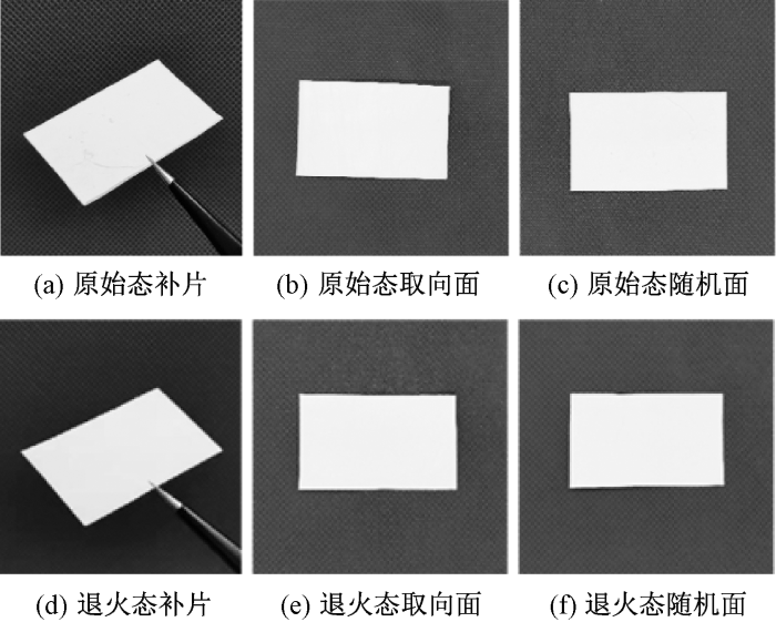
图1
原始态补片与退火态补片宏观形貌
Fig.1
Macroscopic morphologies of as-received and as-annealed patches. (a) As-received patch; (b) As-received oriented layer;(c) As-received isotropic layer; (d) As-annealed patch; (e) As-annealed oriented layer; (f) As-annealed isotropic layer
图2示出原始态补片和退火态补片在干态条件下的扫描电镜照片。由图可知,原始态补片与退火态补片均呈现连续且完整的纤维网状结构。2类样品的纤维平均直径分别为(0.966±0.241)、(0.967±0.228) μm,其直径离散度均较大,这与I型胶原成分的复合添加有关,天然类聚合物的存在使得静电纺中的射流鞭动概率增多。尽管如此,同时存在的微纳尺度纤维有益于补片对肩袖组织的生物学仿生。压汞法测定结果也进一步说明了该补片的结构优势,无论是退火态补片的孔隙率(80.92 ± 3.56)%,还是原始态补片的(81.21 ± 2.72)%,均大于80%。经统计分析可知,采用恒应力退火的处理工艺并未对补片的纤维尺度及膜孔隙结构产生明显影响,二者间差异无显著性(p>0.05),表明维持了原有的三维多孔微纳网络特征。
图2
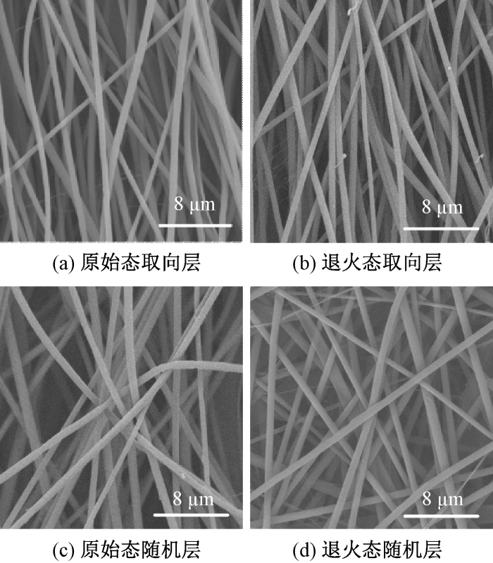
图2
干态条件下原始态补片与退火态补片电镜照片
Fig.2
SEM images of as-received and as-annealed patches under dry conditions. (a) As-received oriented layer; (b) As-annealed oriented layer; (c) As-received isotropic layer; (d) As-annealed isotropic layer
2.1.2 化学组成与胶原结构稳定性分析
图3为不同样品的红外光谱图。在原始态补片与退火态补片的红外光谱中,1 750 cm-1处可见归属于PDLLA分子内酯羰基(C=O)伸缩振动的特征吸收峰;同时,在1 645 cm-1(酰胺I带,C=O伸缩振动)、1 540 cm-1(酰胺II带,N—H弯曲振动耦合C—N伸缩振动)及3 285 cm-1(酰胺A带,N—H伸缩振动)处的特征峰位与I型胶原标准特征峰完全匹配,证实I型胶原组分已成功整合至PDLLA基体中。为进一步确认热退火处理是否会影响I型胶原的结构完整性(这直接关系到其生物活性和补片的最终性能),采用SDS-PAGE对退火前后的I型胶原进行测试分析。
图3
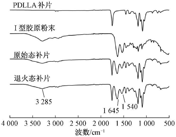
图4示出原始态补片和退火态补片中的I型胶原的SDS-PAGE表征结果。以标准蛋白分子量Marker为参照,可见样品在130~250 ku范围内呈现2条清晰主条带,分别对应于I型胶原特征的α1与α2链;在高于250 ku的区域亦可见2条条带,分别为β链(α链二聚体)与γ链(α链三聚体)。结果显示退火前后胶原的α1、α2、β、γ链与原始态胶原的条带相比,颜色灰度及对比度上无差异。值得注意的是,在α链以下区域未观察到明显低分子量弥散条带。该结果表明,原始态补片与退火态补片中的I型胶原几乎不存在胶原多肽碎片或水解产物,说明其结构完整、纯度较高。由此证明,退火处理并未引起I型胶原主链的显著降解或聚集,较好地维持了其天然三螺旋结构与组分稳定性,说明其具备良好的温度耐受性。
图4
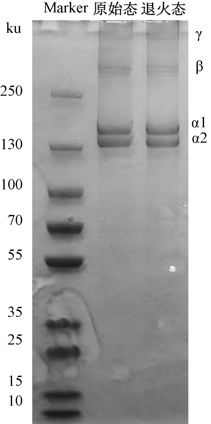
图4
不同样品的聚丙烯酰胺凝胶电泳图
Fig.4
Polyacrylamide gel electrophoresis of different samples
2.1.3 退火诱导的结晶结构变化
为进一步探究退火处理对补片微观结构,尤其是结晶结构的影响,通过XRD分析原始态/退火态补片的晶体结构。结果表明,经退火处理后,退火态补片的结晶度由原始态的23.7%显著提升至27.2%,相对提升了14.8%,充分证明退火处理有效促进了补片内部分子链的重排与有序化,从而形成了更为完善的晶体结构。结晶度提高。为解释退火态补片在湿态环境下展现出更优的力学性能保持率与形态稳定性提供了关键的结构依据。
2.2 补片表面性质与湿态结构稳定性
2.2.1 表面润湿性分析
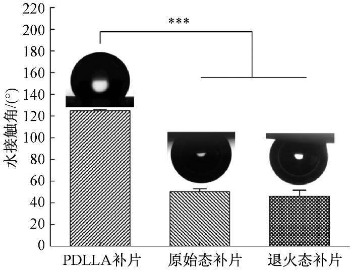
图5示出不同样品水接触角的测试结果。经液滴法测试得到,PDLLA补片表面疏水,水接触角为(124.88±1.36)°;经气泡法测试得到,原始态补片表面水接触角降至(50.28 ± 2.59)°,表现为亲水性;经退火处理后,退火态补片表面水接触角为(45.28±5.68)°,仍保持亲水性,退火前后亲水性能并无显著差异(p>0.05)。上述结果辅助证明了I型胶原的成功引入,且I型胶原组分显著改善了材料的亲水性能。亲水性的提升虽有利于细胞行为,但也意味着补片在生理湿态环境中更易受到水分子渗透与溶胀的挑战。为此,本研究系统评价了补片在湿态环境中长期浸泡的结构稳定性。
图5
2.2.2 湿态环境下的形貌与结构演变
图6示出原始态补片与退火态补片在PBS中培养不同时间后的SEM照片。图7示出不同样品纤维的直径分布图。表1示出PBS培养条件下不同样品的纤维直径变化。图6表明,2种补片在湿态环境下均出现纤维溶胀和取向结构弱化现象。纤维直径的定量统计结果(见图7和表1)进一步显示,随着浸泡时间的延长,原始态补片的直径由初始的(0.966±0.241)μm分别增长至7 d的(1.237±0.158) μm和14 d的(1.602±0.244)μm,14 d内累计增幅达65.79%。相比之下,退火态补片的纤维直径由初始的(0.967±0.228)μm增至7 d的(1.076±0.212)μm,并在14 d时达到(1.219±0.205)μm,累计增幅仅为26.01%,显著低于原始态补片(p<0.001)。以上结果表明,退火处理有效延缓了纤维在湿态环境中的溶胀进程,增强了其长期结构稳定性。
图6
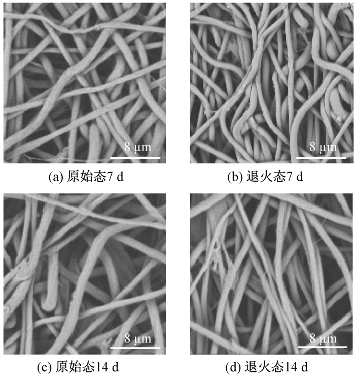
图6
PBS培养条件下原始态补片和退火态补片的电镜照片
Fig.6
SEM images of as-received and as-annealed patches under PBS incubation conditions. (a)7 d-as-received; (b)7 d-as-annealed; (c)14 d-as-received; (d)14 d-as-annealed
图7
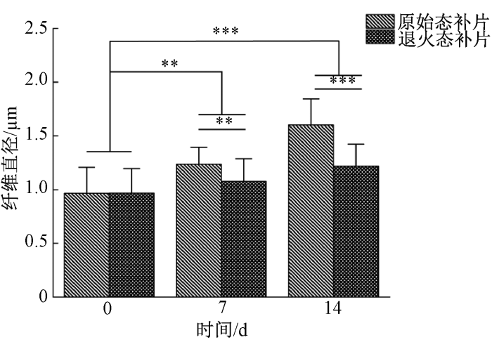
图7
不同样品纤维培养不同时间的直径分布图
Fig.7
Fiber diameter distribution diagram of different samples cultured for different periods
表1 PBS培养条件下不同样品的纤维直径变化
Tab.1
| 取样时间/d | 纤维直径/μm | |
|---|---|---|
| 原始态补片 | 退火态补片 | |
| 0 | 0.966±0.241 | 0.967±0.228 |
| 7 | 1.237±0.158 | 1.076±0.212 |
| 14 | 1.602±0.244 | 1.219±0.205 |
图8示出不同样品纤维的取向分布图。定量分析结果表明:浸泡前,2种补片的纤维取向分布无显著差异;浸泡14 d后,退火态补片的纤维取向保持率较原始态补片提高约50%,其纤维取向结构稳定性显著优于原始态补片(p<0.001)。
图8
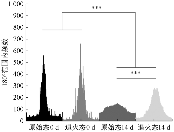
图8
不同样品纤维的取向分布图
Fig.8
Fiber orientation distribution diagram of different samples
上述形态学变化可归因于湿态环境下水分子的渗透与溶胀效应。PDLLA/I型胶原复合体系中,亲水性胶原组分的存在构建了水分快速渗透的通道,水分子首先通过氢键作用与胶原侧链结合,进而扩散至PDLLA非晶区,削弱其链间缠结,引发纤维溶胀。随着浸泡时间延长,溶胀持续累积,最终导致纤维间粘连、取向丧失,甚至结构崩塌。而退火处理显著抑制了这种劣化过程,其原因在于:退火过程中,在恒定应力与温度(65 ℃,高于PDLLA的玻璃化转变温度Tg)的共同作用下,PDLLA分子链获得足够的运动能力发生重排,形成更为完善和稳定的晶体结构。这种结构转变一方面增强了分子链间的相互作用力,提高了抵抗水分子渗透与溶胀变形的能力;另一方面,结晶区的增加有效限制了非晶区链段的运动性,从而更有效地维持了纤维的原有形貌和取向排列。此外,热退火也可促进PDLLA与胶原之间的分子级界面结合,通过氢键或疏水相互作用增强相界面稳定性,这进一步延缓了水分诱导的结构破坏进程。因此,退火处理通过促进链重排、提升结晶度与增强界面结合等多重机制,显著提升了补片在湿态环境下的长期结构稳定性。值得指出的是,力学性能测试中采用的60 min湿态平衡已能诱发显著的性能变化,说明PDLLA/I型胶原复合纤维对水分极为敏感,短时间内即可因水塑化作用导致力学性能衰减。然而,本研究关注的不仅是短时水合响应,更是补片在长期生理环境中的结构维持能力。为此设置了7 d与14 d的湿态培养实验,以更真实地模拟植入后的稳定性需求。
2.2.3 湿态面积收缩行为分析
表2示出PBS培养条件下不同样品在不同时间节点的面积收缩率。结果表明,PDLLA 补片凭借疏水性表现出优异的形态稳定性,14 d内收缩率仅增至(5.74±0.25)%。原始态补片和退火态补片的面积收缩现象显著:原始态补片7 d即达到81.73% 的高收缩率,14 d略升至82.71%;退火态补片收缩程度明显降低,7 d和14 d的收缩率分别为37.09%和43.41%。值得注意的是,2种补片的大部分收缩发生于前7 d,后续变化平缓,且退火处理使补片的面积收缩率降低39.3%(p<0.01),表明退火处理有效提升了补片的形态稳定性。
表2 PBS培养条件下不同样品的面积收缩率
Tab.2
| 时间 | 面积收缩率/% | ||
|---|---|---|---|
| PDLLA补片 | 原始态补片 | 退火态补片 | |
| 30 min | 0 | 0 | 0 |
| 7 d | 3.50±0.28 | 81.73±0.16 | 37.09±0.50 |
| 14 d | 5.74±0.25 | 82.71±0.12 | 43.41±0.78 |
2.3 力学性能及其与湿态稳定性的关联
图9
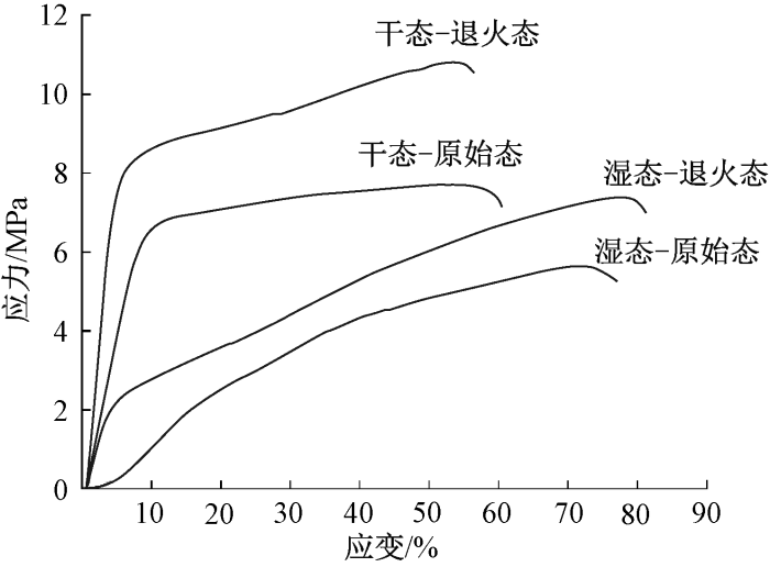
图9
干/湿态条件下不同补片的应力-应变曲线
Fig.9
Stress-strain curves of different samples under dry/wet conditions
表3 干/湿态条件下不同样品力学性能
Tab.3
| 试样名称 | 断裂强度/MPa | 弹性模量/MPa | 断裂伸长率/% |
|---|---|---|---|
| 干态-原始态 | 7.71±0.43 | 87.92±13.89 | 60.52±18.67 |
| 干态-退火态 | 10.79±1.53 | 142.32±25.38 | 56.37±10.73 |
| 湿态-原始态 | 5.64±0.53 | 18.03±2.00 | 77.03±22.76 |
| 湿态-退火态 | 7.40±0.85 | 33.27±1.83 | 81.22±10.51 |
由图9和表3可看出,退火处理显著优化补片在干态条件下的力学性能及在湿条件下的力学完整性:干态下退火态补片断裂强度相比原始态补片提升了39.9%(从(7.71±0.43)MPa增至(10.79±1.53)MPa,p<0.01),弹性模量提升了61.9%(从(87.92±13.89) MPa增至(142.32±25.38) MPa,p<0.01),应力-应变曲线初始斜率陡增印证其刚性强化;而在湿态条件下,退火态补片相比原始态补片能更有效抵抗水塑化效应,断裂强度(从(5.64±0.53)MPa增至(7.40±0.85)MPa,p<0.05)与弹性模量(从(18.03±2.00)MPa增至(33.27±1.83)MPa, p<0.05)分别提高了31.21%和84.53%。该力学强化效应主要源于退火诱导的微观结构优化。首先,结晶度的提升(由23.7%增至27.2%)直接贡献了材料的刚性与强度,晶体区域作为物理交联点,有效传递和分散应力。其次,分子链的取向与重排减少了结构缺陷,使得纤维在拉伸时能承受更高的载荷。在湿态环境下,退火处理形成的致密化结构极大地限制了水分子对聚合物链的塑化作用,减缓了模量与强度的衰减速度,从而使其在充分水合后仍能保持优于原始态的力学完整性。这种结构优势是退火态补片在长期湿态培养中维持更小的纤维溶胀、更高纤维取向保持率和更低面积收缩率的根本原因。尽管60 min的湿态平衡已使2类补片的力学性能出现下降,但长期(7 d、14 d)稳定性实验结果表明,退火处理能显著抑制纤维进一步溶胀与结构崩塌,说明其稳定机制具有持续效应。因此,短时力学测试与长期形态观测相结合,可共同揭示退火处理在提升补片综合性能方面的优势。
2.4 细胞相容性与黏附增殖行为
图10
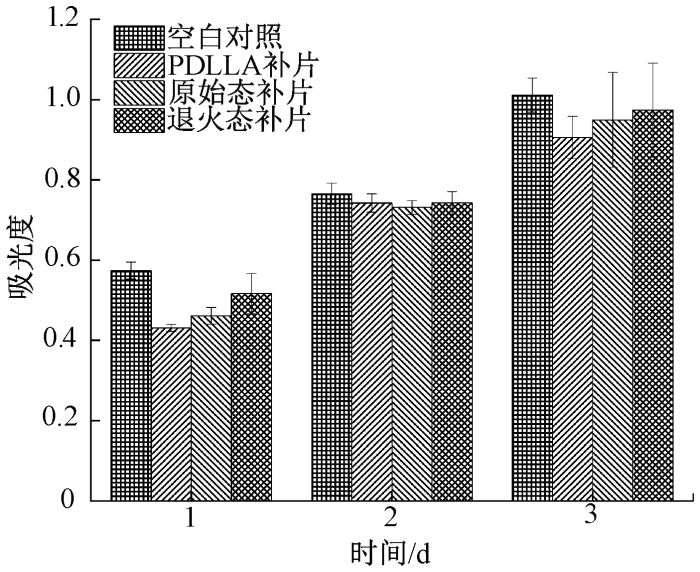
表4 不同样品的相对增殖率
Tab.4
| 样品组别 | 相对增殖率/% | ||
|---|---|---|---|
| 1 d | 2 d | 3 d | |
| PDLLA补片 | 75.09 | 97.00 | 89.55 |
| 原始态补片 | 80.37 | 95.57 | 93.92 |
| 退火态补片 | 90.03 | 97.05 | 96.29 |
表5 细胞在不同基底上的黏附细胞的吸光度
Tab.5
| 样品组别 | 吸光度 | ||||
|---|---|---|---|---|---|
| 4 h | 24 h | 48 h | 72 h | 96 h | |
| 空白对照 | 0.31 | 0.46 | 0.54 | 0.81 | 1.10 |
| PDLLA补片 | 0.22 | 0.33 | 0.38 | 0.64 | 0.69 |
| 原始态补片 | 0.25 | 0.33 | 0.43 | 0.74 | 0.86 |
| 退火态补片 | 0.24 | 0.36 | 0.42 | 0.74 | 0.82 |
表6 细胞在不同样品表面培养后的黏附率
Tab.6
| 样品组别 | 细胞黏附率/% | ||||
|---|---|---|---|---|---|
| 4 h | 24 h | 48 h | 72 h | 96 h | |
| PDLLA补片 | 19.70 | 29.70 | 35.03 | 58.51 | 63.03 |
| 原始态补片 | 22.36 | 30.32 | 39.01 | 67.78 | 78.46 |
| 退火态补片 | 22.03 | 32.92 | 38.28 | 67.68 | 75.18 |
由表6可知,在培养初期(4 h),原始态补片与退火态补片的细胞黏附率分别为22.36%与22.03%,均略高于PDLLA补片(19.70%),表明Ⅰ型胶原的引入在一定程度上促进了细胞早期黏附。随着培养时间延长至24 h与48 h,各组黏附率均呈上升趋势,且原始态补片与退火态补片组始终优于PDLLA补片组,进一步证实胶原组分对细胞行为的积极影响。在培养后期(72 h与96 h),各组细胞黏附率均显著提高。值得注意的是,原始态补片与退火态补片在72 h时的黏附率分别达到67.78%与67.68%,显著高于PDLLA补片组(58.51%);至96 h时,原始态补片组黏附率进一步提高至78.46%,退火态补片为75.18%,均明显高于PDLLA补片组(63.03%)。以上结果表明,I型胶原的引入有效增强了补片表面对细胞的亲和力,促进了细胞的长期黏附与扩展。尽管退火处理在部分时间点(如24 h)显示出略高于原始态的黏附率,但在整体趋势上,原始态补片与退火态补片之间的细胞黏附行为未表现出显著差异。这说明退火处理在维持补片湿态稳定性与力学性能的同时,并未对I型胶原所赋予的生物活性表面产生不利影响,补片仍保持良好的细胞相容性与促黏附能力。
3 结论
本研究针对静电纺丝生物医用纤维补片在湿态应用环境中易发生力学性能衰减和结构收缩、难以长期稳定服役的问题,构建了一种基于PDLLA/I型胶原共混体系并结合恒应力热退火处理的微纳米纤维膜增强策略。研究结果表明,通过在PDLLA基体中引入牛跟腱来源的Ⅰ型胶原,并在受控恒应力条件下进行热退火处理,可显著改善纤维膜在湿态环境中的形态保持能力和力学性能保持率。与未处理样品相比,经退火处理的共混纤维膜在湿态拉伸过程中表现出更高的模量保持水平和更小的尺寸变化幅度,表明该策略能够有效缓解高分子纤维在吸水条件下因链段松弛和结构重排引起的性能劣化问题。
PDLLA/胶原共混结合恒应力热退火的策略具有工艺可控性强、材料体系成熟、易于放大制备等优势,不仅可用于肩袖修复等腱骨界面相关补片的性能优化,也为其它需要在湿态环境中长期承载的生物医用纤维支架提供了新的设计思路。该研究为通过后处理工艺提升静电纺丝纤维结构稳定性提供了实验依据和理论参考,具有良好的推广潜力和应用前景。
参考文献
Advanced nanofiber-based scaffolds for Achilles tendon regenerative engineering
[J].
DOI:10.3389/fbioe.2022.897010
URL
[本文引用: 1]

The Achilles tendon (AT) is responsible for running, jumping, and standing. The AT injuries are very common in the population. In the adult population (21–60 years), the incidence of AT injuries is approximately 2.35 per 1,000 people. It negatively impacts people’s quality of life and increases the medical burden. Due to its low cellularity and vascular deficiency, AT has a poor healing ability. Therefore, AT injury healing has attracted a lot of attention from researchers. Current AT injury treatment options cannot effectively restore the mechanical structure and function of AT, which promotes the development of AT regenerative tissue engineering. Various nanofiber-based scaffolds are currently being explored due to their structural similarity to natural tendon and their ability to promote tissue regeneration. This review discusses current methods of AT regeneration, recent advances in the fabrication and enhancement of nanofiber-based scaffolds, and the development and use of multiscale nanofiber-based scaffolds for AT regeneration.
Biomaterials based strategies for rotator cuff repair
[J].DOI:10.1016/j.colsurfb.2017.06.004 URL [本文引用: 1]
Histopathology of rotator cuff tears
[J].
DOI:10.1097/JSA.0b013e318213bccb
PMID:21822106
[本文引用: 1]
The pathogenesis of rotator cuff tears is multifactorial. Tendon abnormalities of the rotator cuff include alteration of collagen fiber structure, tenocytes, cellularity, and vascularity. Ruptured tendons show marked collagen degeneration and disordered arrangement of collagen fibers. Fibroblast population decreases as the size of the tear in the rotator cuff increases. The larger fibroblast population seen in the smaller tears is also actively proliferating and is part of an active reparative process. Inflammatory cell infiltrate correlates inversely to rotator cuff tear size in the torn supraspinatus tendon samples, with larger tears showing a marked reduction in all cell types. As tear size increase, there is also a progressive decrease in the number of blood vessels. Whether rotator cuff tear heals spontaneously is an important pathologic and clinical question. Histologic changes indicative of repair and inflammation lead to consider biological options in addition to biomechanical treatment of the rotator cuff tears.
Epidemiology, natural history, and indications for treatment of rotator cuff tears
[J].
DOI:10.1016/j.csm.2012.07.001
PMID:23040548
[本文引用: 1]
The etiology of rotator cuff disease is likely multifactorial, including age-related degeneration and microtrauma and macrotrauma. The incidence of rotator cuff tears increases with aging with more than half of individuals in their 80s having a rotator cuff tear. Smoking, hypercholesterolemia, and genetics have all been shown to influence the development of rotator cuff tearing. Substantial full-thickness rotator cuff tears, in general, progress and enlarge with time. Pain, or worsening pain, usually signals tear progression in both asymptomatic and symptomatic tears and should warrant further investigation if the tear is treated conservatively. Larger (>1-1.5 cm) symptomatic full-thickness cuff tears have a high rate of tear progression and, therefore, should be considered for earlier surgical repair in younger patients if the tear is reparable and there is limited muscle degeneration to avoid irreversible changes to the cuff, including tear enlargement and degenerative muscle changes. Smaller symptomatic full-thickness tears have been shown to have a slower rate of progression, similar to partial-thickness tears, and can be considered for initial nonoperative treatment due to the limited risk for rapid tear progression. In both small full-thickness tears and partial-thickness tears, increasing pain should alert physicians to obtain further imaging as it can signal tear progression. Natural history data, along with information on factors affecting healing after rotator cuff repair, can help guide surgeons in making appropriate decisions regarding the treatment of rotator cuff tears. The management of rotator cuff tears should be considered in the context of the risks and benefits of operative versus nonoperative treatment. Tear size and acuity, the presence of irreparable changes to the rotator cuff or glenohumeral joint, and patient age should all be considered in making this decision. Initial nonoperative care can be safely undertaken in older patients (>70 years old) with chronic tears; in patients with irreparable rotator cuff tears with irreversible changes, including significant atrophy and fatty infiltration, humeral head migration, and arthritis; in patients of any age with small (<1 cm) full-thickness tears; or in patients without a full-thickness tear. Early surgical treatment can be considered in significant (>1 cm-1.5 cm) acute tears or young patients with full-thickness tears who have a significant risk for the development of irreparable rotator cuff changes.Copyright © 2012. Published by Elsevier Inc.
Tendon tissue engineering: an overview of biologics to promote tendon healing and repair
[J].
DOI:10.1177/20417314231196275
URL
[本文引用: 1]
Tendons are dense connective tissues with a hierarchical polarized structure that respond to and adapt to the transmission of muscle contraction forces to the skeleton, enabling motion and maintaining posture. Tendon injuries, also known as tendinopathies, are becoming more common as populations age and participation in sports/leisure activities increases. The tendon has a poor ability to self-heal and regenerate given its intrinsic, constrained vascular supply and exposure to frequent, severe loading. There is a lack of understanding of the underlying pathophysiology, and it is not surprising that disorder-targeted medicines have only been partially effective at best. Recent tissue engineering approaches have emerged as a potential tool to drive tendon regeneration and healing. In this review, we investigated the physiochemical factors involved in tendon ontogeny and discussed their potential application in vitro to reproduce functional and self-renewing tendon tissue. We sought to understand whether stem cells are capable of forming tendons, how they can be directed towards the tenogenic lineage, and how their growth is regulated and monitored during the entire differentiation path. Finally, we showed recent developments in tendon tissue engineering, specifically the use of mesenchymal stem cells (MSCs), which can differentiate into tendon cells, as well as the potential role of extracellular vesicles (EVs) in tendon regeneration and their potential for use in accelerating the healing response after injury.
Biomimetic patch with gradient-induced regeneration for tendon-bone interface to repair rotator cuff injury
[J].DOI:10.1016/j.cej.2025.159985 URL [本文引用: 1]
Nanofiber-based matrices for rotator cuff regenerative engineering
[J].DOI:10.1016/j.actbio.2019.05.041 URL [本文引用: 2]
Electrospun scaffolds for tissue engineering: a review
[J].
Recent progress in biomedical scaffold fabricated via electrospinning: design, fabrication and tissue engineering application
[J].DOI:10.1002/adfm.v35.1 URL [本文引用: 1]
Biomimetic natural biomaterials for tissue engineering and regenerative medicine: new biosynthesis methods, recent advances, and emerging applications
[J].
DOI:10.1186/s40779-023-00448-w
PMID:36978167
[本文引用: 1]
Biomimetic materials have emerged as attractive and competitive alternatives for tissue engineering (TE) and regenerative medicine. In contrast to conventional biomaterials or synthetic materials, biomimetic scaffolds based on natural biomaterial can offer cells a broad spectrum of biochemical and biophysical cues that mimic the in vivo extracellular matrix (ECM). Additionally, such materials have mechanical adaptability, microstructure interconnectivity, and inherent bioactivity, making them ideal for the design of living implants for specific applications in TE and regenerative medicine. This paper provides an overview for recent progress of biomimetic natural biomaterials (BNBMs), including advances in their preparation, functionality, potential applications and future challenges. We highlight recent advances in the fabrication of BNBMs and outline general strategies for functionalizing and tailoring the BNBMs with various biological and physicochemical characteristics of native ECM. Moreover, we offer an overview of recent key advances in the functionalization and applications of versatile BNBMs for TE applications. Finally, we conclude by offering our perspective on open challenges and future developments in this rapidly-evolving field.© 2023. The Author(s).
Electrospinning and electrospun nanofibers: methods, materials, and applications
[J].
DOI:10.1021/acs.chemrev.8b00593
PMID:30916938
[本文引用: 1]
Electrospinning is a versatile and viable technique for generating ultrathin fibers. Remarkable progress has been made with regard to the development of electrospinning methods and engineering of electrospun nanofibers to suit or enable various applications. We aim to provide a comprehensive overview of electrospinning, including the principle, methods, materials, and applications. We begin with a brief introduction to the early history of electrospinning, followed by discussion of its principle and typical apparatus. We then discuss its renaissance over the past two decades as a powerful technology for the production of nanofibers with diversified compositions, structures, and properties. Afterward, we discuss the applications of electrospun nanofibers, including their use as "smart" mats, filtration membranes, catalytic supports, energy harvesting/conversion/storage components, and photonic and electronic devices, as well as biomedical scaffolds. We highlight the most relevant and recent advances related to the applications of electrospun nanofibers by focusing on the most representative examples. We also offer perspectives on the challenges, opportunities, and new directions for future development. At the end, we discuss approaches to the scale-up production of electrospun nanofibers and briefly discuss various types of commercial products based on electrospun nanofibers that have found widespread use in our everyday life.
A hierarchical, stretchable and stiff fibrous biotemplate engineered using stagger-electrospinning for augmentation of rotator cuff tendon-healing
[J].
DOI:10.1039/c4tb01642d
PMID:32261978
[本文引用: 1]
The regeneration of fibrocartilage at the tendon-bone insertion site in rotator cuff tears (RCTs) is challenging due to the complexity of its composition and mechanical properties. In this study, hierarchical, stretchable and stiff fibrous scaffolds composed of microfibers of poly(ε-caprolactone) (PCL) and nanofibers of chitosan (CS) were fabricated using stagger-electrospinning for the augmentation of RCT-healing. It was found that the composite PCL-CS scaffolds had significantly improved strength and failure strain compared to the control CS scaffolds and increased stiffness compared to the control PCL scaffolds. These scaffolds also showed enhanced hydrophilicity, water absorption and a faster degradation rate compared to the PCL scaffolds. Moreover, they demonstrated better fibroblast attachment and proliferation compared to the PCL scaffolds. Radiological and histological analysis revealed that the PCL-CS scaffolds enhanced new bone formation (mineralization) and collagen and glycosaminoglycan expression (major components of extracellular matrix) compared to the PCL scaffolds. Furthermore, the torn tissues at the tendon-bone insertion site regenerated with the PCL-CS scaffolds showed higher strength and failure strain as well as stiffness compared to those repaired using only the PCL scaffolds. The above mentioned results suggest that the hierarchical, stretchable and stiff fibrous scaffolds engineered using stagger-electrospinning have great potential for the augmentation of RCT-healing.
静电纺丝聚己内酯/Ⅰ型胶原纳米纤维取向性补片用于肩袖修复
[J].
Electrospun polycaprolactone/collagen type Ⅰ nanofibers oriented patch for rotator cuff repairing
[J].
DOI:10.7507/1002-1892.201811034
PMID:31090359
[本文引用: 1]
Electrospinning technique was used to manufacture polycaprolactone (PCL)/collagen typeⅠ nanofibers orientated patches and to study their physical and chemical characterization, discussing their feasibility as synthetic patches for rotator cuff repairing.PCL patches were prepared by electrospinning with 10% PCL electrospinning solution (control group) and PCL/collagen typeⅠorientated nanofibers patches were prepared by electrospinning with PCL electrospinning solution with 25% collagen type Ⅰ(experimental group). The morphology and microstructure of the two patches were observed by gross and scanning electron microscopy, and the diameter and porosity of the fibers were measured; the mechanical properties of the patches were tested by uniaxial tensile test; the composition of the patches was analyzed by Fourier transform infrared spectroscopy; and the contact angle of the patch surface was measured. Two kinds of patch extracts were co-cultured with the third generation of rabbit tendon stem cells. Cell counting kit 8 (CCK-8) was used to detect the toxicity and cell proliferation of the materials. Normal cultured cells were used as blank control group. Rabbit tendon stem cells were co-cultured with the two patches and stained with dead/living cells after 3 days of culture, and laser confocal scanning microscopy was used to observe the cell adhesion and activity on the patch.Gross and scanning electron microscopy showed that the two patch fibers were arranged in orientation. The diameter of patch fibers in the experimental group was significantly smaller than that in the control group ( =26.907, =0.000), while the porosity in the experimental group was significantly larger than that in the control group ( =2.506, =0.032). The tensile strength and Young's modulus of the patch in the experimental group were significantly higher than those in the control group ( =3.705, =0.029; =4.064, =0.034). Infrared spectrum analysis showed that PCL and collagen type Ⅰ were successfully mixed in the experimental group. The surface contact angle of the patch in the experimental group was (73.88±4.97)°, which was hydrophilic, while that in the control group was (128.46±5.10) °, which was hydrophobic. There was a significant difference in the surface contact angle between the two groups ( =21.705, =0.002). CCK-8 test showed that with the prolongation of culture time, the cell absorbance ( ) value increased gradually in each group, and there was no significant difference between the experimental group and the control group at each time point ( >0.05). Laser confocal scanning microscopy showed that rabbit tendon stem cells could adhere and grow on the surface of both patches after 3 days of culture. The number of cells adhered to the surface of the patches in the experimental group was more than that in the control group, and the activity was better.PCL/ collagen type Ⅰ nanofibers orientated patch prepared by electrospinning technology has excellent physical and chemical properties, cell adhesion, and no cytotoxicity. It can be used as an ideal scaffold material in tendon tissue engineering for rotator cuff repair in the future.
Multilayered polycaprolactone/gelatin fiber-hydrogel composite for tendon tissue engineering
[J].
DOI:10.1016/j.actbio.2016.03.004
PMID:26945631
[本文引用: 1]
Regeneration of injured tendon and ligament (T&L) remains a clinical challenge due to their poor intrinsic healing capacity. Tissue engineering provides a promising alternative treatment approach to facilitate T&L healing and regeneration. Successful tendon tissue engineering requires the use of three-dimensional (3D) biomimetic scaffolds that possess the physical and biochemical features of native tendon tissue. We report here the development and characterization of a novel composite scaffold fabricated by co-electrospinning of poly-ε-caprolactone (PCL) and methacrylated gelatin (mGLT). We found that photocrosslinking retained mGLT, resulted in a uniform distribution of mGLT throughout the depth of scaffold and also preserved scaffold mechanical strength. Moreover, photocrosslinking was able to integrate stacked scaffold sheets to form multilayered constructs that mimic the structure of native tendon tissues. Importantly, cells impregnated into the constructs remained responsive to topographical cues and exogenous tenogenic factors, such as TGF-β3. The excellent biocompatibility and highly integrated structure of the scaffold developed in this study will allow the creation of a more advanced tendon graft that possesses the architecture and cell phenotype of native tendon tissues.The clinical challenges in tendon repair have spurred the development of tendon tissue engineering approaches to create functional tissue replacements. In this study, we have developed a novel composite scaffold as a tendon graft consisting of aligned poly-ε-caprolactone (PCL) microfibers and methacrylated gelatin (mGLT). Cell seeding and photocrosslinking between scaffold layers can be performed simultaneously to create cell impregnated multilayered constructs. This cell-scaffold construct combines the advantages of PCL nanofibrous scaffolds and photocrosslinked gelatin hydrogels to mimic the structure, mechanical anisotropy, and cell phenotype of native tendon tissue. The scaffold engineered here as a building block for multilayer constructs should have applications beyond tendon tissue engineering in the fabrication of tissue grafts that consist of both fibrous and hydrogel components.Copyright © 2016 Acta Materialia Inc. Published by Elsevier Ltd. All rights reserved.
A novel electrospun-aligned nanoyarn-reinforced nanofibrous scaffold for tendon tissue engineering
[J].DOI:10.1016/j.colsurfb.2014.06.061 URL [本文引用: 1]
Biomimetic scaffolds with dual gradients of biological effectors for tendon-to-bone repair
[J].DOI:10.1002/adhm.v14.28 URL [本文引用: 1]
Design challenges in polymeric scaffolds for tissue engineering
[J].
DOI:10.3389/fbioe.2021.617141
URL
[本文引用: 1]
Numerous surgical procedures are daily performed worldwide to replace and repair damaged tissue. Tissue engineering is the field devoted to the regeneration of damaged tissue through the incorporation of cells in biocompatible and biodegradable porous constructs, known as scaffolds. The scaffolds act as host biomaterials of the incubating cells, guiding their attachment, growth, differentiation, proliferation, phenotype, and migration for the development of new tissue. Furthermore, cellular behavior and fate are bound to the biodegradation of the scaffold during tissue generation. This article provides a critical appraisal of how key biomaterial scaffold parameters, such as structure architecture, biochemistry, mechanical behavior, and biodegradability, impart the needed morphological, structural, and biochemical cues for eliciting cell behavior in various tissue engineering applications. Particular emphasis is given on specific scaffold attributes pertaining to skin and brain tissue generation, where further progress is needed (skin) or the research is at a relatively primitive stage (brain), and the enumeration of some of the most important challenges regarding scaffold constructs for tissue engineering.
A review of biomaterial degradation assessment approaches employed in the biomedical field
[J].
DOI:10.1038/s41529-024-00487-1
[本文引用: 1]
The biological response to biomaterials plays a crucial role in selecting suitable materials for the formulation and development of tissue engineering platforms. Biodegradation is one of the properties that is considered in selecting appropriate biomaterials for biomedical applications. Biodegradation is the process of breaking down large molecules into smaller molecules with/without the aid of catalytic enzymes. The biodegradation process is crucial in the chemical absorption, distribution, metabolism, excretion, and toxicity (ADMET) process of biomaterials and small molecules in the body. Degradation of biomaterials can be followed by assessing the physical, mechanical, and chemical attributes of biomaterials. There are several techniques/parameters that can be targeted when studying the degradation of biomaterials, with gravimetric analysis, surface erosion, and morphological changes being the largely employed techniques. However, the techniques present a few limitations, such as technical errors and material solubility being mistaken for degradation, and these techniques can infer but not confirm degradation as they do not provide the chemical composition of fragmenting/fragmented molecules. The American Society for Testing and Materials (ASTM) guidelines provide techniques and parameters for assessing biodegradation. However, the ASTM guidelines for degradation assessment approaches and techniques need to be updated to provide sufficient evidence to draw conclusive decisions regarding the degradation of biomaterials. In this review, the degradation assessment approaches and techniques are critically reviewed about their advantages and disadvantages, and to provide suggestions on how they can still play a role in assessing the degradation of biomaterials. This review could assist researchers employ cost-effective, efficient, and multiple degradation assessment techniques to evaluate and provide sufficient information about the degradation of biomaterials. Suggested future ASTM guidelines for assessing biodegradation should include measuring parameters (such as chemical, mechanical, or physical attributes of biomaterials) in real-time, employing non-invasive, continuous, and automated processes.
Thermal annealing optimization for improved mechanical performance of PLA parts produced via 3D printing
[J].DOI:10.1016/j.polymertesting.2025.108735 URL [本文引用: 1]
Mechanical and thermal characterization of annealed oriented PAN nanofibers
[J].
DOI:10.3390/polym15153287
URL
[本文引用: 1]
Polyacrylonitrile (PAN) nanofibers have extensive applications as filters in various fields, including air and water filtration, biofluid purification, and the removal of toxic compounds and hazardous pollutants from contaminated water. This research focuses on investigating the impacts of annealing on the mechanical and thermal characteristics of oriented PAN nanofibers produced through the electrospinning of a PAN solution. The nanofiber mats were subjected to annealing temperatures ranging from 70 °C to 350 °C and characterized using a tensile test machine, thermogravimetry, differential scanning calorimetry, and scanning electron microscopy (SEM). The study aimed to examine the tensile strength in the transverse and longitudinal directions, Young’s modulus, and glass transition temperatures of PAN nanofiber mats. The results indicate that, upon annealing, the diameter of the nanofibers decreased by approximately 20%, while the tensile strength increased in the longitudinal and transverse directions by 32% and 23%, respectively. Furthermore, the annealing temperature influenced the glass transition temperature of the nanofiber mats, which exhibited a 6% decrease at 280 °C, while the degradation temperature showed a slight increase of 3.5% at 280 °C. The findings contribute to a better understanding of the effects of annealing on PAN nanofiber mats, facilitating their potential for various filtration applications.
Biomanufacturing organized collagen-based microfibers as a tissue engineered device (TEND) for tendon regeneration
[J].
DOI:10.1088/1748-605X/abb875
[本文引用: 1]
Approximately 800, 000 surgical repairs are performed annually in the U.S. for debilitating injuries to ligaments and tendons of the foot, ankle, knee, wrist, elbow and shoulder, presenting a significant healthcare burden. To overcome current treatment shortcomings and advance the treatment of tendon and ligament injuries, we have developed a novel electrospun Tissue ENgineered Device (TEND), comprised of type I collagen and poly(D,L-lactide) (PDLLA) solubilized in a benign solvent, dimethyl sulfoxide (DMSO). TEND fiber alignment, diameter and porosity were engineered to enhance cell infiltration leading to promote tissue integration and functional remodeling while providing biomechanical stability. TEND rapidly adsorbs blood and platelet-rich-plasma (PRP), and gradually releases growth factors over two weeks. TEND further supported cellular alignment and upregulation of tenogenic genes from clinically relevant human stem cells within three days of culture. TEND implanted in a rabbit Achilles tendon injury model showed new in situ tissue generation, maturation, and remodeling of dense, regularly oriented connective tissue in vivo. In all, TEND’s organized microfibers, biological fluid and cell compatibility, strength and biocompatiblility make significant progress towards clinically translating electrospun collagen-based medical devices for improving the clinical outcomes of tendon injuries.

 京公网安备11010502044800号


