



为此,本研究融合熔体微分静电纺丝、熔体静电纺直写及导电改性技术,以“防渗漏基底构建-力学各向异性设计-导电功能赋予”为技术主线,构建层级化聚己内酯/碳纳米管(PCL/CNTs)复合心脏补片。首先通过熔体微分静电纺丝技术制备微孔基底膜,阻隔细胞渗漏;进而利用熔体静电纺直写技术成形菱形拓扑结构,精准调控力学各向异性;最后通过超声分散碳纳米管实现导电功能。三者协同,旨在实现心肌组织结构与功能的仿生整合,为心肌细胞定向排列与电信号传导提供多维调控。该技术路线有望突破传统心脏补片在结构仿生与功能集成方面的瓶颈,为心肌梗死后的心肌再生提供新策略。
1 实验部分
1.1 材料与仪器
材料:聚己内酯(PCL,牌号CAPA6500,重均分子量50 ku,熔体流动速率28 g/(10 min),密度1.1 g/cm3),购自美国Perstorp公司;多壁碳纳米管(CNTs,型号NC700,直径9.5 nm、长度1.5 μm),购自上海析研科技服务中心;大鼠H9c2心肌细胞株、杜尔贝科改良伊格尔培养基(DMEM培养基),均由中国科学院分子细胞科学卓越创新中心提供;最低必需培养基(MEM培养基),购自Corning公司;胎牛血清(FBS)、胰酶消化液、细胞计数试剂盒-8(CCK-8试剂盒)、4',6-二脒基-2-苯基吲哚(DAPI)细胞核染料及4%多聚甲醛固定液,购自北京拜尔迪生物科技有限公司;红色鬼笔环肽(Actin-Tracker Red-594),购自上海碧云天生物技术有限公司;磷酸盐缓冲液(PBS),购自深圳逗点生物科技有限公司;十二烷基磺酸钠(SDS,货号S105389),购自上海阿拉丁生化科技股份有限公司;大鼠骨髓间充质干细胞(CP-R131)及其完全培养基(CM-R131),购自武汉普诺赛生命科技有限公司。
仪器:熔体微分静电纺丝设备[7]、熔体静电纺直写系统[8](自主研发);MAIA3型扫描电子显微镜(泰思肯贸易上海有限公司);3H-2000PB型通孔孔径分析仪(贝士德仪器科技北京有限公司);YG001B型单纤维拉伸试验机(常州新纺检测仪器设备有限公司);UT58C型双探针数显万用表(优利德科技(中国)股份有限公司);DZF-6020型真空干燥箱(上海精其仪器有限公司);CX31型金相显微镜(日本奥林巴斯株式会社);MCO-18AC型CO2培养箱(山东博科科学仪器有限公司);BBS-DDC型超净工作台(济南来宝医疗器械有限公司);微量移液器(德国艾本德股份公司);TD5型台式低温超速离心机(上海笃特科学仪器有限公司);48孔培养板(上海荣泰健康科技股份有限公司);ReadMax 1200型酶联免疫检测仪(上海闪谱生物科技有限公司);BX53型荧光显微镜(日本奥林巴斯株式会社)。
1.2 复合补片制备
复合补片的具体制备流程如图1所示。通过熔体微分静电纺丝技术将PCL制成纤维膜,经热压处理使膜表面平整;随后将该纤维膜铺于氧化铟锡导电玻璃基板,采用熔体静电纺直写技术制备特定菱形支架,形成“基底膜-取向支架”双层结构;再将所得补片浸入CNTs(质量分数0.1%)和十二烷基磺酸钠(质量分数0.5% )分散剂的混合水溶液中超声处理1 h,干燥后即得PCL/CNTs复合补片。制备前将PCL原料在40 ℃下干燥4 h。
图1
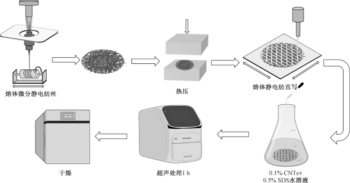
PCL熔体微分静电纺丝工艺参数为:电压35 kV,温度200 ℃,挤出机转速3 r/min,辊子转速1 000 r/min;所得纤维膜热压温度为60 ℃。PCL熔体静电纺直写工艺参数为:电压2.5 kV,温度160 ℃,气压17.24 kPa,接收距离3 mm,喷头移动速度10 mm/s,空气相对湿度控制在37%~45%之间。通过该工艺制得30 mm×30 mm的菱形心脏补片支架,同时制备30 mm×30 mm的方形心脏补片支架作为对照,打印路径如图2所示。
图2
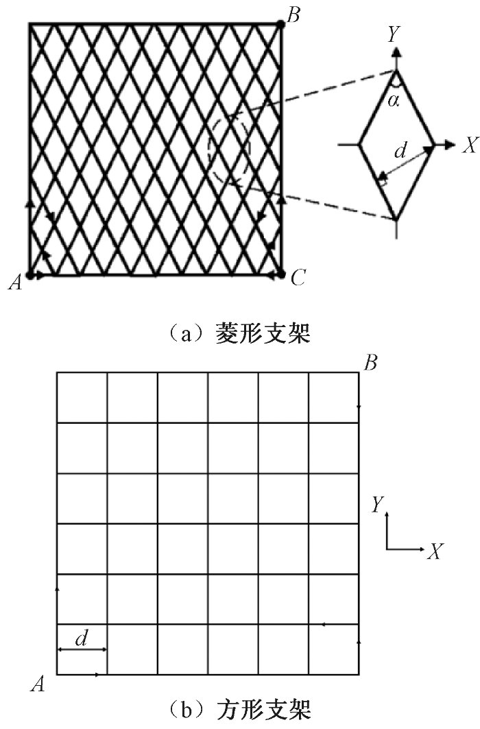
图2
补片支架形状与打印路径示意图
注:α为网格角度;d为纤维间距;X代表横向;Y代表纵向。
Fig.2
Schematic diagrams of patch stents shape and printing path. (a) Diamond-shaped; (b) Square-shaped
1.3 测试与表征
微观形貌观察:采用扫描电镜对复合支架的微观形貌进行观察。
力学性能测试:采用单纤维拉伸仪测试样品纵向和横向上的应力-应变曲线,设定拉伸速率为5 mm/min。根据应力-应变曲线初始直线段的斜率,分别计算样品在纵向和横向上的弹性模量,二者的比值即为各向异性弹性模量比,简称各向异性模量比。
采用台式双探针数显万用表测试样品的导电性能:将探针平行固定于样品两端,沿纵向多点测量10次取均值;随后旋转样品90°以同法测定横向数据。按照下式计算电导率:
式中:G为电导率,S/m;ρ为电阻率,Ω·m;L为导线长度,m;R为电阻,Ω;S为横截面积,m2。
孔径分布测试:采用通孔孔径分析仪(基于泡压法原理),将30 mm×30 mm的方形纤维膜经无水乙醇完全浸润后进行测试。每组测试3次,结果取平均值。
细胞存活率测试:采用CCK-8法进行测试。将补片与大鼠骨髓间充质干细胞共培养,细胞密度为2×10⁴个/mL;培养结束后用PBS清洗3次,加入10% CCK-8溶液,37 ℃下孵育 2 h。分别对培养1 d和3 d的样品,采用酶标仪测试450 nm波长下的吸光度。按照下式计算细胞存活率:
式中:R为细胞存活率,%;De为实验组的吸光度;Dc为空白组(未添加任何细胞培养基的48孔板本身)的吸光度。
鬼笔环肽细胞骨架染色:将补片与大鼠H9c2心肌细胞共培养在48孔板上,细胞密度为2×10⁴个/mL,每孔1 mL。染色时,用4%多聚甲醛固定1 h,用PBS清洗3次;随后采用鬼笔环肽染液与DAPI染液避光孵育30 min。分别对培养1 d和3 d的样品,在荧光显微镜下观察并拍摄;利用ImageJ 2X 软件对不同荧光通道图像进行合成与结果分析。
2 结果与讨论
2.1 支架结构调控与力学性能分析
2.1.1 网格角度对支架力学性能的影响
图3示出支架在纵横方向上的力学性能。对于90°网格支架,纤维均呈伸直状态,受力即刻响应,导致应力随应变线性激增。而随着网格角度倾斜,应力-应变曲线呈现出典型的“J”型非线性特征,这归因于拉伸初期的几何重排缓冲效应。数据表明,减小网格角度能显著增强纵向性能,同时削弱横向弹性模量(横向强度波动较小)。所有组别的弹性应变范围均覆盖了天然心肌的需求(大于25%)。当网络角度降至50°时,各向异性最为显著:纵向抗拉强度(0.65 MPa)与弹性模量(3.08 MPa)达到峰值,而横向性能则降至最低点(强度为0.23 MPa,弹性模量为0.24 MPa)。
图3
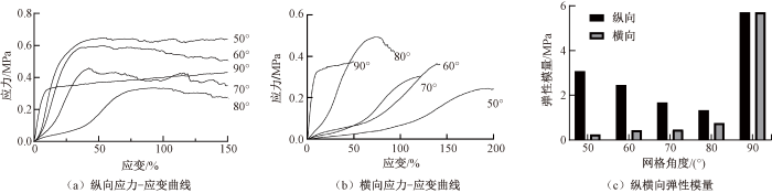
图3
50°~90°网格支架的力学性能
Fig.3
Mechanical properties of stent with grid angle of 50°-90°. (a) Longitudinal stress-strain curve; (b) Transverse stress-strain curve; (c) Longitudinal and transverse elastic modulus
支架的各向异性模量比如图4所示。其中,灰色区域表示天然心肌的允许范围(1.9~3.9)。当网格角度在67°~78°范围内时,支架模量比满足心肌仿生要求。
图4
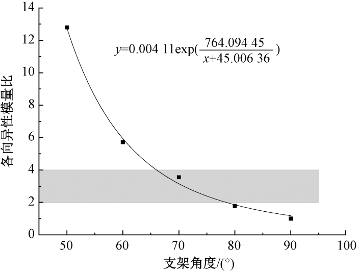
图4
不同网格角度支架的各向异性模量比
Fig.4
Anisotropic modulus ratios corresponding to stents of different grid angles
2.1.2 纤维间距对支架力学性能的影响
在保持70°取向及15层堆叠结构不变的条件下,对比了4种规格(间距300~600 μm)支架的纵向拉伸性能,结果如图5所示。数据显示,随着网格变得稀疏,材料的抗拉强度与弹性模量均呈现下降趋势。其中,300 μm间距组表现出最高的力学值(强度0.45 MPa,弹性模量1.67 MPa),而600 μm组则降至最低(强度0.3 MPa,弹性模量1.35 MPa)。这种性能衰减归因于实体材料占比的变化:在相同测试条件下,间距越宽,内部实际承担载荷的纤维根数越少,导致支架抵抗拉伸变形的能力随之下降。
图5
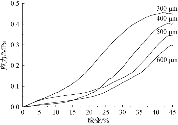
图5
不同纤维间距下支架的力学性能
Fig.5
Mechanical properties of stents with different fiber spacings
2.2 导电性及稳定性分析
2.2.1 支架导电性
图6示出PCL/CNTs超声波处理后支架的光学显微照片。可见,CNTs均匀包覆在纤维表面。
图6
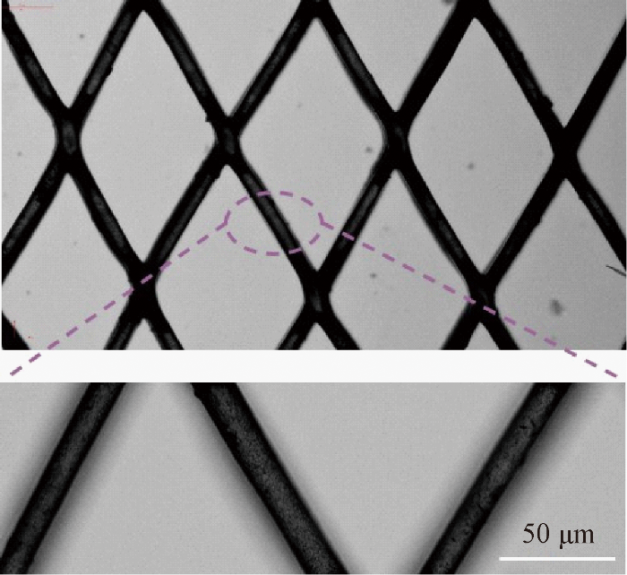
图6
PCL/CNTs超声处理后支架的光学显微镜照片
Fig.6
Optical microscope image of PCL/CNTs ultrasonic treated stent
测量了网格角度为70°及90°(各向同性)、纤维间距为300 μm、15层支架的纵向电导率。结果显示:当网格角度为90°时,支架电导率为1.36×10⁻³ S/cm;而当网格角度为70°时,支架纵向与横向电导率均低于90°支架,分别为1.15×10-3、8.67×10-4 S/cm。可见,该支架的电导率与天然心肌组织(纵向电导率为1.6×10-3 S/cm,横向电导率为5×10-5 S/cm)接近。
2.2.2 时间对支架电导率衰减的影响
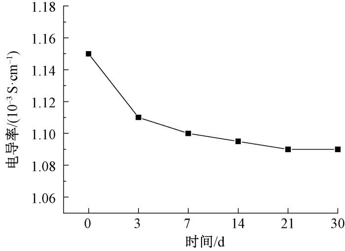
图7示出支架在模拟生理环境(水浴环境、37 ℃、湿度50%)下的纵向电导率演变规律。从整体曲线来看,其导电性能呈现先下降后趋于稳定的变化趋势。在浸泡初期(0~3 d),由于部分结合松散的CNTs发生流失,电导率出现一定程度的衰减,于3 d时降至1.11×10⁻³ S/cm。此后(3~21 d),电导率进入平台期,21 d时测定值为1.09×10⁻³ S/cm,基本达到平衡状态。综上,该支架在通常的细胞培养周期内电导率损耗极小,具备优异的湿环境稳定性,可确保实验的可靠性。
图7
2.3 基底膜孔径分析
图8
2.4 体外生物评价
2.4.1 大鼠H9c2心肌细胞的铺展与黏附情况
为评估不同结构支架 (PCL、PCL/CNTs菱形及方形支架)对细胞行为的影响,将大鼠H9c2心肌细胞接种于各组样品表面,并在培养1 d及3 d后进行鬼笔环肽/DAPI双重荧光染色,以观测细胞的黏附铺展状态,测试结果如图9所示。其中红色标记F-actin骨架,蓝色标记细胞核。
图9
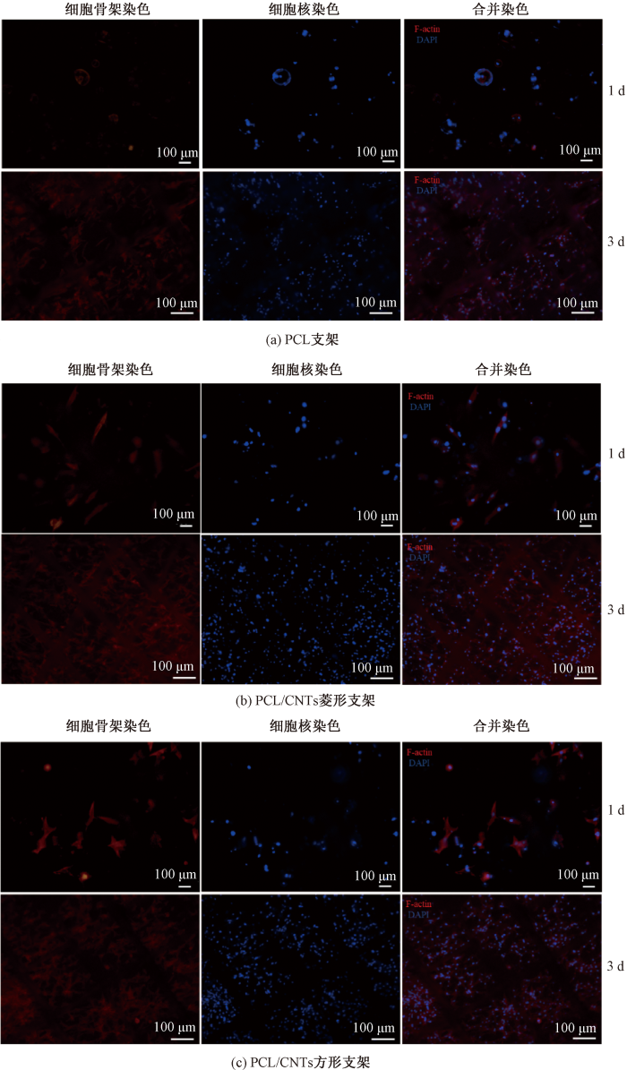
图9
荧光显微镜下大鼠H9c2心肌细胞在不同支架的骨架染色情况
Fig.9
Skeleton staining of rat H9c2 cardiomyocytes on different stents by fluorescence microscopy. (a) PCL stents; (b) PCL/CNTs diamond-shaped stent; (c) PCL/CNTs square-shaped stent
由图9可知,共培养24 h后,PCL组细胞大都仍维持未铺展的圆形;相比之下,2组掺杂CNTs的复合支架上,部分细胞已呈现出仿生心肌特征的长梭形形态,表明CNTs的引入有助于诱导细胞极性,加速细胞成熟。随着培养时间延长至第3 d,各组细胞增殖显著,骨架伸展更为充分。其中,PCL/CNTs复合组的细胞铺展面积显著优于纯PCL组。尤为关键的是,菱形支架组的H9c2细胞展现出沿着直写纤维定向排列的趋势,证实了菱形拓扑结构能提供有效的“接触导向”线索,诱导心肌细胞实现各向异性生长。
2.4.2 复合补片的生物相容性
采用CCK-8试剂盒测定大鼠骨髓间充质干细胞在纯PCL、PCL/CNTs方形及菱形支架上的活性,以评估各组材料在培养1 d及3 d的细胞毒性,测试结果如图10所示。由图可知,接种1 d后,PCL对照组与方形支架组表现出相似的细胞相容性,存活率均维持在90%左右;相比之下,菱形支架组的数据略低(87%)。随着培养时间延长至3 d,3组样品的细胞活性均呈稳定增长趋势且无显著差异,其中菱形组的回升幅度最大,由87%跃升至94%。鉴于所有实验组在各检测节点的细胞存活率始终高于80%,证实了3类支架均未表现出明显的细胞毒性,均具备优异的细胞相容性。
图10
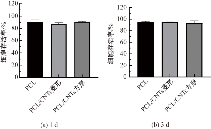
图10
不同支架在细胞培养1 d和3 d的细胞存活率
Fig.10
Cell survival rates of different stents cultured for 1 d (a) and 3 d (b)
3 结论
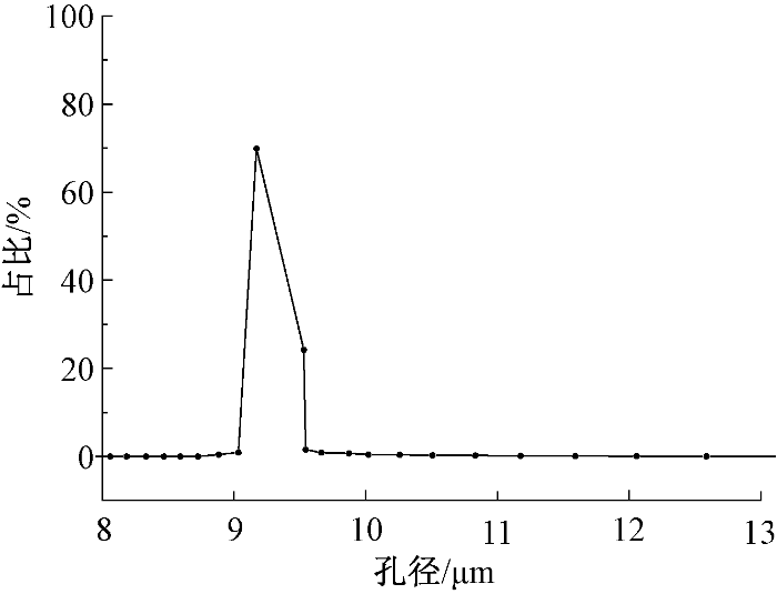
本研究以生物可降解聚己内酯(PCL)为基体,导电碳纳米管(CNTs)为功能填料,结合熔体微分静电纺丝与熔体静电纺直写技术,成功构建了一种具有层级结构的导电各向异性心脏补片。研究结果表明,熔体静电纺直写技术可实现菱形PCL支架力学各向异性的精准调控,70°结构的纵横向弹性模量比为3.55,与天然心肌相匹配;CNTs后处理使支架电导率达到10-3 S/cm量级,熔体微分静电纺丝制备的超细纤维基底膜平均孔径为9.36 μm,可有效防止细胞渗漏;体外培养3 d结果显示,细胞存活率大于90%,且补片可促进心肌细胞定向排列。该复合制备技术为仿生心脏补片的开发提供了新思路,未来工作将进一步开展动物体内实验,将补片植入心肌梗死模型动物体内,系统评估其组织整合性、血管化程度及心功能恢复效果;同时,通过长期随访监测材料的降解动力学与电传导稳定性,可优化参数以适配体内环境,为推进补片临床前研究提供参考。
参考文献
Global, regional, and national burden of cardiovascular diseases for 10 causes, 1990 to 2015
[J].
DOI:S0735-1097(17)37244-3
PMID:28527533
[本文引用: 1]

The burden of cardiovascular diseases (CVDs) remains unclear in many regions of the world.The GBD (Global Burden of Disease) 2015 study integrated data on disease incidence, prevalence, and mortality to produce consistent, up-to-date estimates for cardiovascular burden.CVD mortality was estimated from vital registration and verbal autopsy data. CVD prevalence was estimated using modeling software and data from health surveys, prospective cohorts, health system administrative data, and registries. Years lived with disability (YLD) were estimated by multiplying prevalence by disability weights. Years of life lost (YLL) were estimated by multiplying age-specific CVD deaths by a reference life expectancy. A sociodemographic index (SDI) was created for each location based on income per capita, educational attainment, and fertility.In 2015, there were an estimated 422.7 million cases of CVD (95% uncertainty interval: 415.53 to 427.87 million cases) and 17.92 million CVD deaths (95% uncertainty interval: 17.59 to 18.28 million CVD deaths). Declines in the age-standardized CVD death rate occurred between 1990 and 2015 in all high-income and some middle-income countries. Ischemic heart disease was the leading cause of CVD health lost globally, as well as in each world region, followed by stroke. As SDI increased beyond 0.25, the highest CVD mortality shifted from women to men. CVD mortality decreased sharply for both sexes in countries with an SDI >0.75.CVDs remain a major cause of health loss for all regions of the world. Sociodemographic change over the past 25 years has been associated with dramatic declines in CVD in regions with very high SDI, but only a gradual decrease or no change in most regions. Future updates of the GBD study can be used to guide policymakers who are focused on reducing the overall burden of noncommunicable disease and achieving specific global health targets for CVD.Copyright © 2017 The Authors. Published by Elsevier Inc. All rights reserved.
Constructing biomimetic cardiac tissues: a review of scaffold materials for engineering cardiac patches
[J].DOI:10.1007/s42247-019-00046-4 [本文引用: 2]
Next generation of heart regenerative therapies: progress and promise of cardiac tissue engineering
[J].
DOI:10.1038/s41536-021-00140-4
PMID:34075050
[本文引用: 1]
The adult heart is a vital and highly specialized organ of the human body, with limited capability of self-repair and regeneration in case of injury or disease. Engineering biomimetic cardiac tissue to regenerate the heart has been an ambition in the field of tissue engineering, tracing back to the 1990s. Increased understanding of human stem cell biology and advances in process engineering have provided an unlimited source of cells, particularly cardiomyocytes, for the development of functional cardiac muscle, even though pluripotent stem cell-derived cardiomyocytes poorly resemble those of the adult heart. This review outlines key biology-inspired strategies reported to improve cardiomyocyte maturation features and current biofabrication approaches developed to engineer clinically relevant cardiac tissues. It also highlights the potential use of this technology in drug discovery science and disease modeling as well as the current efforts to translate it into effective therapies that improve heart function and promote regeneration.
Stem cell therapy: a novel approach for myocardial infarction
[J].
DOI:10.1002/jcp.28381
PMID:30811054
[本文引用: 1]
Myocardial infarction (MI) is a major cause of morbidity and mortality worldwide. Until recently, it was thought that myocardium was not able to repair itself, but studies have now shown that resident cardiac stem cells have regenerative capacity, and stem cell therapy may be a novel approach for cardiac muscle repair and regeneration. Stem cell-derived paracrine factors have been shown to regulate ventricular remodeling, inflammation, apoptosis, cardiomyocytes regeneration, and neovascularization in regions of infarcted cardiac tissue. In this review, we summarize the evidence from cellular, animal, and clinical studies supporting the potential clinical significance of stem cell therapy as a novel therapeutic approach for the treatment of MI.© 2019 Wiley Periodicals, Inc.
Current advances in biodegradable synthetic polymer based cardiac patches
[J].
DOI:10.1002/jbm.a.36874
PMID:31895482
[本文引用: 1]
The number of people affected by heart disease such as coronary artery disease and myocardial infarction increases at an alarming rate each year. Currently, the methods to treat these diseases are restricted to lifestyle change, pharmaceuticals, and eventually heart transplant if the condition is severe enough. While these treatment options are the standard for caring for patients who suffer from heart disease, limited regenerative ability of the heart restricts the effectiveness of treatment and may lead to other heart-related health problems in the future. Because of the increasing need for more effective therapeutic technologies for treating diseased heart tissue, cardiac patches are now a large focus for researchers. The cardiac patches are designed to be integrated into the patients' natural tissue to introduce mechanical support and healing to the damaged areas. As a promising alternative, synthetic biodegradable polymer based biomaterials can be easily manipulated to customize material properties, as well as possess certain desired characteristics for cardiac patch use. This comprehensive review summarizes recent works on synthetic biodegradable cardiac patches implanted into infarcted animal models. In addition, this review describes the basic requirements that should be met for cardiac patch development, and discusses the inspirations to designing new biomaterials and technologies for cardiac patches.© 2020 Wiley Periodicals, Inc.
Biomaterials based cardiac patches for the treatment of myocardial infarction
[J].
熔体微分静电纺丝纳米纤维高效绿色制备技术
[J].
DOI:10.13543/j.bhxbzr.2018.05.014
[本文引用: 1]
纳米纤维在生物医疗、高效过滤及生化防护等领域有着广泛的应用前景。静电纺丝被学术界及工业界认为是最具产业化制备纳米纤维的前景技术之一,其中熔体静电纺丝无需使用溶剂,相比溶液静电纺丝,避免了有毒溶剂残留、回收及处理等问题,是聚合物纳米纤维绿色制造的发展方向。然而,受到装备复杂、工艺滞后的影响,熔体静电纺丝始终未能突破纤维细化难、制备效率低的瓶颈。为此,团队创新提出了熔体微分静电纺丝新方法,经过十余年探索,围绕其工艺、装备、材料及应用等进行了系统的研究,率先实现了500 nm范围内熔体电纺纳米纤维的工业化制备,并建立了世界上第一套熔体微分静电纺丝纳米纤维工业化生产线。本文将从熔体微分静电纺丝的机理、关键技术、纳米纤维批量绿色制造及应用三个方面介绍熔体微分静电纺丝的研究成果及最新进展。
Nanofiber preparation technology by melt differential electrospinning with high efficiency in the absence of a solvent
[J].
熔体电纺直写技术的成型工艺精度
[J].
Precision of molding process of melt electrospinning direct writing technology
[J].
Fibroblast-myocyte coupling in the heart: potential relevance for therapeutic interventions
[J].
DOI:10.1016/j.yjmcc.2016.01.010
PMID:26774702
[本文引用: 1]
Cardiac myocyte-fibroblast electrotonic coupling is a well-established fact in vitro. Indirect evidence of its presence in vivo exists, but few functional studies have been published. This review describes the current knowledge of fibroblast-myocyte electrical signaling in the heart. Further research is needed to understand the frequency and extent of heterocellular interactions in vivo in order to gain a better understanding of their relevance in healthy and diseased myocardium. It is hoped that associated insight into myocyte-fibroblast coupling in the heart may lead to the discovery of novel therapeutic targets and the development of agents for improving outcomes of myocardial scarring and fibrosis. Copyright © 2016 Elsevier Ltd. All rights reserved.

 京公网安备11010502044800号


