



为此,本研究聚焦于CS/PCL纳米纤维膜的结构调控及其物理引导作用,通过系统探究静电纺丝参数(接收距离、推注速度、接收滚轮转速)对CS/PCL纳米纤维膜形貌结构的影响,优化制备工艺,并评估了所制备的纤维膜对骨髓间充质干细胞(MSCs)的定向生长、增殖及神经向分化等方面的物理引导作用,旨在为基于物理结构调控的神经组织工程支架的设计提供实验依据。
1 实验部分
1.1 材料与仪器
聚己内酯(PCL,数均分子量为80 000,上海易恩化学技术有限公司);壳聚糖(CS,脱乙酰度≥95%,上海阿拉丁生化科技股份有限公司);冰乙酸(国药集团化学试剂有限公司);α-最小必需培养基(α-MEM)、胎牛血清(FBS)、胰蛋白酶、青霉素/链霉素双抗,美国赛默飞公司;4',6-二脒基-2-苯基吲哚(DAPI)、磷酸盐缓冲溶液(PBS),北京索莱宝科技有限公司;4%多聚甲醛通用型组织固定液(北京兰杰柯科科技有限公司);2.5%戊二醛固定液(上海阿拉丁生化科技股份有限公司);曲拉通X-100(Triton X-100,上海源叶生物科技有限公司);细胞计数试剂盒-8(CCK-8,上海翌圣生物科技股份有限公司);活/死细胞染色试剂盒(江苏凯基生物技术股份有限公司);异硫氰酸荧光素标记的鬼笔环肽(鬼笔环肽-FITC,美国AAT Bioquest公司);大鼠骨髓间充质干细胞(MSCs,中科院细胞库)。
TL-Pro-BM-H型静电纺丝机,深圳市通力微纳科技有限公司;DZF-6030B型真空干燥箱,上海析宇仪器有限公司;SU8010型扫描电子显微镜,日本日立公司;YG(B)026H-500型电子织物强力机,温州市大荣纺织仪器有限公司;Multiskan Sky型酶标仪,美国赛默飞公司;Ti-S型倒置荧光显微镜,日本尼康公司。
1.2 CS/PCL纳米纤维膜的制备
与前期研究侧重仿生复合神经支架整体性能不同,本文聚焦于静电纺丝关键参数对纤维微观结构的调控规律,并系统评估高取向结构对MSCs的物理引导机制。CS/PCL取向纳米纤维膜的制备方法参照文献[24]。具体制备过程如下:为兼顾材料的性能,采用CS与PCL质量比为3∶7的共混体系,以90%乙酸为溶剂,制备质量分数为10%的纺丝前驱体液。采用静电纺丝装置,在温度为25 ℃、相对湿度为30%的环境中,以21G金属针头为喷丝口,施加29 kV电压,通过接收滚轮收集纤维。
在此基础上,本研究系统考察了接收距离(15、16、17 cm)、推注速度(0.6、0.8、1.0 mL/h)及接收滚轮转速(1 500、2 000、2 500、3 000 r/min)对纳米纤维膜形貌结构的影响。
1.3 材料表征与性能测试
1.3.1 微观形貌与力学性能
纳米纤维膜经真空干燥后,裁剪为0.5 cm × 0.5 cm大小的样品,喷金处理,采用扫描电子显微镜观察其表面形貌。
依据扫描电镜照片,采用Image J软件对纤维的直径及取向进行统计分析。绘制纤维直径分布直方图并计算纤维的平均直径。绘制纤维取向分布直方图,将纤维取向分布的峰值方向定义为主轴方向,并计算取向因子f。
式中:θ为单根纤维与主轴方向的夹角;<cos2θ>为cos2θ的平均值。f=0表示纤维呈完全随机分布,f=1表示纤维均平行于主轴方向。
将纳米纤维膜分别沿取向与非取向方向裁剪为3 cm×1 cm大小的样品,用电子织物强力机以100 mm/min的速度对样品进行拉伸,以评估其力学性能。
1.3.2 生物相容性
采用MSCs评估纳米纤维膜的生物相容性,且均选用第3~10代细胞。完全培养基由89% α-MEM、10% FBS和1%双抗配制而成。样品经灭菌处理后置于24孔板,将MSCs以1×10⁴ 个/孔进行接种,并在37 ℃、5% CO2条件下培养。作为对照组的空白组除不放置样品外,其它条件与实验组完全一致。
在培养1、3、5 d后进行CCK-8实验。在避光条件下配制工作液(90%α-MEM+10% CCK-8),以500 μL/孔加入后孵育2 h,使用酶标仪测试450 nm波长下的吸光度值,根据公式计算细胞相对增殖率(R),即实验组与对照组在450 nm波长下吸光度值的百分比。
为评估细胞存活状态,在培养1、3 d后进行活/死细胞染色。具体操作如下:在避光条件下配制工作液,以500 μL/孔加入,于室温避光孵育30 min,PBS清洗3次,使用荧光显微镜观察。
1.3.3 CS/PCL纳米纤维膜对MSCs的调控作用
为可视化细胞骨架重排行为,在培养3 d后对MSCs进行免疫荧光染色。具体步骤如下:加入4%多聚甲醛于室温固定10 min以稳定细胞结构,随后用PBS冲洗。其次,采用0.5% Triton X-100溶液对细胞膜进行5 min透化处理,以增强染料渗透性,透化后再次用PBS冲洗,加入鬼笔环肽-FITC工作液,于避光条件下静置30 min,以标记肌动蛋白纤维。最后,使用DAPI对细胞核进行复染5 min,经PBS清洗后使用荧光显微镜采集图像。
在培养3 d后,通过梯度脱水法观察细胞在纳米纤维膜上的黏附与延伸情况。采用2.5%戊二醛固定3 h以充分稳定细胞结构,用PBS清洗3遍。为避免水相蒸发导致的结构塌陷,配制6个浓度梯度的乙醇溶液(30%、50%、70%、90%、95%、100%)逐步置换细胞内水分。脱水完成后,将样品置于37 ℃下干燥,随后采用扫描电子显微镜观察。
2 结果与讨论
2.1 CS/PCL纳米纤维膜的形貌结构优化
2.1.1 接收距离与推注速度
在固定纺丝电压为29 kV、接收滚轮转速为2 500 r/min条件下,探究接收距离与推注速度对纳米纤维膜形貌结构的影响,将不同参数下的样品分别编号为1#~9#,具体纺丝参数见表1。
表1 CS/PCL纳米纤维膜的静电纺丝参数
Tab.1
| 样品 编号 | 接收滚轮转速/ (r·min-1) | 接收 距离/cm | 推注速度/ (mL·h-1) |
|---|---|---|---|
| 1# | 2 500 | 15 | 0.6 |
| 2# | 2 500 | 15 | 0.8 |
| 3# | 2 500 | 15 | 1.0 |
| 4# | 2 500 | 16 | 0.6 |
| 5# | 2 500 | 16 | 0.8 |
| 6# | 2 500 | 16 | 1.0 |
| 7# | 2 500 | 17 | 0.6 |
| 8# | 2 500 | 17 | 0.8 |
| 9# | 2 500 | 17 | 1.0 |
图1
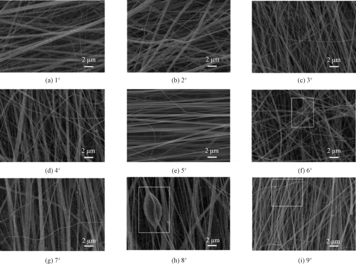
图1
不同接收距离与推注速度下CS/PCL纳米纤维膜的SEM照片
Fig.1
SEM images of CS/PCL nanofiber membranes prepared under conditions of different receiving distances and outflow velocities
在接收距离为16 cm时,推注速度(1.0 mL/h)过高,输送的溶液量过多,导致溶液在有限飞行时间内未能被充分拉伸,此时电场力不足以有效克服溶液的表面张力和黏性阻力,从而造成纤维牵伸不足,出现局部融合与缠结现象。
在接收距离增大至17 cm时,由于接收距离变远,射流的飞行路径更长,溶剂挥发更充分,溶液黏度增大。在此条件下,推注速度(≥0.8 mL/h)过高,推出的液体所受到的牵伸力不足,易形成串珠结构或发生局部液滴堆积,从而影响纤维形貌结构。
图2
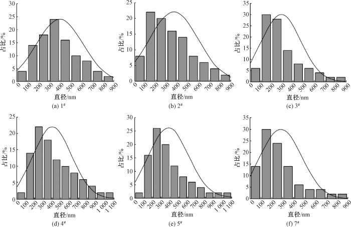
图2
不同接收距离与推注速度下CS/PCL纤维直径分布
Fig.2
Diameter distribution of CS/PCL fibers prepared under conditions of different receiving distances and outflow velocities
图3
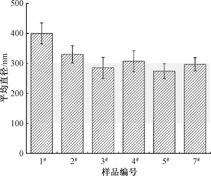
图3
接收距离与推注速度对CS/PCL纤维平均直径的影响
Fig.3
Influence of receiving distance and outflow velocity on average diameter of CS/PCL fibers
通过系统对比不同参数组合下所制备的纤维的平均直径及其分布发现,3组参数(15 cm,1 mL/h)、(16 cm,0.8 mL/h)、(17 cm,0.6 mL/h)均可获得平均直径在100~300 nm范围内、形貌均一的CS/PCL纳米纤维膜,符合细胞黏附的理想尺寸。
进一步对3组优化参数下CS/PCL纳米纤维膜中纤维的取向进行分析,结果如图4所示。对比发现,当接收距离为16 cm,推注速度为0.8 mL/h时,所得纤维的平均直径为274 nm,且取向因子最高(f=0.88),表明在该参数下,所制备的纤维取向度最高,为本阶段的最佳工艺组合。
图4
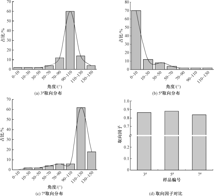
图4
不同接收距离与推注速度下CS/PCL纤维取向分布与取向因子
Fig.4
Orientation distributions and orientation factors of CS/PCL fibers under conditions of different receiving distances and outflow velocities. (a) Orientation distribution of 3#; (b) Orientation distribution of 5#; (c) Orientation distribution of 7#; (d) Comparison of orientation factors
2.1.2 接收滚轮转速
在接收距离为16 cm,推注速度为0.8 mL/h条件下,探究接收滚轮转速对CS/PCL纳米纤维膜的纤维取向度的影响。设置转速为1 500、2 000、2 500、3 000 r/min,所制备的纳米纤维膜分别命名为A、B、C、D。
图5示出接收滚轮转速对CS/PCL纳米纤维膜的纤维取向因子的影响。实验显示,随着转速提升,纤维受到更强的力学牵引力,排列逐渐趋于有序,取向度显著提升,然而,当转速提升至3 000 r/min时,纤维中出现纺锤状结构,且排列紊乱。该现象由以下3个因素共同导致:1)过高的滚轮转速产生过大的力学应力,使纤维在沉积过程中发生过度拉伸、断裂甚至缠绕;2)CS/PCL纺丝液的弹性响应滞后,引发局部应力集中,诱发纺锤状结构;3)长时间高速纺丝使金属滚筒表面积累静电荷,易形成局部反向电场,干扰射流的飞行轨迹与沉积路径,导致纤维分布不均、取向紊乱甚至局部堆积。综上,增大滚轮转速虽可提升纤维取向度;但过高的转速会损害纤维形貌,因此,为兼顾取向性与结构均一性,接收滚轮转速应控制在3 000 r/min以下,最佳值为2 500 r/min。
图5
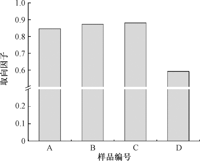
图5
接收滚轮转速对CS/PCL纤维取向因子的影响
Fig.5
Influence of receiving roller speeds on orientation factor of CS/PCL fibers
对样品C(接收滚轮转速2 500 r/min)进行力学性能测试,结果如图6所示。CS/PCL纳米纤维膜在取向方向的力学性能明显优于非取向方向,且断裂伸长率在12%以上,满足神经修复对支架材料力学性能的基本要求。
图6
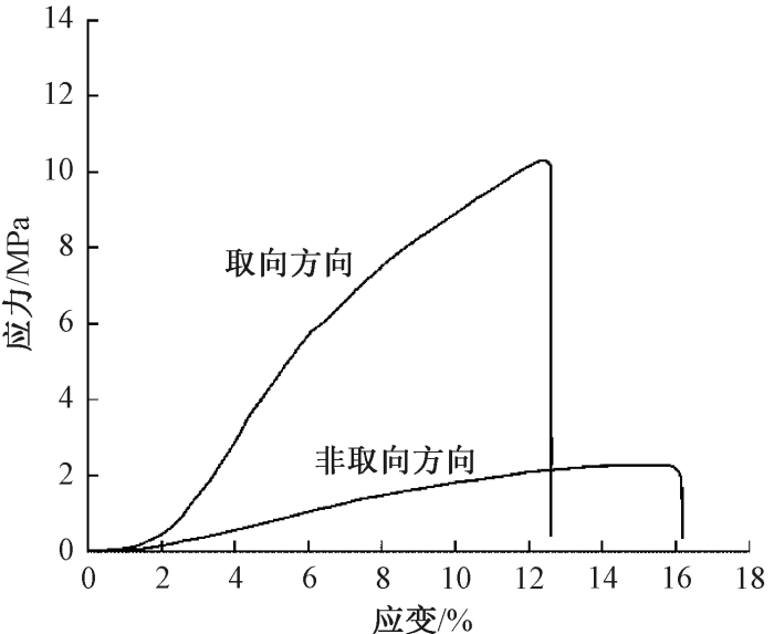
图6
CS/PCL纳米纤维膜的应力-应变曲线
Fig.6
Stress-strain curves of CS/PCL nanofiber membrane
2.2 CS/PCL纳米纤维膜的生物相容性
图7
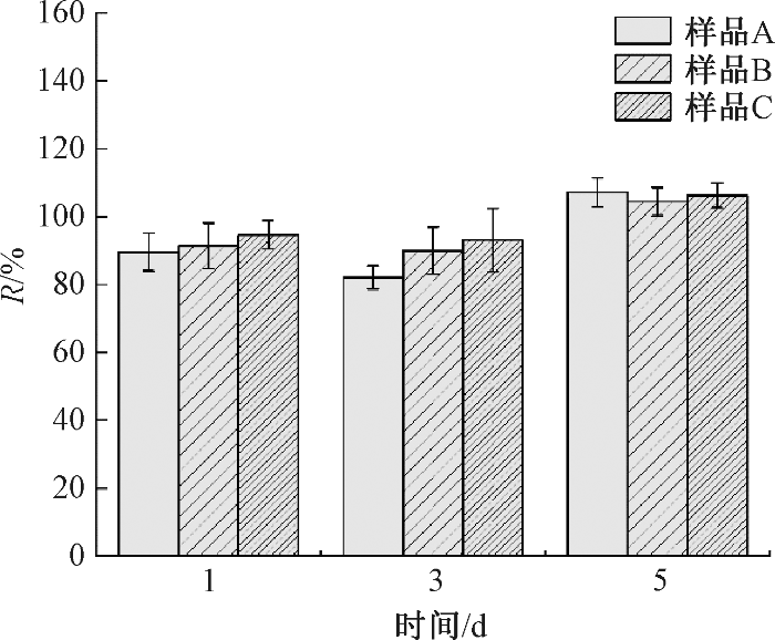
图7
CS/PCL纳米纤维膜的细胞相对增殖率
Fig.7
Relation growth rate of CS/PCL nanofiber membranes
由图7可知,培养1、3、5 d,所有样品的R值均在80%以上,根据GB/T 16886.5—2017《医疗器械生物学评价 第5部分:体外细胞毒性试验》,表明样品无细胞毒性。此外,所有样品第5 d的R值均超过100%,说明该材料对MSCs具有一定的促增殖作用。该结果可归因于CS的引入。首先,CS分子链上富含氨基和羟基,利于细胞初始黏附与铺展;其次,CS作为一种天然阳离子多糖,不仅具有优异的生物相容性,还具备广谱抗菌活性,可有效抑制局部微生物污染,有助于构建利于MSCs增殖的微环境。
活/死细胞染色图像(见图8)显示,细胞主要被标记为绿色(活细胞),仅微量是红色(死细胞)。
图8
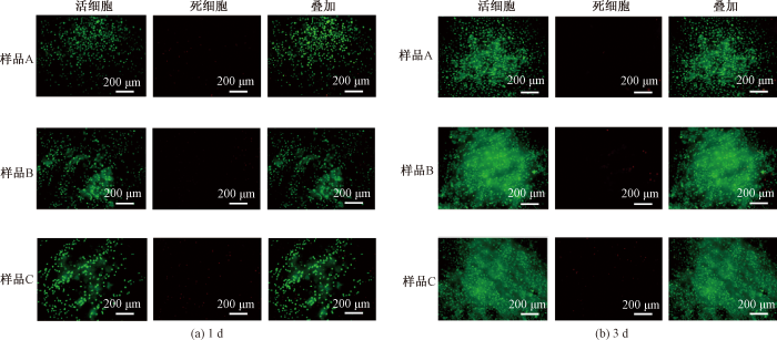
图8
MSCs在CS/PCL纳米纤维膜上共培养1、3 d的活/死细胞染色图像
Fig.8
Live/dead cell stained images of MSCs on CS/PCL nanofiber membranes after 1 d and 3 d co-culture
进一步采用Image J软件对图像进行计数,以定量分析活/死细胞比例,结果如图9所示。所有样品组活细胞数量占比均超过95%,进一步证实了材料无细胞毒性。
图9
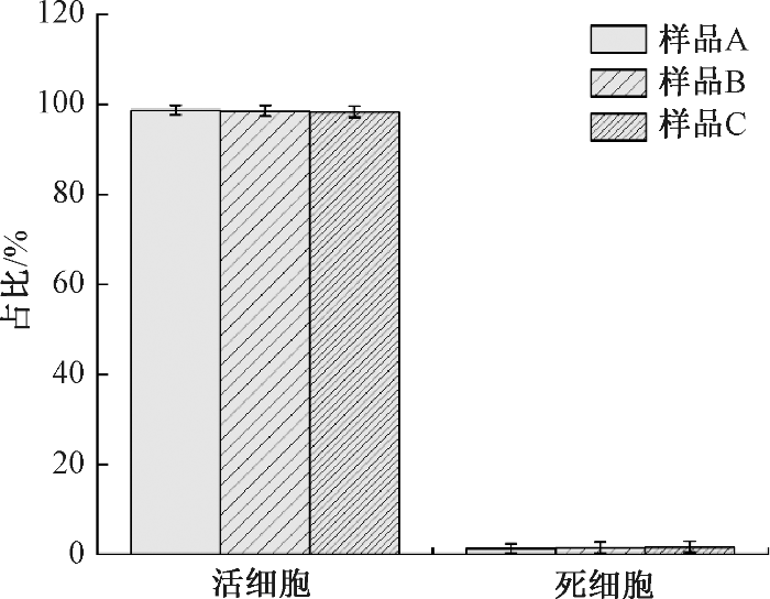
图9
MSCs在CS/PCL纳米纤维膜上共培养3 d的活/死细胞占比
Fig.9
Proportion of live/dead cells of MSCs on CS/PCL nanofiber membranes after 3 d co-culture
2.3 CS/PCL纤维膜对MSCs物理引导作用
研究证实,细胞的形态和行为易受到材料理化性质的调控,其中材料的物理结构是影响细胞骨架重排和定向排列的关键因素[31]。
为探究CS/PCL纳米纤维膜的纤维取向度对MSCs形态的影响,采用鬼笔环肽-FITC和DAPI对MSCs进行染色,并通过荧光显微镜观察细胞的形貌特征,结果如图10所示。在低取向度纤维膜上,MSCs随机分布,生长方向无序。随着纤维取向度的提升,MSCs形态发生显著变化,且细胞骨架沿纤维轴向排列;当纤维取向因子达0.88时,MSCs展现出显著的极化特征,其形态学特征与神经元轴突相似,且细胞沿纤维轴向伸展,表现出显著的定向生长趋势。该结果表明高取向度的纳米纤维膜能有效调控MSCs的黏附、铺展、定向排列与向神经样细胞分化。
图10

图10
MSCs在CS/PCL纳米纤维膜上培养3 d免疫荧光图像
Fig.10
Immunofluorescence images of MSCs on CS/PCL nanofiber membranes after 3 d co-culture
为直观评估MSCs在CS/PCL纳米纤维膜上的黏附与延伸情况,利用梯度脱水法结合扫描电镜进行表征,结果如图11所示。由图可知,当取向度较低时,纤维膜表面的纤维随机排列,细胞分布无明显规律,且形态更趋向于未分化梭形;随着取向度的提升,细胞沿纤维轴向铺展,表现出定向延伸趋势。该结果直观证实了高取向的纳米纤维膜能够为MSCs提供方向性引导,有效促进细胞的定向黏附与铺展。
图11
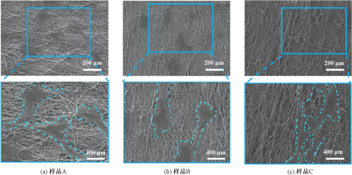
图11
MSCs在CS/PCL纳米纤维膜上共培养3 d的SEM照片
Fig.11
SEM images of MSCs on CS/PCL nanofiber membranes after 3 d co-culture. (a) Sample A; (b) Sample B; (c) Sample C
3 结论
本研究系统探究了静电纺丝参数对壳聚糖(CS)/聚己内酯(PCL)纳米纤维膜形貌结构的调控作用。通过优化接收距离、推注速度和接收滚轮转速,成功制备了平均直径为274 nm、取向因子达0.88的CS/PCL纳米纤维膜。该纤维膜具有良好的生物相容性,且对骨髓间充质干细胞(MSCs)具有一定的促增殖作用。结果表明,高取向度的纳米纤维膜可以有效调控MSCs的黏附、铺展、定向排列与向神经样细胞分化。本研究为物理信号调控干细胞行为用于神经修复提供了坚实的实验基础。
参考文献
MSC-derived exosomes-based therapy for peripheral nerve injury: a novel therapeutic strategy
[J].
Macrophage-specific RhoA knockout delays Wallerian degeneration after peripheral nerve injury in mice
[J].
DOI:10.1186/s12974-021-02292-y
PMID:34654444
[本文引用: 1]

Plenty of macrophages are recruited to the injured nerve to play key roles in the immunoreaction and engulf the debris of degenerated axons and myelin during Wallerian degeneration, thus creating a conducive microenvironment for nerve regeneration. Recently, drugs targeting the RhoA pathway have been widely used to promote peripheral axonal regeneration. However, the role of RhoA in macrophage during Wallerian degeneration and nerve regeneration after peripheral nerve injury is still unknown. Herein, we come up with the hypothesis that RhoA might influence Wallerian degeneration and nerve regeneration by affecting the migration and phagocytosis of macrophages after peripheral nerve injury.Immunohistochemistry, Western blotting, H&E staining, and electrophysiology were performed to access the Wallerian degeneration and axonal regeneration after sciatic nerve transection and crush injury in the Lyz; RhoA (cKO) mice or Lyz2 (Cre) mice, regardless of sex. Macrophages' migration and phagocytosis were detected in the injured nerves and the cultured macrophages. Moreover, the expression and potential roles of ROCK and MLCK were also evaluated in the cultured macrophages.1. RhoA was specifically knocked out in macrophages of the cKO mice; 2. The segmentation of axons and myelin, the axonal regeneration, and nerve conduction in the injured nerve were significantly impeded while the myoatrophy was more severe in the cKO mice compared with those in Cre mice; 3. RhoA knockout attenuated the migration and phagocytosis of macrophages in vivo and in vitro; 4. ROCK and MLCK were downregulated in the cKO macrophages while inhibition of ROCK and MLCK could weaken the migration and phagocytosis of macrophages.Our findings suggest that RhoA depletion in macrophages exerts a detrimental effect on Wallerian degeneration and nerve regeneration, which is most likely due to the impaired migration and phagocytosis of macrophages resulted from disrupted RhoA/ROCK/MLCK pathway. Since previous research has proved RhoA inhibition in neurons was favoring for axonal regeneration, the present study reminds us of that the cellular specificity of RhoA-targeted drugs is needed to be considered in the future application for treating peripheral nerve injury.© 2021. The Author(s).
Advances in biomaterial-based tissue engineering for peripheral nerve injury repair
[J].
DOI:10.1016/j.bioactmat.2024.12.005
PMID:39760068
[本文引用: 1]
Peripheral nerve injury is a common clinical disease. Effective post-injury nerve repair remains a challenge in neurosurgery, and clinical outcomes are often unsatisfactory, resulting in social and economic burden. Particularly, the repair of long-distance nerve defects remains a challenge. The existing nerve transplantation strategies show limitations, including donor site morbidity and immune rejection issues. The multiple studies have revealed the potential of tissue engineering strategies based on biomaterials in the repair of peripheral nerve injuries. We review the events of regeneration after peripheral nerve injury, evaluates the efficacy of existing nerve grafting strategies, and delves into the progress in the construction and application strategies of different nerve guidance conduits. A spotlight is cast on the materials, technologies, seed cells, and microenvironment within these conduits to facilitate optimal nerve regeneration. Further discussion was conducted on the approve of nerve guidance conduits and potential future research directions. This study anticipates and proposes potential avenues for future research, aiming to refine existing strategies and uncover innovative approaches in biomaterial-based nerve repair. This study endeavors to synthesize the collective insights from the fields of neuroscience, materials science, and regenerative medicine, offering a multifaceted perspective on the role of biomaterials in advancing the frontiers of peripheral nerve injury treatment.© 2024 The Authors.
Peripheral nerve regeneration and muscle reinnervation
[J].
DOI:10.3390/ijms21228652
URL
[本文引用: 1]
Injured peripheral nerves but not central nerves have the capacity to regenerate and reinnervate their target organs. After the two most severe peripheral nerve injuries of six types, crush and transection injuries, nerve fibers distal to the injury site undergo Wallerian degeneration. The denervated Schwann cells (SCs) proliferate, elongate and line the endoneurial tubes to guide and support regenerating axons. The axons emerge from the stump of the viable nerve attached to the neuronal soma. The SCs downregulate myelin-associated genes and concurrently, upregulate growth-associated genes that include neurotrophic factors as do the injured neurons. However, the gene expression is transient and progressively fails to support axon regeneration within the SC-containing endoneurial tubes. Moreover, despite some preference of regenerating motor and sensory axons to “find” their appropriate pathways, the axons fail to enter their original endoneurial tubes and to reinnervate original target organs, obstacles to functional recovery that confront nerve surgeons. Several surgical manipulations in clinical use, including nerve and tendon transfers, the potential for brief low-frequency electrical stimulation proximal to nerve repair, and local FK506 application to accelerate axon outgrowth, are encouraging as is the continuing research to elucidate the molecular basis of nerve regeneration.
Polymer scaffolds for peripheral nerve injury repair
[J].DOI:10.1016/j.pmatsci.2025.101497 URL [本文引用: 1]
A responsive cascade drug delivery scaffold adapted to the therapeutic time window for peripheral nerve injury repair
[J].
DOI:10.1039/d3mh01511d
PMID:38073476
[本文引用: 1]
Peripheral nerve injury (PNI) is a common clinical challenge, requiring timely and orderly initiation of synergistic anti-inflammatory and reparative therapy. Although the existing cascade drug delivery system can realize sequential drug release through regulation of the chemical structure of drug carriers, it is difficult to adjust the release kinetics of each drug based on the patient's condition. Therefore, there is an urgent need to develop a cascade drug delivery system that can dynamically adjust drug release and realize personalized treatment. Herein, we developed a responsive cascade drug delivery scaffold (RCDDS) which can adapt to the therapeutic time window, in which Vitamin B12 is used in early controllable release to suppress inflammation and nerve growth factor promotes regeneration by cascade loading. The RCDDS exhibited the ability to modulate the drug release kinetics by hierarchically opening polymer chains triggered by ultrasound, enabling real-time adjustment of the anti-inflammatory and neuroregenerative therapeutic time window depending on the patient's status. In the rat sciatic nerve injury model, the RCDDS group was able to achieve neural repair effects comparable to the autograft group in terms of tissue structure and motor function recovery. The development of the RCDDS provides a useful route toward an intelligent cascade drug delivery system for personalized therapy.
Editorial: surgical treatment of peripheral neuropathic pain, peripheral nerve tumors, and peripheral nerve injury
[J].DOI:10.3389/fneur.2023.1266638 URL [本文引用: 1]
Toward a new generation of bio-scaffolds for neural tissue engineering: challenges and perspectives
[J].DOI:10.3390/pharmaceutics15061750 URL [本文引用: 1]
Injectable hydrogel guides neurons growth with specific directionality
[J].
DOI:10.3390/ijms24097952
URL
[本文引用: 1]
Visual disabilities affect more than 250 million people, with 43 million suffering from irreversible blindness. The eyes are an extension of the central nervous system which cannot regenerate. Neural tissue engineering is a potential method to cure the disease. Injectability is a desirable property for tissue engineering scaffolds which can eliminate some surgical procedures and reduce possible complications and health risks. We report the development of the anisotropic structured hydrogel scaffold created by a co-injection of cellulose nanofiber (CNF) solution and co-polypeptide solution. The positively charged poly (L-lysine)-r-poly(L-glutamic acid) with 20 mol% of glutamic acid (PLLGA) is crosslinked with negatively charged CNF while promoting cellular activity from the acid nerve stimulate. We found that CNF easily aligns under shear forces from injection and is able to form hydrogel with an ordered structure. Hydrogel is mechanically strong and able to support, guide, and stimulate neurite growth. The anisotropy of our hydrogel was quantitatively determined in situ by 2D optical microscopy and 3D X-ray tomography. The effects of PLLGA:CNF blend ratios on cell viability, neurite growth, and neuronal signaling are systematically investigated in this study. We determined the optimal blend composition for stimulating directional neurite growth yielded a 16% increase in length compared with control, reaching anisotropy of 30.30% at 10°/57.58% at 30°. Using measurements of calcium signaling in vitro, we found a 2.45-fold increase vs. control. Based on our results, we conclude this novel material and unique injection method has a high potential for application in neural tissue engineering.
Engineering a wirelessly self-powered and electroconductive scaffold to promote peripheral nerve regeneration
[J].DOI:10.1016/j.nanoen.2022.108145 URL [本文引用: 1]
Magnetoresponsive anisotropic fiber-integrating hydrogels for neural tissue regeneration
[J].DOI:10.1002/sstr.v5.11 URL [本文引用: 1]
再生丝素蛋白/聚乙烯醇共混取向纳米纤维膜的制备与性能
[J].
Preparation and properties of regenerated silk fibroin/polyvinyl alcohol blended nanofiber membranes with predesigned orientation
[J].
Electrospun scaffold with bioactive polyurethane shell infused with propolis and starch-hyaluronic acid core: an advanced therapeutic platform for skin tissue engineering
[J].DOI:10.1016/j.ijbiomac.2024.138745 URL [本文引用: 1]
Controllable fiber orientation and nonlinear elasticity of electrospun nanofibrous small diameter tubular scaffolds for vascular tissue engineering
[J].DOI:10.1088/1748-605X/ab07f1 [本文引用: 1]
Advances in nanotechnology and nanomaterials based strategies for neural tissue engineering
[J].DOI:10.1016/j.jddst.2020.101617 URL [本文引用: 1]
Gum tragacanth/poly(L-lactic acid) nanofibrous scaffolds for application in regeneration of peripheral nerve damage
[J].DOI:10.1016/j.carbpol.2015.12.012 URL [本文引用: 1]
Directional submicrofiber hydrogel composite scaffolds supporting neuron differentiation and enabling neurite alignment
[J].
DOI:10.3390/ijms231911525
URL
[本文引用: 1]
Cell cultures aiming at tissue regeneration benefit from scaffolds with physiologically relevant elastic moduli to optimally trigger cell attachment, proliferation and promote differentiation, guidance and tissue maturation. Complex scaffolds designed with guiding cues can mimic the anisotropic nature of neural tissues, such as spinal cord or brain, and recall the ability of human neural progenitor cells to differentiate and align. This work introduces a cost-efficient gelatin-based submicron patterned hydrogel–fiber composite with tuned stiffness, able to support cell attachment, differentiation and alignment of neurons derived from human progenitor cells. The enzymatically crosslinked gelatin-based hydrogels were generated with stiffnesses from 8 to 80 kPa, onto which poly(ε-caprolactone) (PCL) alignment cues were electrospun such that the fibers had a preferential alignment. The fiber–hydrogel composites with a modulus of about 20 kPa showed the strongest cell attachment and highest cell proliferation, rendering them an ideal differentiation support. Differentiated neurons aligned and bundled their neurites along the aligned PCL filaments, which is unique to this cell type on a fiber–hydrogel composite. This novel scaffold relies on robust and inexpensive technology and is suitable for neural tissue engineering where directional neuron alignment is required, such as in the spinal cord.
高亲水性壳聚糖纳米纤维膜的制备及性能
[J].
Preparation and performance of highly hydrophilic chitosan nanofiber membranes
[J].Medical dressings are a common medical device, playing a significant role in the medical realm. It has a wide range of applications in the treatment, healing, and infection control of trauma and surgical wounds. Research has found that wounds can heal better and faster in a moist environment. Hence, an ideal medical dressing should maintain optimal moisture levels at the wound site and effectively absorb exudates. Serving as a barrier against microorganisms, medical dressings must possess robust mechanical properties, flexibility to conform to diverse wound shapes, and effective antibacterial qualities to expedite the healing process.<br>Electrospinning, a technique that utilizes a high-voltage electrostatic field to produce ultrafine fibers, has attracted much attention due to the highly similar structure of the nanofiber membrane to the natural extracellular matrix of the human body. Electrospinning usually uses natural or synthetic polymer materials as raw materials, most of which have good biocompatibility, can be compatible with human tissues, and reduce allergic reactions to patients. By modifying the membrane's surface, adjusting the electrospun material, or incorporating drugs, we can create specialized functional dressings tailored to diverse application scenarios, thus offering convenience to both medical professionals and patients Consequently, further investigation and utilization of nanofiber membrane medical dressings hold substantial clinical importance and promise for the future<br>Chitosan (CS), a multifunctional biomaterial, is renowned for its non-toxicity, biodegradability, and excellent biocompatibility. In addition, materials based on CS also have immune stimulation and antibacterial performance, and are often used in fields such as wound healing, hemostasis, tissue engineering, and medical textiles. Nonetheless, CS suffers from poor electrospinning performance and insufficient mechanical strength, often necessitating crosslinking or blending with other polymers to enhance these properties. Polyvinyl alcohol (PVA), a water-soluble polymer, is extensively employed in the biomedical field, particularly in tissue engineering and wound dressings, owing to its favorable biocompatibility and biodegradability. PVA also has excellent film-forming and fiber forming properties, making it widely used in the field of electrospinning. Glycerol (GL) is a transparent, colorless, odorless, viscous, and hygroscopic liquid produced by the hydrolysis of triGLcerides. It is often used as a wetting agent in the food industry (plasticizer, stabilizer, and emulsifier) and cosmetic formulations.<br>To meet the requirements of high hydrophilicity and antibacterial properties for medical trauma dressings, PVA/CS/GL nanofiber membranes were prepared by using CS, polyvinyl alcohol (PVA), and GL as raw materials through electrospinning technology. The morphology and structure of the nanofiber membranes were characterized, and their thermal stability, hydrophilicity, swelling degree, mechanical properties, and antibacterial properties were analyzed. The results indicated that the PVA/CS/GL nanofiber membrane exhibited optimal overall performance when the mass fraction of GL was set at 3%, featuring a well-structured network and uniform fiber diameter. The addition of GL improved the poor hydrophilicity and low mechanical strength of chitosan based nanofiber membranes. After adding GL with a mass fraction of 3%, the contact angle of the nanofiber membrane decreased from 53.3 ° to 25.3 °, and the equilibrium swelling rate in PBS buffer increased from 239.5% to 332.1%. The tensile strength and elongation at break increased from 2.68 MPa and 8.2% to 5.83 MPa and 17.9%, respectively. Antibacterial experiments showed that the antibacterial rates of PVA/CS/GL nanofiber membranes against Escherichia coli and Staphylococcus aureus were 94.39% and 89.65%, respectively, showing good antibacterial performance. Therefore, the PVA/CS/GL nanofiber membrane has potential application value in medical trauma dressings. <br>
Chitosan-based scaffolds for bone tissue engineering
[J].Bone defects requiring grafts to promote healing are frequently occurring and costly problems in health care. Chitosan, a biodegradable, naturally occurring polymer, has drawn considerable attention in recent years as scaffolding material in tissue engineering and regenerative medicine. Chitosan is especially attractive as a bone scaffold material because it supports the attachment and proliferation of osteoblast cells as well as formation of mineralized bone matrix. In this review, we discuss the fundamentals of bone tissue engineering and the unique properties of chitosan as a scaffolding material to treat bone defects for hard tissue regeneration. We present the common methods for fabrication and characterization of chitosan scaffolds, and discuss the influence of material preparation and addition of polymeric or ceramic components or biomolecules on chitosan scaffold properties such as mechanical strength, structural integrity, and functional bone regeneration. Finally, we highlight recent advances in development of chitosan-based scaffolds with enhanced bone regeneration capability.
PCL基电纺纤维膜的抑菌、相容及降解性能研究
[J].
Antibacterial, in vitro/vivo biocompatibility and biodegradability of PCL-based electrospun fibers
[J].
静电纺丝技术在神经组织工程中的应用进展
[J].
The application progress of electrospinning technology in nerve tissue engineering
[J].
负载厚朴酚的抗菌纳米纤维膜的制备及其性能
[J].
Preparation and performance of antibacterial nanofiber membrane loaded with magnolol
[J].
DOI:10.1177/004051757604600105
URL
[本文引用: 1]
Relations were studied between skin temperature of the back as well as temperature and humidity inside clothing, and subjective estimates of thermal, humidity, and comfort sensations in 5 subjects wearing cross-country ski dress. The 2-h wear trials were performed in a climatic chamber at −2°C and simulated the actual circumstances under which such a dress is worn by alternating periods of intensive walking (20 min) on a treadmill with periods of resting (5–10 min). Each subject participated in 4 trials wearing alternately one of the 4 experimental, double-layer vests (double-cotton, cotton-polypropylene with cotton layer next to skin, same combination with polypropylene layer next to skin, double-polypropylene).
共聚型聚酰亚胺纳米纤维膜的制备及其性能
[J].
Preparation and performance study of copolymerized polyimide nanofiber membrane
[J].
Dual-regulation biomimetic composite nerve scaffold with oriented structure and conductive function for skin peripheral nerve injury repair
[J].DOI:10.1016/j.colsurfb.2025.114768 URL [本文引用: 1]
静电纺丝参数对丝素蛋白纤维直径的影响及其力学机理研究
[J/OL].
Study on the influence of electrospinning parameters on silk fibroin fiber diameter and mechanical mechanism
[J/OL].
PVDF纳米纤维膜静电纺丝工艺调控分析
[J].
DOI:10.19817/j.cnki.issn1006-3536.2025.08.027
[本文引用: 1]
基于先进的静电纺丝技术,旨在制备出高质量的聚偏氟乙烯(PVDF)纳米纤维膜,并系统地探究了影响纺丝工艺的关键因素。通过调控PVDF质量分数(12%)、溶剂体系中N,N-二甲基甲酰胺(DMF)与丙酮的质量比例(5∶5)、供液流速(1mL/h)、外加电场强度(18kV),以及针尖至接收装置(转辊)的间距(15cm),以确立并验证最佳的纺丝工艺参数组合。结果表明:在上述最优工艺条件下制备的PVDF纳米纤维膜,展现出了优异的微观结构形态,且其β晶相含量高达83.1%,这一发现不仅验证了工艺参数优化的有效性,也为后续纳米材料在特定领域(如过滤、传感、能量存储等)的应用提供了坚实的基础和广阔的前景。
Analysis of electrostatic spinning process regulation for PVDF nanofiber membranes
[J].
DOI:10.19817/j.cnki.issn1006-3536.2025.08.027
[本文引用: 1]
The objective of this study was to prepare high-quality polyvinylidene fluoride (PVDF) nanofibrous membranes using advanced electrostatic spinning technology.The key factors affecting the spinning process were also systematically investigated.The mass concentration of PVDF was set at 12wt%,the mass ratio of <i>N,N</i>-dimethylformamide (DMF) to acetone in the solvent system was fixed at 5∶5,the flow rate of the supply liquid was maintained at 1mL/h,the strength of the applied electric field was set at 18kV,and the distance from the tip of the needle to the receiving device (rotating rollers) was set at 15cm.These parameters were regulated to establish and validate the optimal combination of spinning process parameters.The experimental results demonstrated that the PVDF nanofibrous membranes prepared under the aforementioned optimal process conditions exhibited an excellent microstructural morphology,with a β-crystalline phase content of up to 83.1%.This finding not only validated the effectiveness of the process parameter optimisation but also provided a solid foundation and broad prospects for the subsequent application of the nanomaterials in specific fields,such as filtration,sensing,and energy storage.
Electrospun nanofibers for neural tissue engineering
[J].
DOI:10.1039/b9nr00243j
PMID:20648362
[本文引用: 1]
Biodegradable nanofibers produced by electrospinning represent a new class of promising scaffolds to support nerve regeneration. We begin with a brief discussion on the electrospinning of nanofibers and methods for controlling the structure, porosity, and alignment of the electrospun nanofibers. The methods include control of the nanoscale morphology and microscale alignment of the nanofibers, as well as the fabrication of macroscale, three-dimensional tubular structures. We then highlight recent studies that utilize electrospun nanofibers to manipulate biological processes relevant to nervous tissue regeneration, including stem cell differentiation, guidance of neurite extension, and peripheral nerve injury treatments. The main objective of this feature article is to provide valuable insights into methods for investigating the mechanisms of neurite growth on novel nanofibrous scaffolds and optimization of the nanofiber scaffolds and conduits for repairing peripheral nerve injuries.
The effect of nanofiber alignment on the maturation of engineered meniscus constructs
[J].
DOI:10.1016/j.biomaterials.2007.01.004
PMID:17250888
[本文引用: 1]
The fibrocartilaginous menisci are load-bearing tissues vital to the normal functioning of the knee. Removal of damaged regions of the meniscus subsequent to injury impairs knee function and predisposes patients to osteoarthritis. In this study, we employed biodegradable nanofibrous scaffolds for the tissue engineering of the meniscus. Non-aligned (NA) or fiber-aligned (AL) nanofibrous scaffolds were seeded with meniscal fibrochondrocytes (MFCs) or mesenchymal stem cells (MSCs) to test the hypothesis that fiber-alignment would augment matrix content and organization, resulting in improved mechanical properties. Additionally, we proposed that MSCs could serve as an alternative to MFCs. With time in culture, MSC- and MFC-seeded NA and AL constructs increased in cellularity and extracellular matrix (ECM) content. Counter our initial hypothesis, NA and AL constructs contained comparable amounts of ECM, although a significantly larger increase in mechanical properties was observed for AL compared to NA constructs seeded with either cell type. Cell-seeded NA constructs increased in modulus by approximately 1MPa over 10 weeks while cell-seeded AL construct increased by >7MPa. Additionally, MSC-constructs yielded greater amounts of ECM and demonstrated comparable increases in mechanical properties, thereby confirming the utility of MSCs for meniscus tissue engineering. These results demonstrate that cell-seeded fiber-aligned nanofibrous scaffolds may serve as an instructive micro-pattern for directed tissue growth, reconstituting both the form and function of the native tissue.
Multilineage potential of adult human mesenchymal stem cells
[J].
DOI:10.1126/science.284.5411.143
PMID:10102814
[本文引用: 1]
Human mesenchymal stem cells are thought to be multipotent cells, which are present in adult marrow, that can replicate as undifferentiated cells and that have the potential to differentiate to lineages of mesenchymal tissues, including bone, cartilage, fat, tendon, muscle, and marrow stroma. Cells that have the characteristics of human mesenchymal stem cells were isolated from marrow aspirates of volunteer donors. These cells displayed a stable phenotype and remained as a monolayer in vitro. These adult stem cells could be induced to differentiate exclusively into the adipocytic, chondrocytic, or osteocytic lineages. Individual stem cells were identified that, when expanded to colonies, retained their multilineage potential.
Mesenchymal stem cells: a new trend for cell therapy
[J].
DOI:10.1038/aps.2013.50
PMID:23736003
[本文引用: 1]
Mesenchymal stem cells (MSCs), the major stem cells for cell therapy, have been used in the clinic for approximately 10 years. From animal models to clinical trials, MSCs have afforded promise in the treatment of numerous diseases, mainly tissue injury and immune disorders. In this review, we summarize the recent opinions on methods, timing and cell sources for MSC administration in clinical applications, and provide an overview of mechanisms that are significant in MSC-mediated therapies. Although MSCs for cell therapy have been shown to be safe and effective, there are still challenges that need to be tackled before their wide application in the clinic.
Conductive hydrogels for tissue repair
[J].
DOI:10.1039/d3sc00145h
PMID:36970088
[本文引用: 1]
Conductive hydrogels (CHs) combine the biomimetic properties of hydrogels with the physiological and electrochemical properties of conductive materials, and have attracted extensive attention in the past few years. In addition, CHs have high conductivity and electrochemical redox properties and can be used to detect electrical signals generated in biological systems and conduct electrical stimulation to regulate the activities and functions of cells including cell migration, cell proliferation, and cell differentiation. These properties give CHs unique advantages in tissue repair. However, the current review of CHs is mostly focused on their applications as biosensors. Therefore, this article reviewed the new progress of CHs in tissue repair including nerve tissue regeneration, muscle tissue regeneration, skin tissue regeneration and bone tissue regeneration in the past five years. We first introduced the design and synthesis of different types of CHs such as carbon-based CHs, conductive polymer-based CHs, metal-based CHs, ionic CHs, and composite CHs, and the types and mechanisms of tissue repair promoted by CHs including anti-bacterial, antioxidant and anti-inflammatory properties, stimulus response and intelligent delivery, real-time monitoring, and promoted cell proliferation and tissue repair related pathway activation, which provides a useful reference for further preparation of bio-safer and more efficient CHs used in tissue regeneration.This journal is © The Royal Society of Chemistry.

 京公网安备11010502044800号


