纺织学报 ›› 2026, Vol. 47 ›› Issue (03): 9-17.doi: 10.13475/j.fzxb.20250903701
刘金枝1, 赵回汇1, 吴焕友2, 张建明2, 高晶1( )
)
LIU Jinzhi1, ZHAO Huihui1, WU Huanyou2, ZHANG Jianming2, GAO Jing1()
摘要:
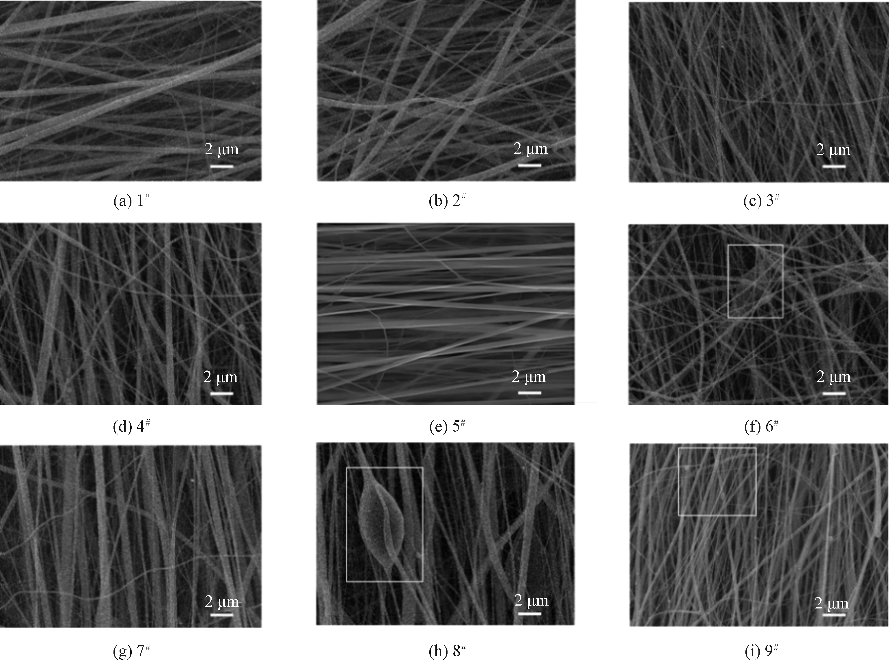
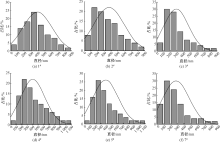
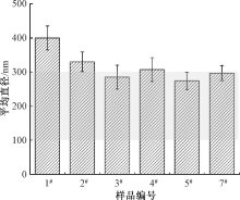
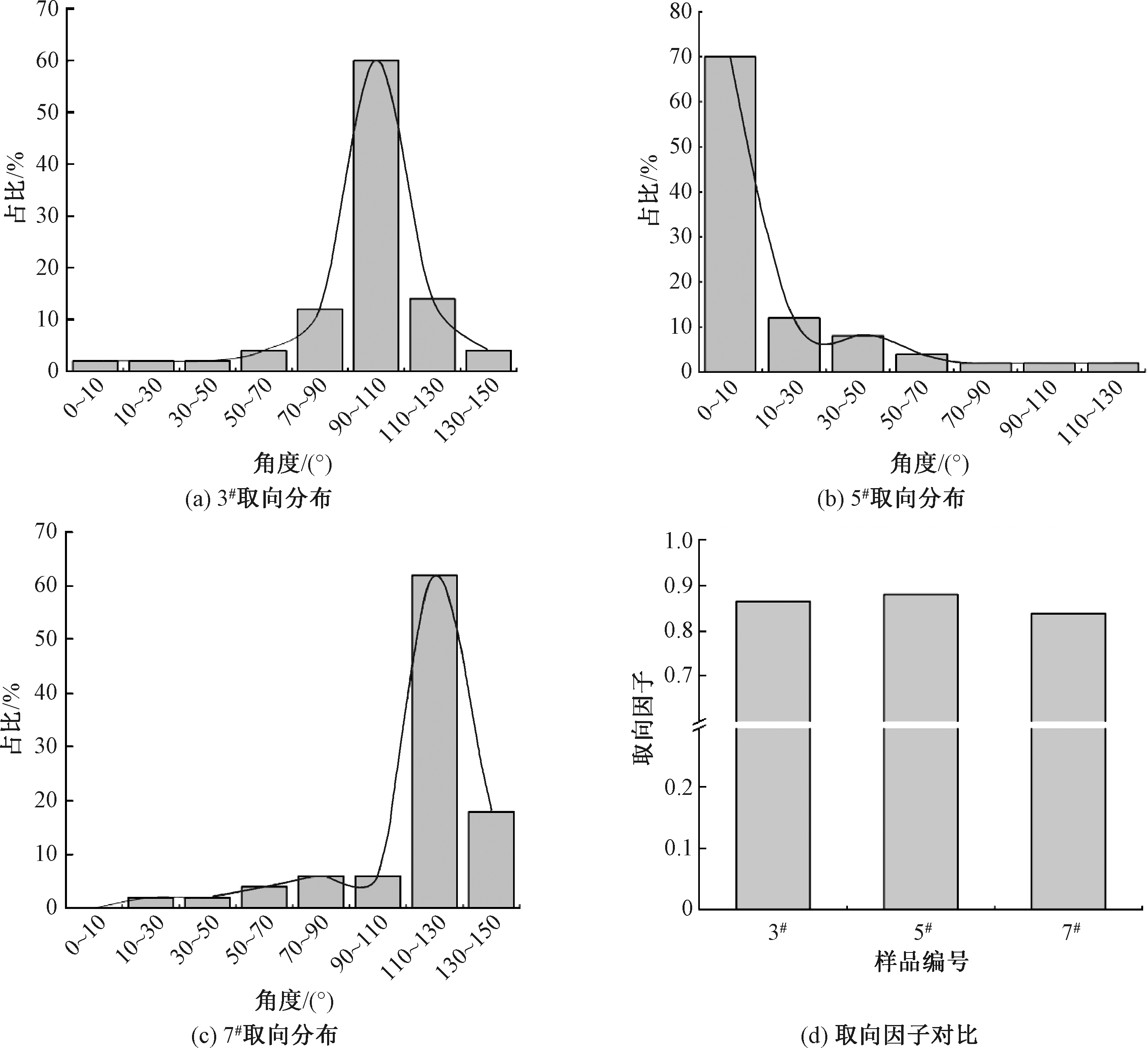
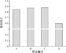
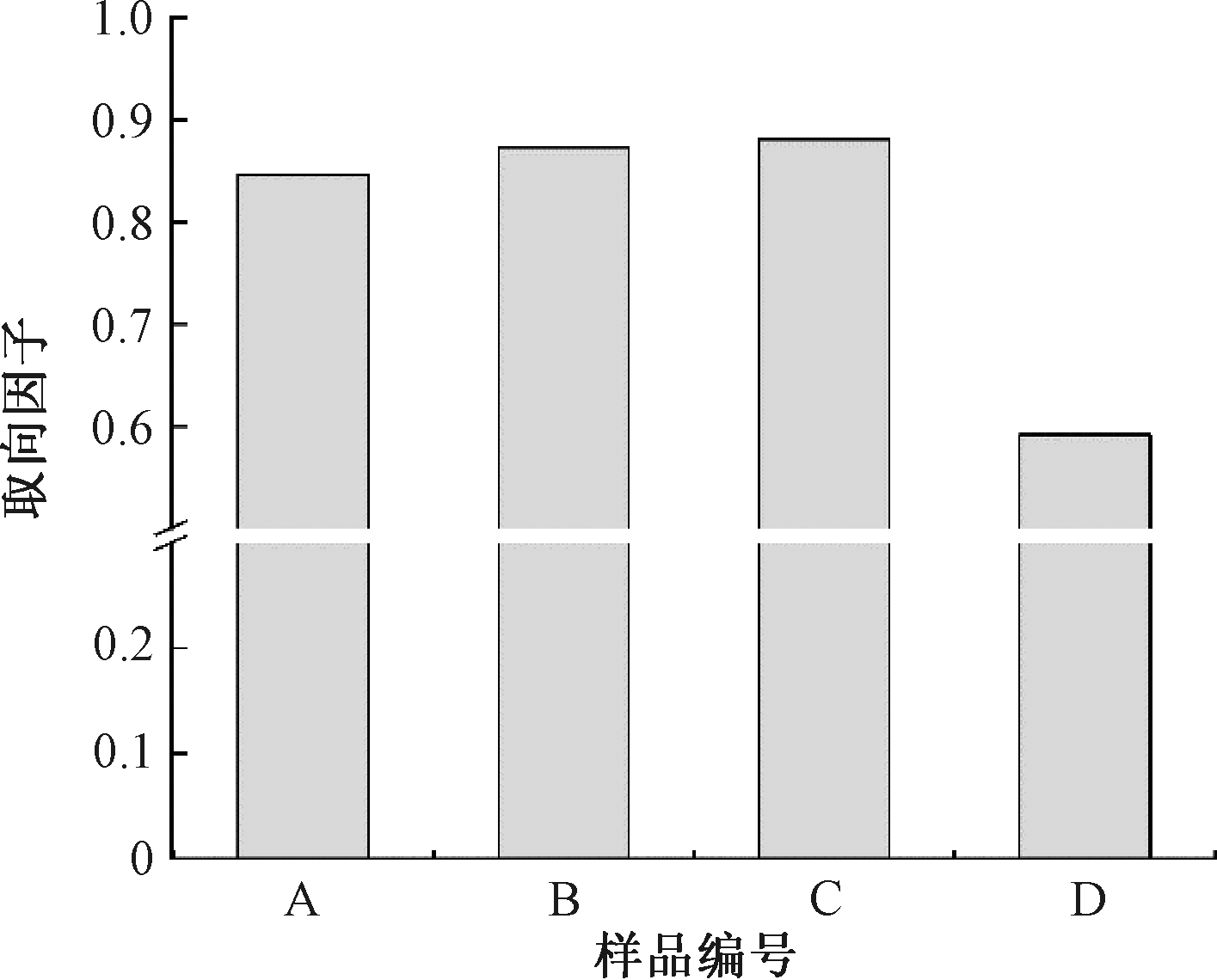
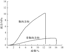
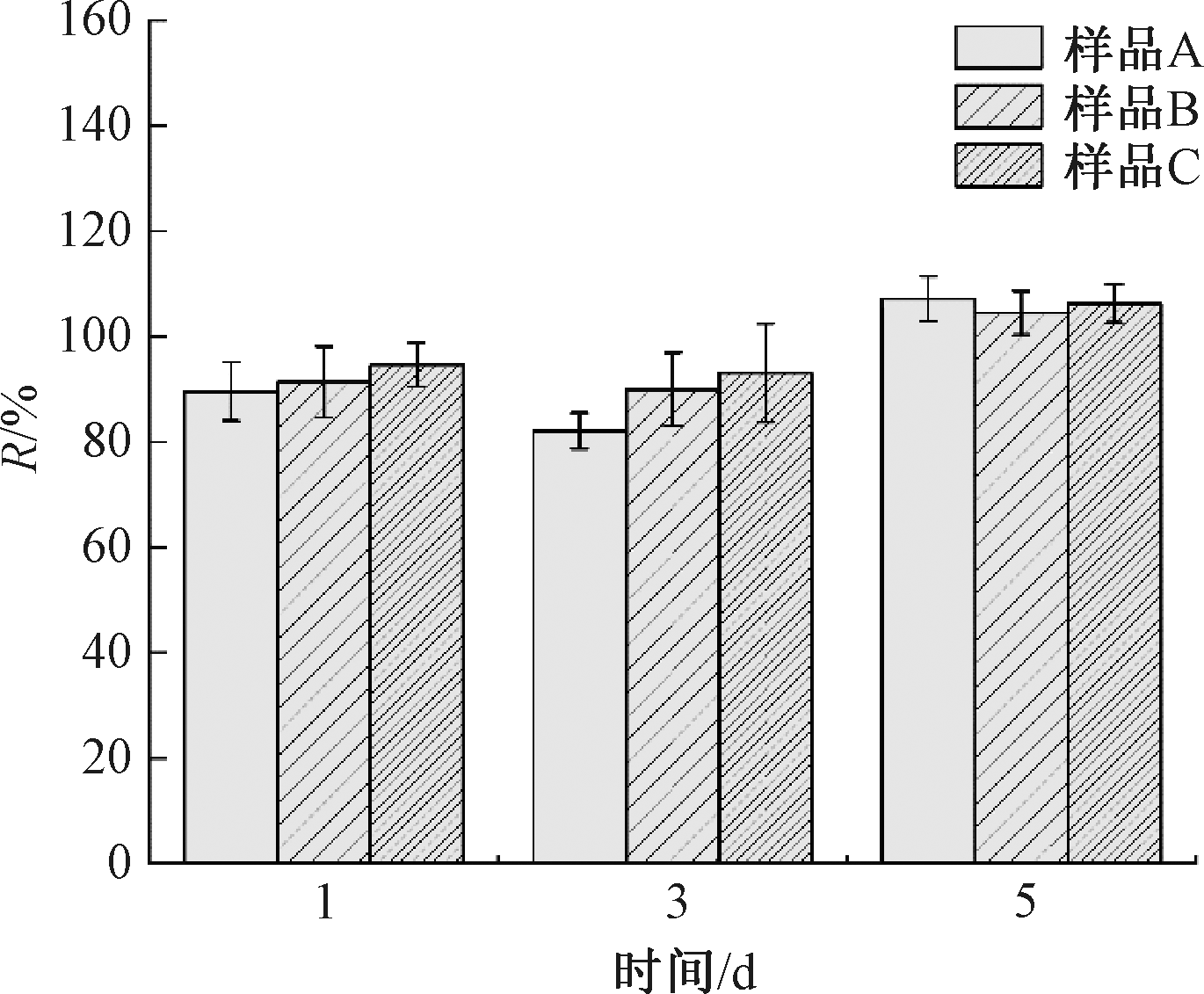
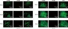
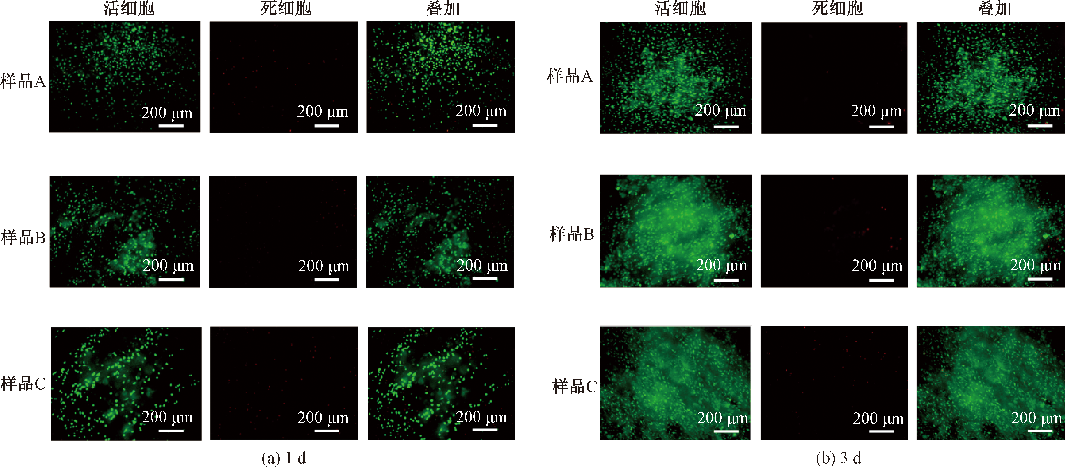
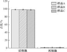
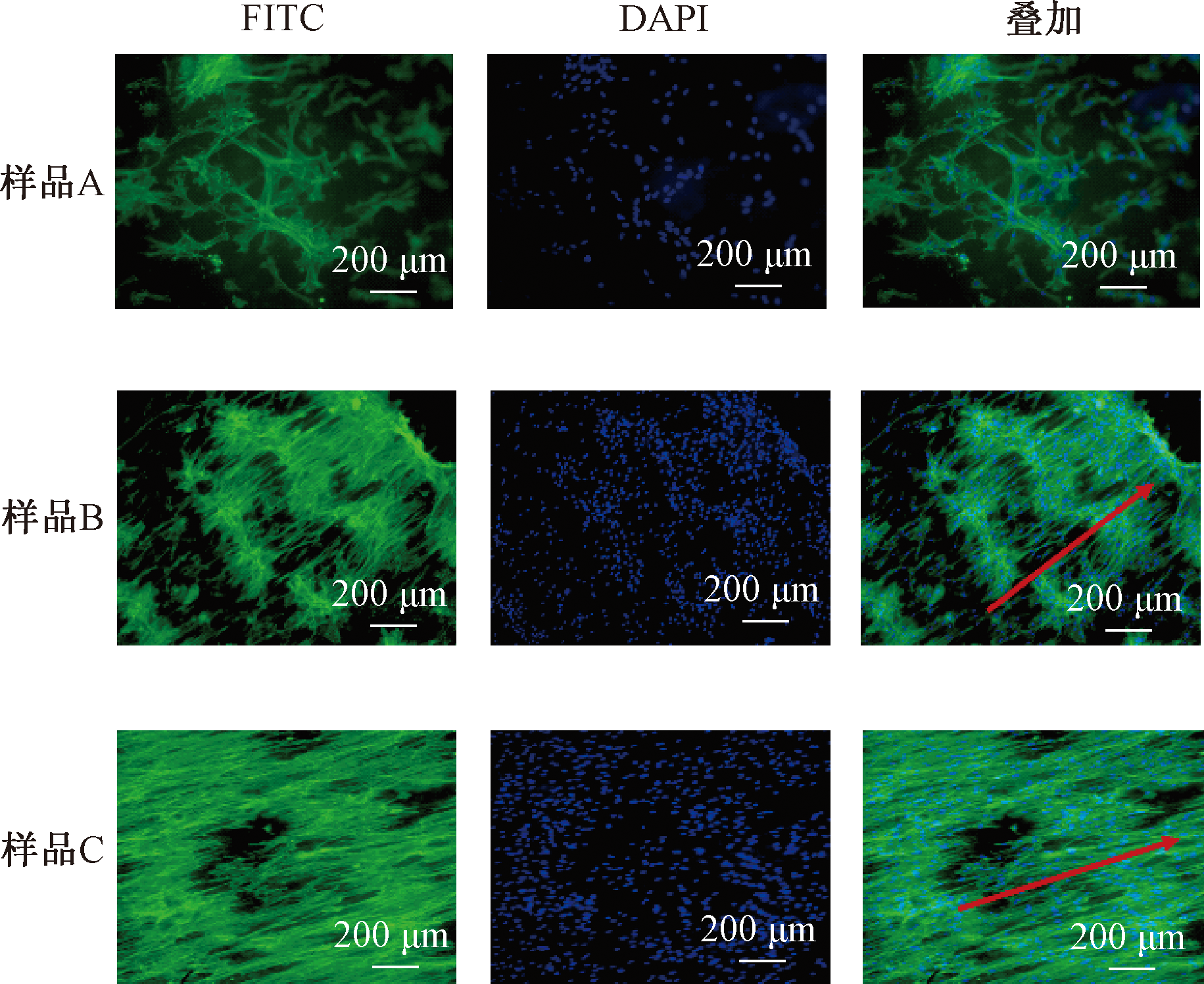
为精准调控取向纳米纤维膜的物理结构并探究其对骨髓间充质干细胞(MSCs)的物理引导作用,通过调控静电纺丝参数,制备了壳聚糖/聚己内酯(CS/PCL)取向纳米纤维膜,系统考察了接收距离、推注速度和接收滚轮转速对纳米纤维膜形貌结构的影响。结果表明,当接收距离为16 cm、推注速度为0.8 mL/h时,可获得形貌均一的纤维;在2 500 r/min滚轮转速条件下,纤维呈现高取向性(取向因子达0.88)。该取向纳米纤维膜表现出良好的生物相容性,且对MSCs具有一定的促增殖作用。更重要的是,高取向的纳米纤维膜可有效引导MSCs沿纤维方向定向排列,并呈现神经样细长形态,表明其具有促进MSCs神经向分化的潜力。研究证实,通过调控静电纺丝参数可成功制备出具有显著物理引导作用的CS/PCL取向纳米纤维膜,为基于物理结构调控的神经组织工程支架设计提供实验依据。
中图分类号:
| [1] | DONG R Q, LIU Y M, YANG Y X, et al. MSC-derived exosomes-based therapy for peripheral nerve injury: a novel therapeutic strategy[J]. BioMed Research International, 2019, 2019: 6458237. |
| [2] |
XU J W, WEN J K, FU L Y, et al. Macrophage-specific RhoA knockout delays Wallerian degeneration after peripheral nerve injury in mice[J]. Journal of Neuroinflammation, 2021, 18(1): 234.
doi: 10.1186/s12974-021-02292-y pmid: 34654444 |
| [3] |
YAO X L, XUE T, CHEN B Q, et al. Advances in biomaterial-based tissue engineering for peripheral nerve injury repair[J]. Bioactive Materials, 2025, 46: 150-172.
doi: 10.1016/j.bioactmat.2024.12.005 pmid: 39760068 |
| [4] |
GORDON T. Peripheral nerve regeneration and muscle reinnervation[J]. International Journal of Molecular Sciences, 2020, 21(22): 8652.
doi: 10.3390/ijms21228652 |
| [5] |
YANG S H, CHEN L, BAI C H, et al. Polymer scaffolds for peripheral nerve injury repair[J]. Progress in Materials Science, 2025, 153: 101497.
doi: 10.1016/j.pmatsci.2025.101497 |
| [6] |
SHAN Y Z, XU L L, CUI X, et al. A responsive cascade drug delivery scaffold adapted to the therapeutic time window for peripheral nerve injury repair[J]. Materials Horizons, 2024, 11(4): 1032-1045.
doi: 10.1039/d3mh01511d pmid: 38073476 |
| [7] |
DONG Y Z, LU H. Editorial: surgical treatment of peripheral neuropathic pain, peripheral nerve tumors, and peripheral nerve injury[J]. Frontiers in Neurology, 2023, 14: 1266638.
doi: 10.3389/fneur.2023.1266638 |
| [8] |
VILLANUEVA-FLORES F, GARCIA-ATUTXA I, SANTOS A, et al. Toward a new generation of bio-scaffolds for neural tissue engineering: challenges and perspectives[J]. Pharmaceutics, 2023, 15(6): 1750.
doi: 10.3390/pharmaceutics15061750 |
| [9] |
TSENG Y H, MA T L, TAN D H, et al. Injectable hydrogel guides neurons growth with specific directionality[J]. International Journal of Molecular Sciences, 2023, 24(9): 7952.
doi: 10.3390/ijms24097952 |
| [10] |
YANG Y F, YIN X, WANG H D, et al. Engineering a wirelessly self-powered and electroconductive scaffold to promote peripheral nerve regeneration[J]. Nano Energy, 2023, 107: 108145.
doi: 10.1016/j.nanoen.2022.108145 |
| [11] |
SOUSA J P M, MONTEIRO C F, DEUS I A, et al. Magnetoresponsive anisotropic fiber-integrating hydrogels for neural tissue regeneration[J]. Small Structures, 2024, 5(11): 2400213.
doi: 10.1002/sstr.v5.11 |
| [12] | 姚双双, 付少举, 张佩华, 等. 再生丝素蛋白/聚乙烯醇共混取向纳米纤维膜的制备与性能[J]. 纺织学报, 2023, 44(9): 11-19. |
| YAO Shuangshuang, FU Shaoju, ZHANG Peihua, et al. Preparation and properties of regenerated silk fibroin/polyvinyl alcohol blended nanofiber membranes with predesigned orientation[J]. Journal of Textile Research, 2023, 44(9): 11-19. | |
| [13] |
HAJIPOUR F P, FEYZBAKHSH A, MALEKNIA L, et al. Electrospun scaffold with bioactive polyurethane shell infused with propolis and starch-hyaluronic acid core: an advanced therapeutic platform for skin tissue engineering[J]. International Journal of Biological Macromolecules, 2025, 288: 138745.
doi: 10.1016/j.ijbiomac.2024.138745 |
| [14] |
NIU Z W, WANG X F, MENG X, et al. Controllable fiber orientation and nonlinear elasticity of electrospun nanofibrous small diameter tubular scaffolds for vascular tissue engineering[J]. Biomedical Materials, 2019, 14(3): 035006.
doi: 10.1088/1748-605X/ab07f1 |
| [15] |
KUMAR R, AADIL K R, RANJAN S, et al. Advances in nanotechnology and nanomaterials based strategies for neural tissue engineering[J]. Journal of Drug Delivery Science and Technology, 2020, 57: 101617.
doi: 10.1016/j.jddst.2020.101617 |
| [16] |
RANJBAR-MOHAMMADI M, PRABHAKARAN M P, BAHRAMI S H, et al. Gum tragacanth/poly(L-lactic acid) nanofibrous scaffolds for application in regeneration of peripheral nerve damage[J]. Carbohydrate Polymers, 2016, 140: 104-112.
doi: 10.1016/j.carbpol.2015.12.012 |
| [17] |
MUNGENAST L, ZÜGER F, SELVI J, et al. Directional submicrofiber hydrogel composite scaffolds supporting neuron differentiation and enabling neurite alignment[J]. International Journal of Molecular Sciences, 2022, 23(19): 11525.
doi: 10.3390/ijms231911525 |
| [18] | 戈亚锋, 王琰, 徐楚琪, 等. 高亲水性壳聚糖纳米纤维膜的制备及性能[J]. 现代纺织技术, 2024, 32(10): 11-19. |
| GE Yafeng, WANG Yan, XU Chuqi, et al. Preparation and performance of highly hydrophilic chitosan nanofiber membranes[J]. Advanced Textile Technology, 2024, 32(10): 11-19. | |
| [19] |
LEVENGOOD S L, ZHANG M Q. Chitosan-based scaffolds for bone tissue engineering[J]. Journal of Materials Chemistry B, 2014, 2(21): 3161-3184.
pmid: 24999429 |
| [20] | 石锐, 薛佳佳, 黎广平, 等. PCL基电纺纤维膜的抑菌、相容及降解性能研究[J]. 生物骨科材料与临床研究, 2015, 12(1): 6-11. |
| SHI Rui, XUE Jiajia, LI Guangping, et al. Antibacterial, in vitro/vivo biocompatibility and biodegradability of PCL-based electrospun fibers[J]. Orthopaedic Biomechanics Materials and Clinical Study, 2015, 12(1): 6-11. | |
| [21] | 宋文杰, 杨琪苑, 阚诗琦, 等. 静电纺丝技术在神经组织工程中的应用进展[J]. 中国美容医学, 2024, 33(11): 185-188. |
| SONG Wenjie, YANG Qiyuan, KAN Shiqi, et al. The application progress of electrospinning technology in nerve tissue engineering[J]. Chinese Journal of Aesthetic Medicine, 2024, 33(11): 185-188. | |
| [22] | 吴乐然, 吴霓欢, 李林耿, 等. 负载厚朴酚的抗菌纳米纤维膜的制备及其性能[J]. 纺织学报, 2025, 46(10): 30-38. |
|
WU Leran, WU Nihuan, LI Lingeng, et al. Preparation and performance of antibacterial nanofiber membrane loaded with magnolol[J]. Journal of Textile Research, 2025, 46(10): 30-38.
doi: 10.1177/004051757604600105 |
|
| [23] | 贾琳, 杨奥杰, 张芳铖, 等. 共聚型聚酰亚胺纳米纤维膜的制备及其性能[J]. 纺织学报, 2025, 46(8): 37-44. |
| JIA Lin, YANG Aojie, ZHANG Fangcheng, et al. Preparation and performance study of copolymerized polyimide nanofiber membrane[J]. Journal of Textile Research, 2025, 46(8): 37-44. | |
| [24] |
LIU J Z, LIN Z Y, WU H Y, et al. Dual-regulation biomimetic composite nerve scaffold with oriented structure and conductive function for skin peripheral nerve injury repair[J]. Colloids and Surfaces B: Biointerfaces, 2025, 253: 114768.
doi: 10.1016/j.colsurfb.2025.114768 |
| [25] | 张泽天, 陈艳梅, 侯宗姊, 等. 静电纺丝参数对丝素蛋白纤维直径的影响及其力学机理研究[J/OL]. 化工新型材料, 2026.DOI:10.19817/j.cnki.issn1006-3536.2026.04.015. |
| ZHANG Zetian, CHEN Yanmei, HOU Zongzi, et al. Study on the influence of electrospinning parameters on silk fibroin fiber diameter and mechanical mechanism[J/OL]. New Chemical Materials, 2026.DOI:10.19817/j.cnki.issn1006-3536.2026.04.015. | |
| [26] |
朱志超, 李阳, 谯云, 等. PVDF纳米纤维膜静电纺丝工艺调控分析[J]. 化工新型材料, 2025, 53(8): 91-96.
doi: 10.19817/j.cnki.issn1006-3536.2025.08.027 |
|
ZHU Zhichao, LI Yang, QIAO Yun, et al. Analysis of electrostatic spinning process regulation for PVDF nanofiber membranes[J]. New Chemical Materials, 2025, 53(8): 91-96.
doi: 10.19817/j.cnki.issn1006-3536.2025.08.027 |
|
| [27] |
XIE J W, MACEWAN M R, SCHWARTZ A G, et al. Electrospun nanofibers for neural tissue engineering[J]. Nanoscale, 2010, 2(1): 35-44.
doi: 10.1039/b9nr00243j pmid: 20648362 |
| [28] |
BAKER B M, MAUCK R L. The effect of nanofiber alignment on the maturation of engineered meniscus constructs[J]. Biomaterials, 2007, 28(11): 1967-1977.
doi: 10.1016/j.biomaterials.2007.01.004 pmid: 17250888 |
| [29] |
PITTENGER M F, MACKAY A M, BECK S C, et al. Multilineage potential of adult human mesenchymal stem cells[J]. Science, 1999, 284(5411): 143-147.
doi: 10.1126/science.284.5411.143 pmid: 10102814 |
| [30] |
WEI X, YANG X, HAN Z P, et al. Mesenchymal stem cells: a new trend for cell therapy[J]. Acta Pharmacologica Sinica, 2013, 34(6): 747-754.
doi: 10.1038/aps.2013.50 pmid: 23736003 |
| [31] |
LIANG Y P, QIAO L P, QIAO B W, et al. Conductive hydrogels for tissue repair[J]. Chemical Science, 2023, 14(12): 3091-3116.
doi: 10.1039/d3sc00145h pmid: 36970088 |
| [1] | 郭一铭, 喻爽, 赵帆, 王富军. 血管监测用纤维基压电传感器的构建及性能评价[J]. 纺织学报, 2026, 47(03): 118-128. |
| [2] | 林晓静, 毛迎, 陈文兴, 吕汪洋. 载姜黄素静电纺丝纤维膜的制备及其抗菌与抗氧化性能[J]. 纺织学报, 2026, 47(03): 217-224. |
| [3] | 易珊, 王丽芳, 陈黎, 邱虹, 唐一卡, 张国清, 王美英, 高艳春, 葛秀敏, 刘丽芳. 纳米纤维素基pH响应型抗菌抗氧化伤口敷料的制备及其性能[J]. 纺织学报, 2026, 47(03): 26-34. |
| [4] | 陈泳良, 杨潇, 王朝荣, 黄俊鸿, 李彦, 王璐. 静电纺丝-恒应力退火协同构建的湿态稳定型聚乳酸/I型胶原肩袖补片[J]. 纺织学报, 2026, 47(03): 60-69. |
| [5] | 李好义, 田鑫哲, 张毅, 牟文英, 张超, 赵千龙, 杨卫民. 导电各向异性复合心脏补片的熔体静电纺丝/直写构建及体外评价[J]. 纺织学报, 2026, 47(03): 70-76. |
| [6] | 张曼琦, 孙艳丽, 张晓茹, 李博, 刘哲. 共轭静电纺双模态调温织物的制备及其性能[J]. 纺织学报, 2026, 47(02): 153-161. |
| [7] | 孔珂欣, 张怡帆, 卢哲, 王哲. 载钴钌原子无机微纳米纤维的制备及其电催化水分解性能[J]. 纺织学报, 2026, 47(02): 26-36. |
| [8] | 王世杰, 孙辉, 于斌. 聚乙烯醇/牡丹皮提取物复合纳米静电纺丝膜的制备及其抗菌性能[J]. 纺织学报, 2026, 47(02): 56-64. |
| [9] | 孔艳辉, 张琳萍, 毛志平, 徐红. 甲基丙烯酰化明胶纤维膜的制备及其止血性能[J]. 纺织学报, 2026, 47(01): 1-10. |
| [10] | 赵婧雯, 袁香楠, 高晶, 王璐. 聚丙烯腈-普鲁士蓝/月桂酸/环丙沙星光热响应性抗菌敷料的制备及其性能[J]. 纺织学报, 2026, 47(01): 20-28. |
| [11] | 王世豪, 徐晓禹, 郑挺, 王金星, 姚德刚, 王俊, 叶翔宇, 田慧, 李婷, 朱斐超. 碳纤维非织造材料的研究应用及展望[J]. 纺织学报, 2026, 47(01): 240-249. |
| [12] | 罗家俊, 何耀权, 赵振鸿, 黎锦稻, 赵景, 黄钢, 王先锋. 苯乙烯-乙烯-丁烯-苯乙烯/氟化聚酰亚胺防水透湿纤维膜的制备及其性能[J]. 纺织学报, 2026, 47(01): 38-45. |
| [13] | 凌磊, 陈凯, 高俊, 武丁胜, 汪邓兵, 张春, 凤权. 聚丙烯腈/共价有机框架复合纳米纤维膜的制备及其对Cr(Ⅵ)的吸附性能[J]. 纺织学报, 2026, 47(01): 54-62. |
| [14] | 刘轲, 王雨曦, 程盼, 朱丽萍, 夏明, 梅涛, 向阳, 周丰, 高飞, 王栋. 多孔磺化氢化苯乙烯-丁二烯嵌段共聚物纤维膜制备及其吸附性能[J]. 纺织学报, 2025, 46(12): 1-10. |
| [15] | 王小虎, 包安娜, 魏静雯, 赵晓曼, 韩潇, 洪剑寒. 基于静电纺丝-静电喷涂协同工艺的跨尺度传感纱一步法制备及其应用[J]. 纺织学报, 2025, 46(12): 101-109. |
|
||
 京公网安备11010502044800号