纺织学报 ›› 2026, Vol. 47 ›› Issue (05): 45-55.doi: 10.13475/j.fzxb.20250707801

• 纤维材料 • 上一篇    下一篇

气流辅助静电纺丝聚四氟乙烯血管支架覆膜的抗凝血和促内皮化研究

程尔骕1, 王豪杰1, 刘宇清1,2, 孟凯1,2, 赵荟菁1,2()   

  1. 1 苏州大学 纺织与服装工程学院, 江苏 苏州 215021
    2 现代丝绸国家工程实验室, 江苏 苏州 215123
  • 收稿日期:2025-07-30 修回日期:2026-04-01 出版日期:2026-05-15 发布日期:2026-07-10
  • 通讯作者: 赵荟菁(1981—),女,教授,博士。主要研究方向为生物医用纺织材料。E-mail:zhhj@suda.edu.cn
  • 作者简介:程尔骕(2002—),男,硕士生。主要研究方向为聚四氟乙烯人工血管的构建与功能修饰。
  • 基金资助:
    产学研合作项目(P111505421)

Study on anticoagulant properties and endothelialization promotion of polytetrafluoroethylene vascular stent membranes by airflow-assisted electrospinning

CHENG Ersu1, WANG Haojie1, LIU Yuqing1,2, MENG Kai1,2, ZHAO Huijing1,2()   

  1. 1 College of Textiles and Clothing Engineering, Soochow University, Suzhou, Jiangsu 215021, China
    2 National Engineering Laboratory for Modern Silk(Suzhou), Soochow University, Suzhou, Jiangsu 215123, China
  • Received:2025-07-30 Revised:2026-04-01 Published:2026-05-15 Online:2026-07-10

摘要:

针对传统静电纺丝法制备聚四氟乙烯(PTFE)血管支架覆膜生产效率低、不利于量产的局限性,以及膨体聚四氟乙烯(ePTFE)覆膜材料植入后内皮化不佳、易引起血栓等问题,采用气流辅助静电纺丝技术制备聚四氟乙烯(PTFE)覆膜,并通过等离子体活化、接枝多巴胺/聚乙烯亚胺(DA/PEI)及肝素(HEP)等表面改性技术赋予PTFE支架覆膜抗凝血和促内皮化功能。研究了气流辅助静电纺丝工艺对纤维形貌和生产效率的影响,同时探究了涂层改性后PTFE-DA/PEI-HEP覆膜的形貌、化学成分、抗凝血和促内皮化等性能。结果表明:气流辅助静电纺丝法单位时间产量较传统静电纺丝提升4.13倍,同时纤维直径更细,覆膜断裂强度提升至13.38 MPa;经DA/PEI-HEP涂层后,覆膜的肝素负载量提高4.7倍且实现了缓慢释放;覆膜的凝血指数较未改性PTFE提高了24.97%,血小板黏附密度降低了44.19%;内皮细胞第5天增殖率达到104.13%,较未改性样品提高3.1%。该研究突破了PTFE覆膜生产效率瓶颈,并使PTFE管状覆膜具有抗凝血与促内皮化双重功能,具有临床转化潜力。

关键词: 气流辅助电纺, 聚四氟乙烯血管支架覆膜, 功能化改性, 抗凝血, 促内皮化

Abstract:

Objective Cardiovascular diseases (CVDs) are a class of clinical syndromes characterized by dysfunction of the heart and vascular system, and have become the leading cause of death globally. In clinical practice, stent grafts are widely used for interventional treatment, where the graft membrane serves as a physical barrier and plays a critical role in cases such as aneurysm occlusion, vascular perforation repair, and arterial stenosis management. Currently, expanded polytetrafluoroethylene (ePTFE) has become one of the commonly used membrane materials in clinical practice by virtue of its excellent biocompatibility, stable mechanical properties, and chemical inertness. However, commercial ePTFE grafts often suffer from poor long-term patency due to their chemical inertness, which hinders endothelialization and triggers thrombogenic responses. While electrospinning offers a promising alternative for mimicking the natural extracellular matrix (ECM), conventional electrospinning is severely limited by low production efficiency and jet instability, hindering industrial-scale manufacturing. Therefore, this study aims to address these dual challenges by developing a high-throughput airflow-assisted electrospinning strategy to fabricate PTFE vascular stent membranes. Furthermore, to overcome the bio-inert nature of PTFE, a surface functionalization strategy is proposed to simultaneously endow the grafts with potent anticoagulant properties and the capacity to promote rapid endothelialization, thereby enhancing their clinical translation potential.

Method A novel airflow-assisted electrospinning system utilized a high-velocity air stream to manipulate the polymer jet trajectory and enhance solvent evaporation. The influences of key processing parameters, particularly extrusion rate, on fiber morphology and deposition efficiency were systematically optimized. In order to functionalize the chemically inert PTFE surface, a multi-step modification protocol was employed. First, the membranes underwent air plasma activation to introduce initial reactive groups. Subsequently, a bio-adhesive intermediate layer was constructed by co-depositing dopamine (DA) and polyethyleneimine (PEI) under mild alkaline conditions (Tris-HCl, pH=8.5). Finally, heparin (HEP) was covalently immobilized onto the amine-rich DA/PEI layer by EDC/NHS activation chemistry. Physicochemical properties were characterized using scanning electron microscopy (SEM), Fourier transform infrared spectroscopy(FT-IR), energy dispersive spectroscopy(EDS), and contact angle measurements. Biological performance was evaluated through in vitro heparin release profiles, hemolysis assays, blood clotting index (BBCI) tests, platelet adhesion studies, and endothelial cell (ECs) proliferation assays using CCK-8 and fluorescence imaging.

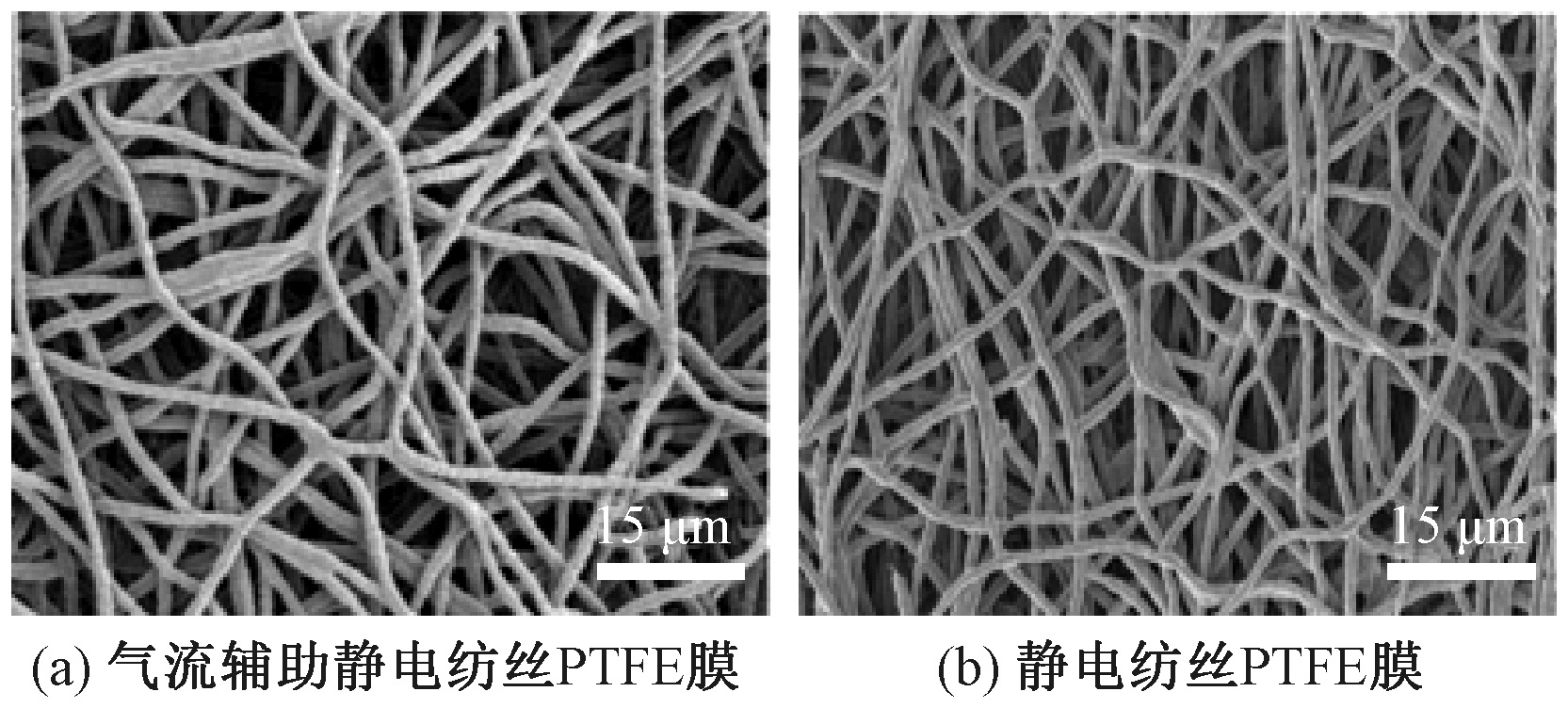
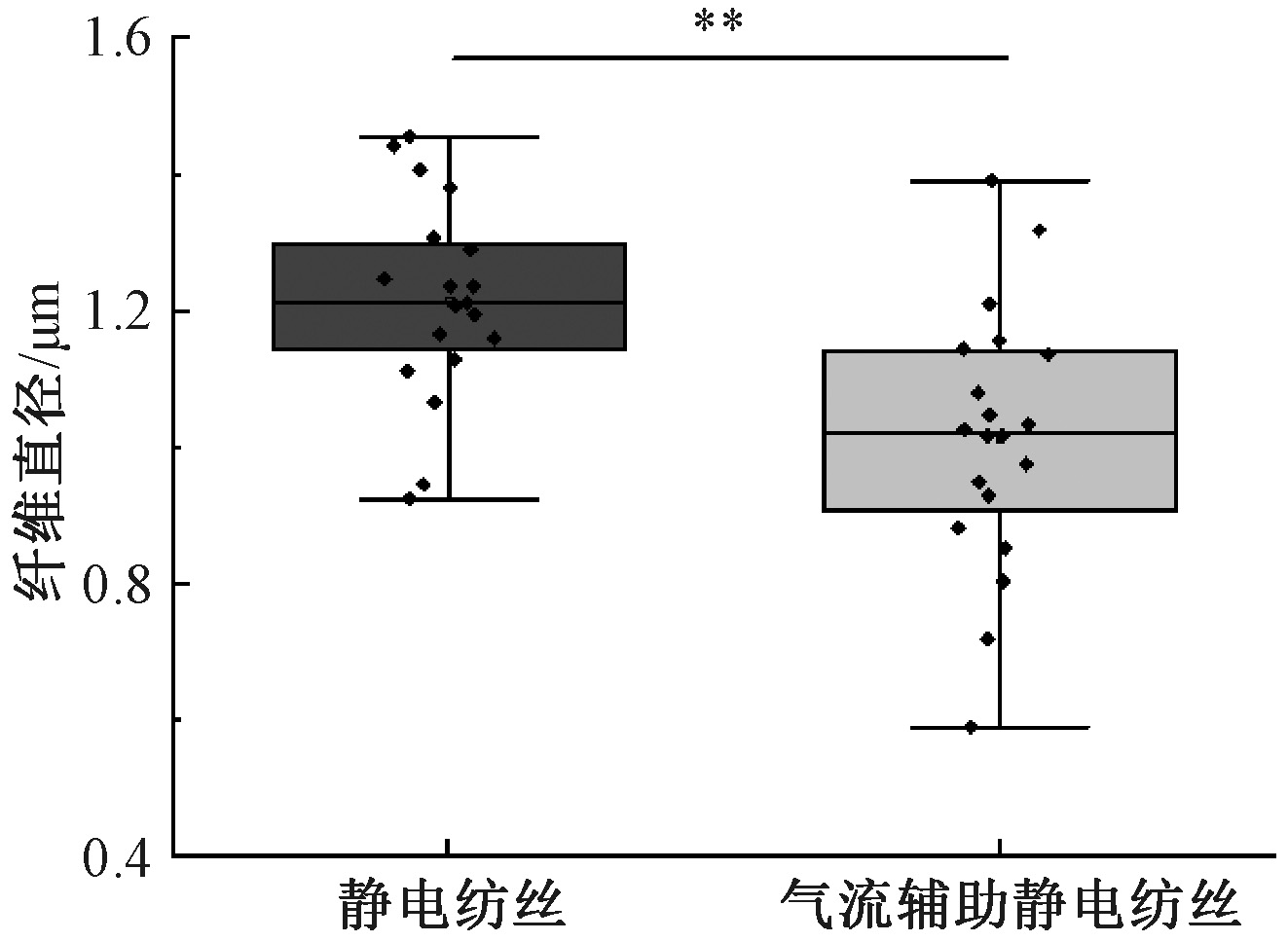
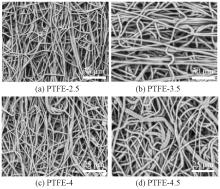
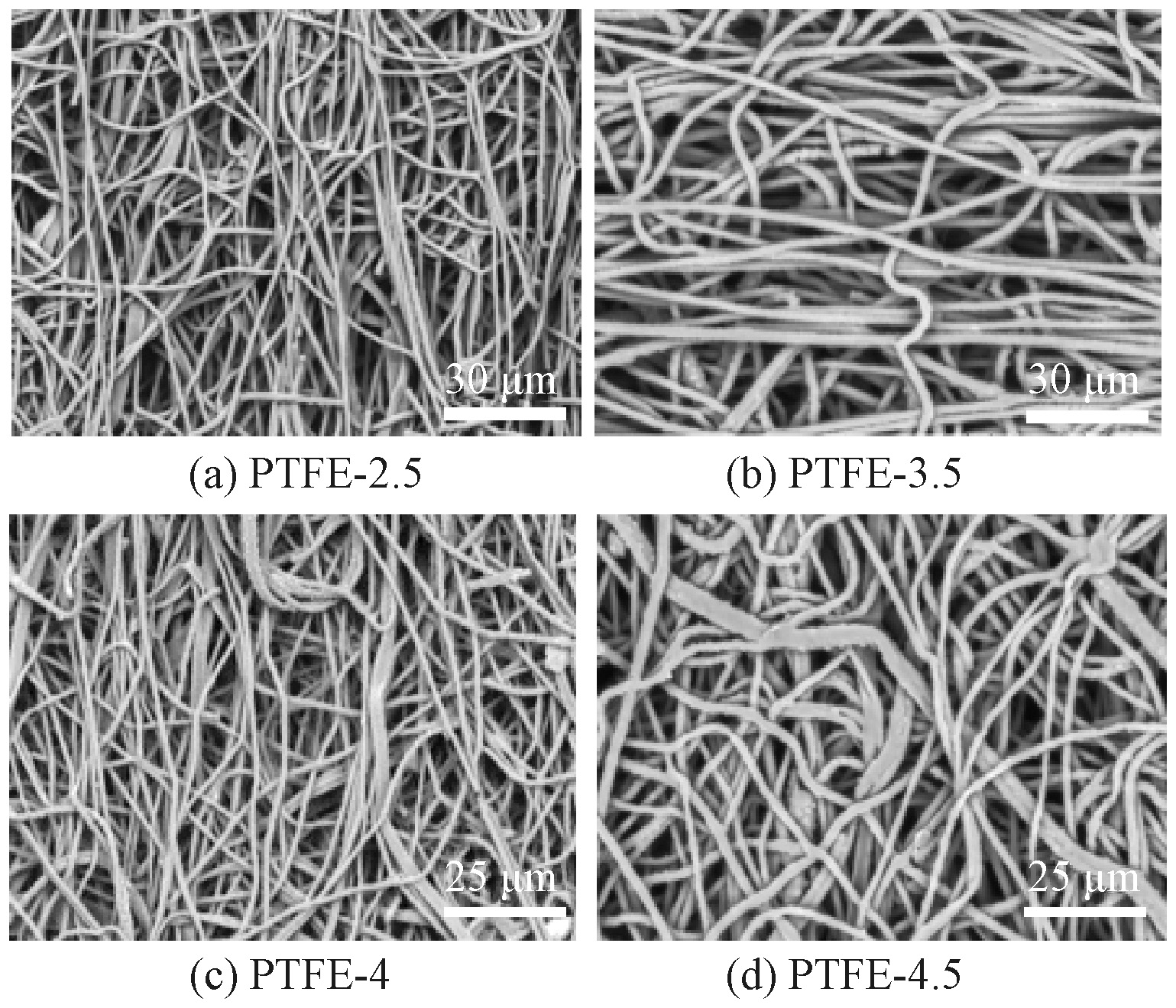
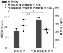
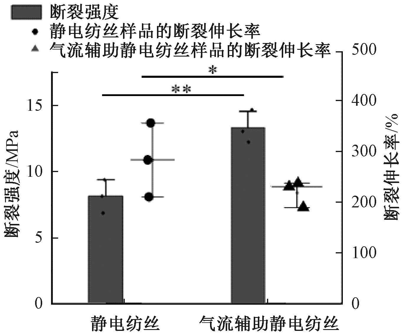
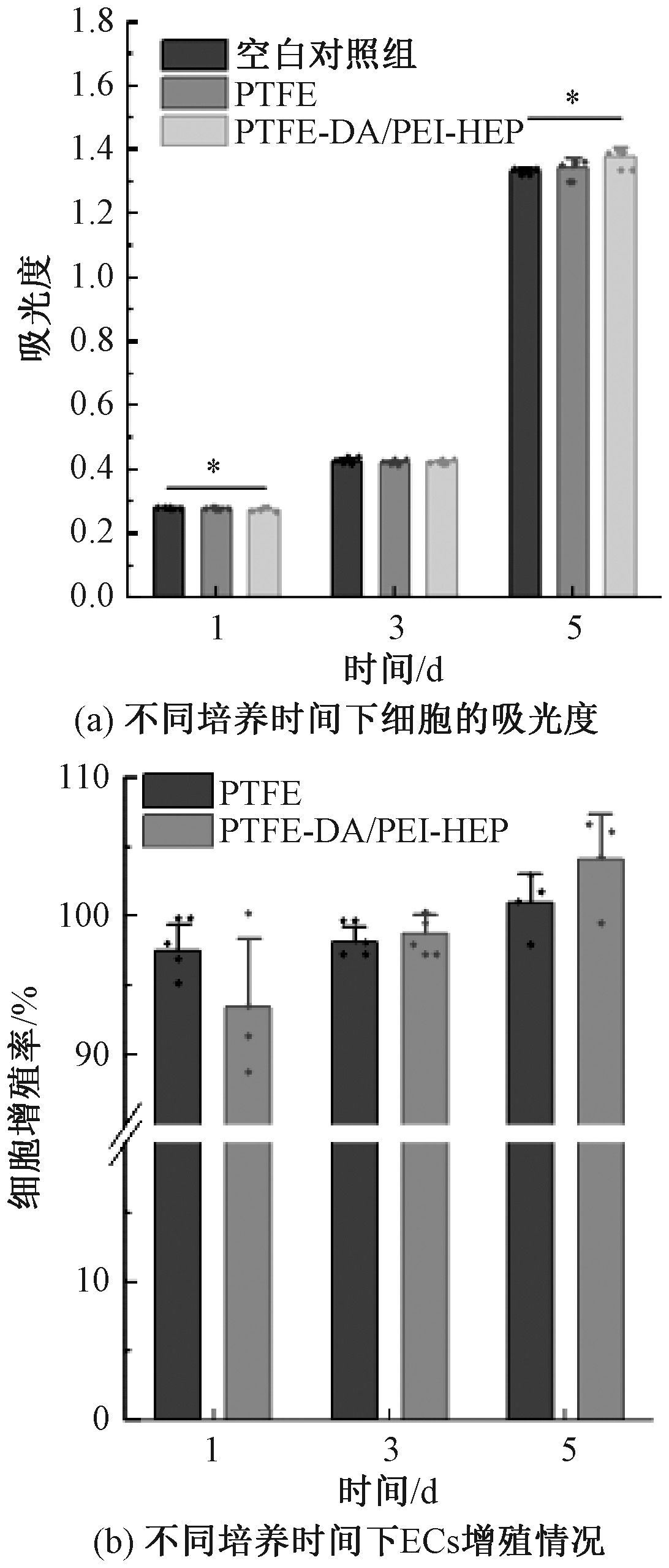
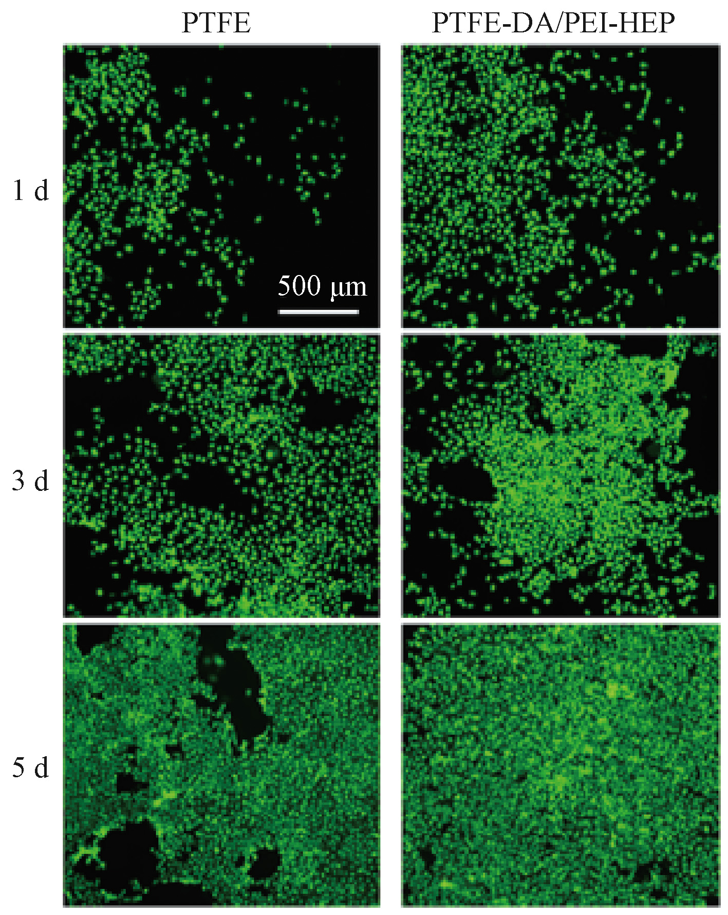
Results Airflow assisting significantly suppressed the whipping instability of the electrospinning jet, resulting in more uniform and finer fibers compared to the conventional method. The airflow-assisted process achieved a unit time production of 0.012 8 g/min, a 4.13 time increase over the 0.003 1 g/min rate of conventional electrospinning. This denser packing improved mechanical performance, with the tensile strength of the PTFE stent membrane increasing significantly to 13.38 MPa. Surface analysis confirmed the successful deposition of the functional coating, with the water contact angle dropping drastically to near 0°, indicating a transition from superhydrophobicity to superhydrophilicity. The DA/PEI intermediate layer is proved highly effective for drug loading; the functionalized PTFE-DA/PEI-HEP surface achieved a heparin density of 32.914 μg/cm2, a 4.7 time increase compared to direct adsorption (P<0.01), and demonstrated a sustained release profile over 7 d. Regarding hemocompatibility, the modified membranes exhibited an extremely low hemolysis ratio of 0.72%, far below the 5% international standard. The BBCI value was improved by 24.97% compared to unmodified PTFE, reaching 74.23%. Furthermore, SEM revealed that platelet adhesion density decreased by 44.19% (to 480 platelets/mm2), with minimal platelet activation observed. Cytocompatibility assays demonstrated that the coating created a favorable microenvironment for endothelial cells; by 5 d, the relative proliferation rate of endothelial cells on the modified surface reached 104.13%, representing a 3.1% enhancement over the unmodified control, with fluorescence imaging confirming a dense, healthy cell monolayer.

Conclusion This study establishes a scalable airflow-assisted electrospinning protocol that overcomes the production efficiency bottleneck of PTFE nanofiber membranes while significantly reducing costs compared to thermal stretching methods. The combination of plasma treatment and DA/PEI-mediated heparin grafting transforms the bio-inert PTFE surface into a bioactive interface. The resulting vascular stent membranes possess excellent mechanical strength, superior hemocompatibility, and the ability to promote endothelialization. These findings suggest that the developed PTFE-DA/PEI-HEP membranes offer a robust solution for replacing ePTFE stent membranes, which is promising for future clinical applications.

Key words: airflow-assisted electrospinning, polytetrafluoroethylene vascular stent membrane, functional modification, anticoagulation, endothelialization promotion

中图分类号: 

  • TS151

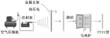
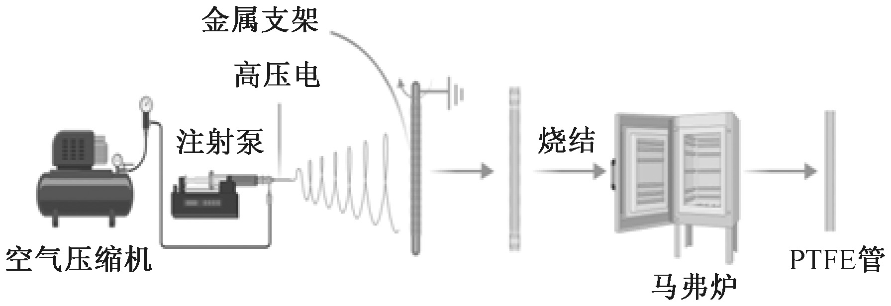
图1

气流辅助静电纺丝法制备PTFE静电纺丝膜的流程示意图"

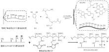
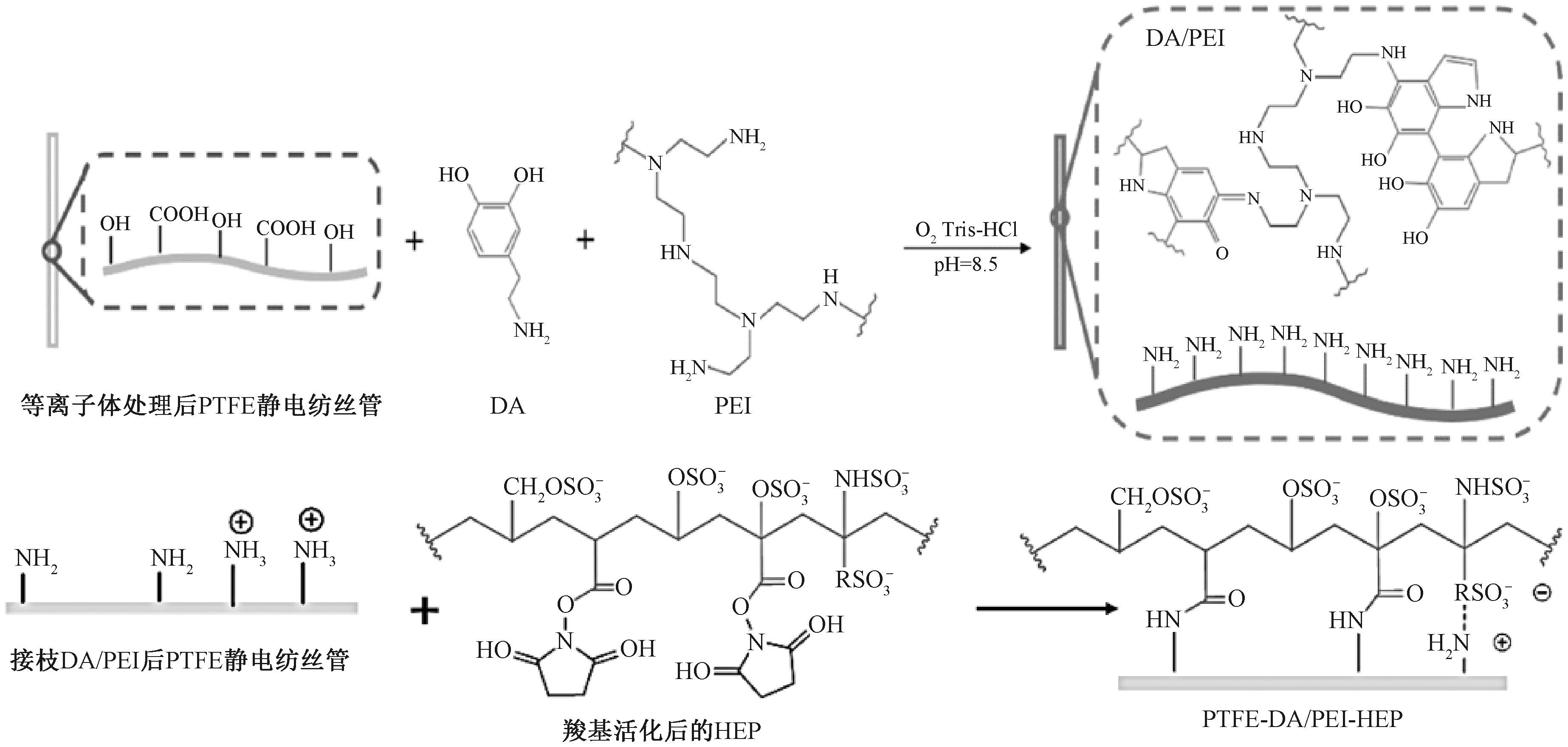
图2

DA/PEI-HEP涂层的反应机制"

图3

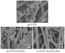
不同方法制备的PTFE膜微观形貌"

图4

不同方法制备的PTFE纤维直径"

图5

不同推料速率的气流辅助静电纺丝PTFE膜微观形貌"

图6

不同样品的力学性能"

图7

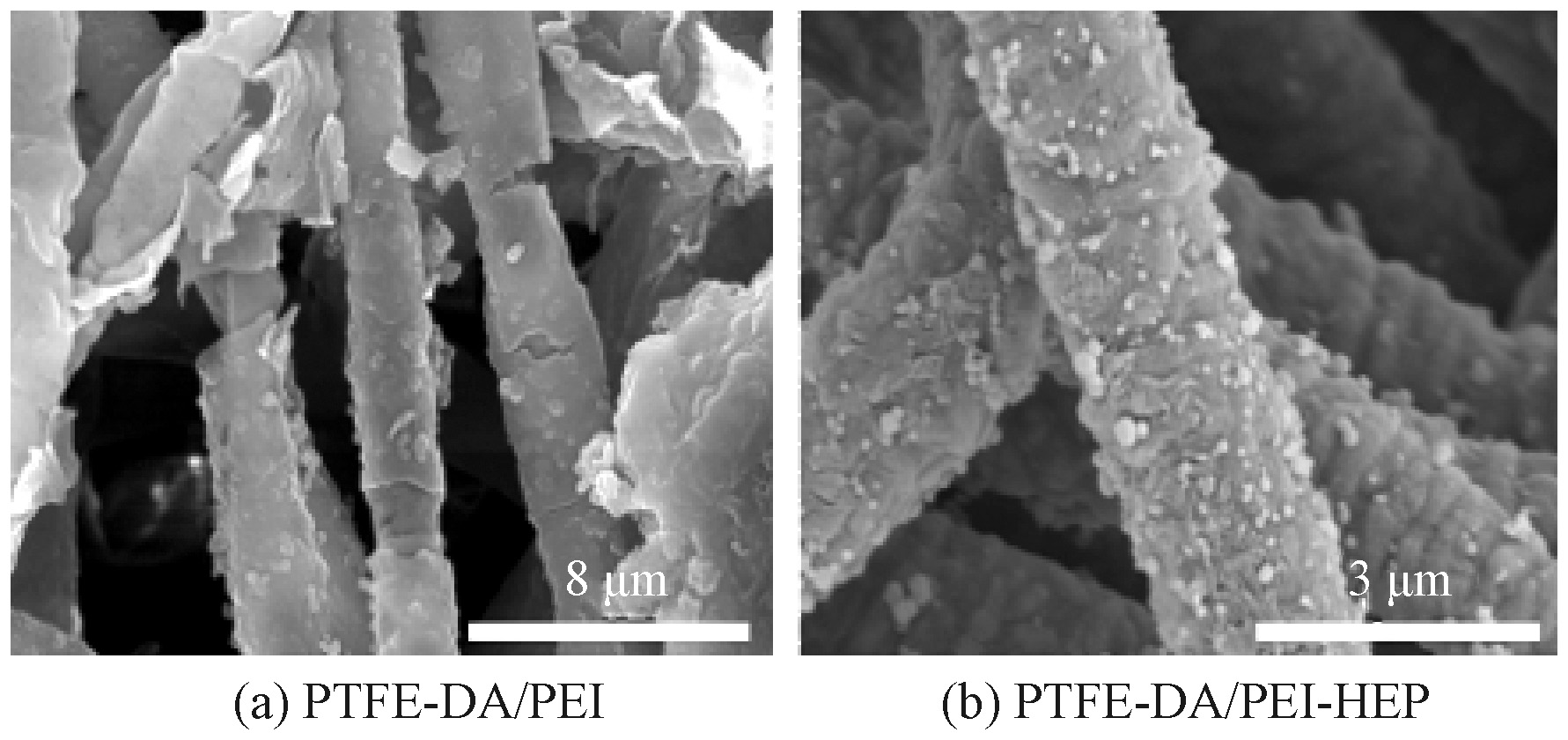
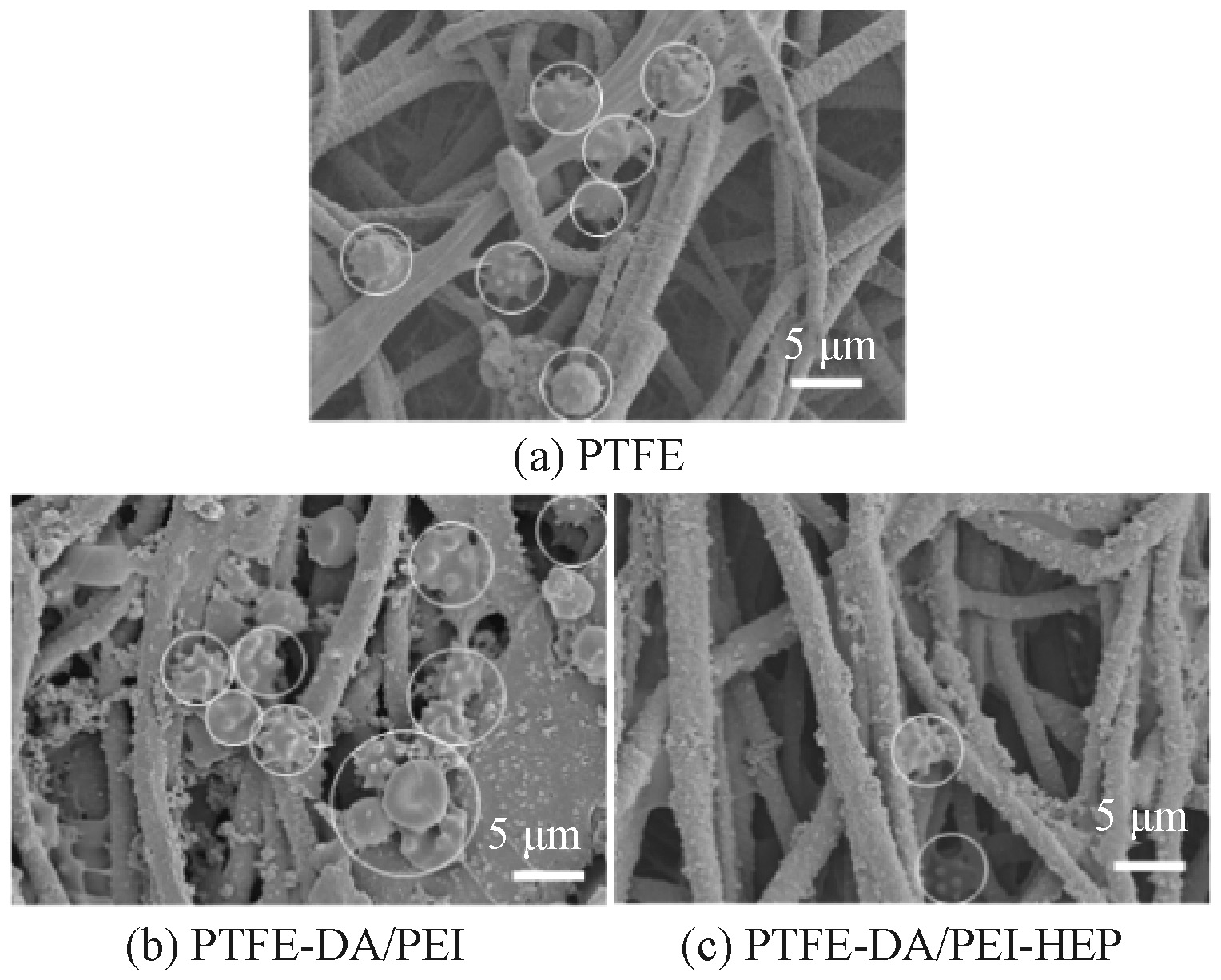
肝素改性前后PTFE膜的微观形貌"

图8

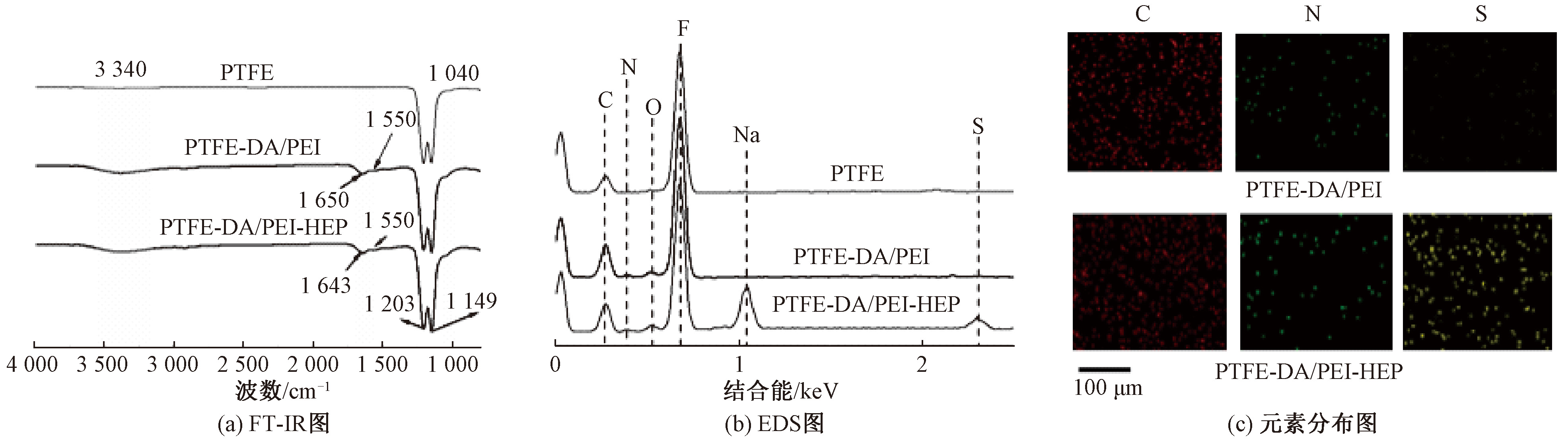
接枝DA/PEI及HEP前后PTFE覆膜的FT-IR、EDS以及元素分布图"

图9

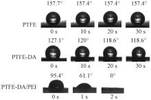
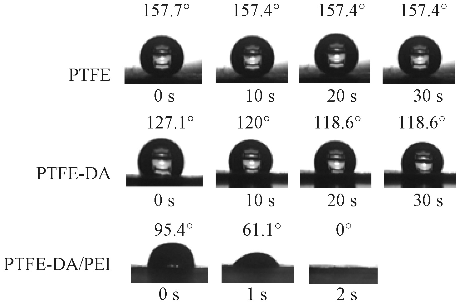
不同涂层的接触角照片"

图10

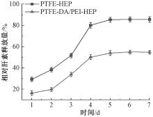
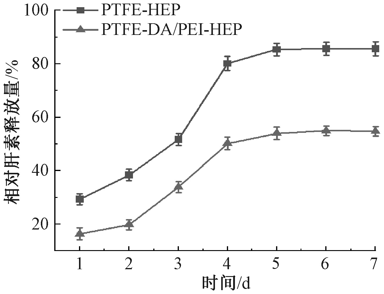
不同样品表面的肝素释放曲线"

图11

不同样品的血小板黏附情况"

图12

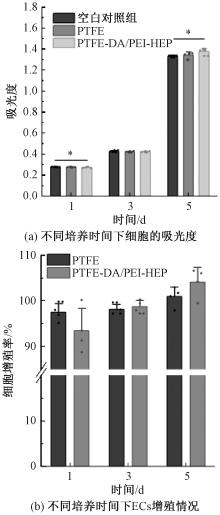
不同样品的吸光度及内皮细胞增殖情况"

图13

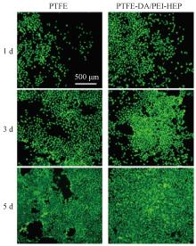
内皮细胞在不同材料上培养1、3、5 d的荧光倒置显微镜图"

[1] SHAMAKI G R, MARKSON F, SOJI-AYOADE D, et al. Peripheral artery disease: a comprehensive updated review[J]. Current Problems in Cardiology, 2022, 47(11): 101082.
doi: 10.1016/j.cpcardiol.2021.101082
[2] BRAUER M, ROTH G A, ARAVKIN A Y, et al. Global burden and strength of evidence for 88 risk factors in 204 countries and 811 subnational locations, 1990-2021: a systematic analysis for the Global Burden of Disease Study 2021[J]. The Lancet, 2024, 403(10440): 2162-2203.
doi: 10.1016/S0140-6736(24)00933-4
[3] PALANIAPPAN L P, ALLEN N B, ALMARZOOQ Z I, et al. 2026 heart disease and stroke statistics: a report of US and global data from the American heart association[J]. Circulation, 2026, 153(9): e275-e906.
[4] THUKKANI A K, KINLAY S. Endovascular intervention for peripheral artery disease[J]. Circulation Research, 2015, 116(9): 1599-1613.
doi: 10.1161/CIRCRESAHA.116.303503 pmid: 25908731
[5] WATSON N W, MOSARLA R C, SECEMSKY E A. Endovascular interventions for peripheral artery disease: a contemporary review[J]. Current Cardiology Reports, 2023, 25(11): 1611-1622.
doi: 10.1007/s11886-023-01973-9 pmid: 37804391
[6] ZHU P, DONG S T, SUN P, et al. Expanded polytetrafluoroethylene (ePTFE)-covered stents versus bare stents for transjugular intrahepatic portosystemic shunt in people with liver cirrhosis[J]. Cochrane Database of Systematic Reviews, 2023, 2023(8): 123-158.
[7] CASSADY A I, HIDZIR N M, GRØNDAHL L. Enhancing expanded poly(tetrafluoroethylene) (ePTFE) for biomaterials applications[J]. Journal of Applied Polymer Science, 2014, 131(15): 40533.
[8] ZHANG L L, HAM S W, WEAVER F A, et al. Performance of gore viabahn VBX compared with atrium iCast as bridging stents during fenestrated endovascular aortic repairs[J]. Journal of Vascular Surgery, 2019, 69(6): e215-e216.
[9] SOUKAS P, BECKER M, STARK K, et al. Three-year results of the GORE VIABAHN endoprosthesis in the superficial femoral artery for in-stent restenosis[J]. Journal of the Society for Cardiovascular Angiography & Interventions, 2023, 2(6): 101183.
[10] CHEN E, LI Z T, TURNG L S. Effects of expansion rate and sintering time on the morphology and mechanical properties of double-expanded polytetrafluoroethylene tubes[J]. Polymer Engineering & Science, 2025, 65(3): 1483-1496.
doi: 10.1002/pen.v65.3
[11] WANG G, FENG Y S, GAO C Y, et al. Biaxial stretching of polytetrafluoroethylene in industrial scale to fabricate medical ePTFE membrane with node-fibril microstructure[J]. Regenerative Biomaterials, 2023, 10: rbad056.
[12] MIURA H, NISHIBE T, YASUDA K, et al. The influence of node-fibril morphology on healing of high-porosity expanded polytetrafluoroethylene grafts[J]. European Surgical Research Europaische Chirurgische Forschung Recherches Chirurgicales Europeennes, 2002, 34(3): 224-231.
[13] GUO Q, HUANG Y, XU M D, et al. PTFE porous membrane technology: a comprehensive review[J]. Journal of Membrane Science, 2022, 664: 121115.
doi: 10.1016/j.memsci.2022.121115
[14] SCHMIDT II G A, LIN Y J, XU Y Y, et al. Viscosity characterization and flow simulation and visualization of polytetrafluoroethylene paste extrusion using a green and biofriendly lubricant[J]. Polymer Engineering & Science, 2021, 61(4): 1050-1065.
doi: 10.1002/pen.v61.4
[15] TOMKOVIC T, HATZIKIRIAKOS S G. Rheology and processing of polytetrafluoroethylene (PTFE) paste[J]. The Canadian Journal of Chemical Engineering, 2020, 98(9): 1852-1865.
doi: 10.1002/cjce.v98.9
[16] WANG H J, XU R, SHE S Y, et al. PTFE stent membrane based on the electrospinning technique and its potential for replacing ePTFE[J]. ACS Applied Bio Materials, 2024, 7(12): 8608-8620.
doi: 10.1021/acsabm.4c01392 pmid: 39601771
[17] HASAN A, MEMIC A, ANNABI N, et al. Electrospun scaffolds for tissue engineering of vascular grafts[J]. Acta Biomaterialia, 2014, 10(1): 11-25.
doi: 10.1016/j.actbio.2013.08.022 pmid: 23973391
[18] ZHANG Y, LIN L N, NIU M Y, et al. Artificial human blood vessels for tissue engineering[J]. ACS Materials Letters, 2025, 7(4): 1626-1645.
doi: 10.1021/acsmaterialslett.5c00038
[19] JIANG S Y, ZHANG A Y, AKHAVAN B, et al. Biofunctionalization of electrospun silk scaffolds with perlecan for vascular tissue engineering[J]. Biomaterials Science, 2025, 13(13): 3598-3616.
doi: 10.1039/D5BM00364D
[20] CASSANO R, PERRI P, ESPOSITO A, et al. Expanded polytetrafluoroethylene membranes for vascular stent coating: manufacturing, biomedical and surgical applications, innovations and case reports[J]. Membranes, 2023, 13(2): 240.
doi: 10.3390/membranes13020240
[21] LUO Y, GONG X S, XU Z W, et al. PTFE electrospun stent graft: preparation, properties and its industrialization prospect[J]. Chemical Research in Chinese Universities, 2021, 37(3): 589-597.
doi: 10.1007/s40242-021-1177-4
[22] 徐溶. PTFE电纺血管支架覆膜的制备及性能优化[D]. 苏州: 苏州大学, 2023:22-26.
XU Rong. Preparation and performance optimization of PTFE electrospun vascular stent covering[D]. Suzhou: Soochow University, 2023:22-26.
[23] XIONG J, HUO P F, KO F K. Fabrication of ultrafine fibrous polytetrafluoroethylene porous membranes by electrospinning[J]. Journal of Materials Research, 2009, 24(9): 2755-2761.
doi: 10.1557/jmr.2009.0347
[1] 戴家木, 聂渡, 李素英, 张瑜, 张伟, 刘蓉. 纤维基人工神经导管的研究进展[J]. 纺织学报, 2022, 43(12): 190-196.
[2] 陈莹 王鸿达 李宥宣. 不同溶剂对聚吡咯/涤纶材料抗凝血性能的影响[J]. 纺织学报, 2018, 39(02): 132-137.
[3] 王曙东;;张幼珠;刘华;王红卫;董智慧. 丝素纳米纤维支架的修饰及其血液相容性[J]. 纺织学报, 2011, 32(3): 10-14.
[4] 张翠玲;赵国樑;王甜甜;王晓春. 热定型温度对涤纶针织物性能的影响[J]. 纺织学报, 2008, 29(9): 78-81.
Viewed
Full text


Abstract

Cited

  Shared   
  Discussed   
No Suggested Reading articles found!