Journal of Textile Research ›› 2021, Vol. 42 ›› Issue (10): 180-189.doi: 10.13475/j.fzxb.20200702810
• Comprehensive Review • Previous Articles Next Articles
LI Feng1, YANG Jiahao1, LAI Gengchang1, WANG Jiannan1,2, XU Jianmei1,2( )
)
CLC Number:
| [1] |
ZHAO J, LI Y S, LIU Z X, et al. Nanosized drug-eluting bead for transcatheter arterial chemoemboliza-tion(ND-TACE)[J]. Journal of Materials Chemistry B, 2020, 8(37):8684-8694.
doi: 10.1039/D0TB01295E |
| [2] | 康卫卫. 5-FU-壳聚糖纳米粒的制备、检测及其对卵巢癌细胞的抑制作用[D]. 西安: 第四军医大学, 2013: 21-55. |
| KANG Weiwei. 5-fluorouracil-chitosan nanoparticles preparation, characterization and a preliminary study on their anticancer effects to human ovary epithelial cancer cell[D]. Xi'an: Fourth Military Medical University, 2013: 21-55. | |
| [3] | 杨春梅, 顾磊, 孙怡, 等. 离子交联法制备氟尿嘧啶壳聚糖微球[J]. 华东理工大学学报(自然科学版), 2010, 36(4):546-549. |
| YANG Chunmei, GU Lei, SUN Yi, et al. Preparation of fluorouracil-loaded chitosan microspheres using ion cross-linking technique[J]. Journal of East China University of Science and Technology (Natural Science Edition), 2010, 36(4):546-549. | |
| [4] | 邵丽, 邓阳全, 吴旭, 等. 载药壳聚糖缓释微球的制备及其释放研究[J]. 功能材料, 2009, 40(6):959-961, 965. |
| SHAO Li, DENG Yangquan, WU Xu, et al. Prepare and release of the chitosan sustained-release drug microspheres[J]. Functional Materials, 2009, 40(6):959-961, 965. | |
| [5] |
ZHANG L P, LIU M, QI T, et al. Preparations and properties of drug-eluting embolization microspheres based on modified gelatin[J]. Soft Materials, 2018, 16(2):117-125.
doi: 10.1080/1539445X.2018.1435552 |
| [6] | HUANG Y, LI A J, QIU C Y, et al. Self-assembled colloidal complexes of polyphenol-gelatin and their stabilizing effects on emulsions[J]. Food & Function, 2017, 8(9):3145-3154. |
| [7] | 叶漫文, 方炜, 石勇, 等. 京尼平交联丝素蛋白-壳聚糖缓释微球的制备与表征[J]. 口腔疾病防治, 2016, 24(2):79-87. |
| YE Manwen, FANG Wei, SHI Yong, et al. Preparation and characterization of genipin-crosslinked silk fibroin/chitosan controlled release microspheres[J]. Prevention and Treatment of Oral Diseases, 2016, 24(2):79-87. | |
| [8] | 张茵, 孟晨, 常俊, 等. 自组装法制备茶多酚-明胶-壳聚糖纳米粒及其性质表征[J]. 中国药科大学学报, 2014, 45(2):178-184. |
| ZHANG Yin, MENG Chen, CHANG Jun, et al. Preparation and characterization of a self-assembled tea polyphenol-gelatin-chitosan nanoparticles[J]. Journal of China Pharmaceutical University, 2014, 45(2):178-184. | |
| [9] | 曲凤华, 栗明献, 陈微, 等. 壳聚糖微球及壳聚糖-明胶复合物微球的制备及缓释性能研究[J]. 化工科技, 2012, 20(3):43-48. |
| QU Fenghua, LI Mingxian, CHEN Wei, et al. A study on the preparation and sustained-releasing property of chitosan microsphere and chitosan-gelatine complex microsphere[J]. Science & Technology in Chemical Industry, 2012, 20(3):43-48. | |
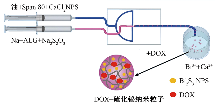
| [10] |
ZOU Q, HOU F L, WANG H, et al. Microfluidic one-step preparation of alginate microspheres encapsulated with in situ-formed bismuth sulfide nanoparticles and their photothermal effect[J]. European Polymer Journal, 2019, 115:282-289.
doi: 10.1016/j.eurpolymj.2019.03.040 |
| [11] |
WENG L H, LE H C, LIN J Y, et al. Doxorubicin loading and eluting characteristics of bioresorbable hydrogel microspheres: in vitro study[J]. International Journal of Pharmaceutics, 2011, 409(1/2) : 185-193.
doi: 10.1016/j.ijpharm.2011.02.058 |
| [12] |
CHOI M H, BLANCO A, STEALEY S, et al. Micro-clotting of platelet-rich plasma upon loading in hydrogel microspheres leads to prolonged protein release and slower microsphere degradation[J]. Polymers, 2020, 12(8):1712.
doi: 10.3390/polym12081712 |
| [13] | 肖文谦, 张静, 李克江, 等. 荔枝状CaCO3@HA/Fe3O4磁性介孔多级微球的制备[J]. 无机材料学报, 2019, 34(9):925-932. |
|
XIAO Wenqian, ZHANG Jing, LI Kejiang, et al. Litchi-like superparamagnetic hydroxyapatite microspheres with hierarchically mesoporous microspheres[J]. Journal of Inorganic Materials, 2019, 34(9):925-932.
doi: 10.15541/jim20180497 |
|
| [14] | QIAO W, LAN X M, TSOI J K H, et al. Biomimetic hollow mesoporous hydroxyapatite microsphere with controlled morphology, entrapment efficiency and degradability for cancer therapy[J]. Royal Society of Chemistry Advances, 2017, 7(71):44788-44798. |
| [15] |
CUI D C, LU W L, SA E A, et al. Poly(acrylic acid) microspheres loaded with lidocaine: preparation and characterization for arterial embolization[J]. International Journal of Pharmaceutics, 2012, 436(1):527-535.
doi: 10.1016/j.ijpharm.2012.07.020 |
| [16] |
WANG A H, CHEN X G, LIU C S, et al. Preparation and characteristics of chitosan microspheres in different acetylation as drug carrier system[J]. Journal of Microencapsulation, 2008, 26(7) : 593-602.
doi: 10.3109/02652040802586167 |
| [17] |
PENG H L, XIONG H, LI J H, et al. Vanillin cross-linked chitosan microspheres for controlled release of resveratrol[J]. Food Chemistry, 2010, 121(1):23-28.
doi: 10.1016/j.foodchem.2009.11.085 |
| [18] |
VENKATESAN C, VIMAL S, HAMEED A S S, et al. Synjournal and characterization of chitosan tripolyphosphate nanoparticles and its encapsulation efficiency containing russell's viper snake venom[J]. Journal of Biochemical and Molecular Toxicology, 2013, 27(8):406-411.
doi: 10.1002/jbt.2013.27.issue-8 |
| [19] |
HOSSEINI S F, ZANDI M, REZAEI M, et al. Two-step method for encapsulation of oregano essential oil in chitosan nanoparticles: preparation, characterization and in vitro release study[J]. Carbohydrate Polymers, 2013, 95(1):50-56.
doi: 10.1016/j.carbpol.2013.02.031 |
| [20] |
WANG L Y, MA G H, SU Z G, et al. Preparation of uniform sized chitosan microspheres by membrane emulsification technique and application as a carrier of protein drug[J]. Journal of Controlled Release, 2005, 106(1):62-75.
doi: 10.1016/j.jconrel.2005.04.005 |
| [21] |
HE P, DAVIS S, IIIUM L, et al. Chitosan microspheres prepared by spray drying[J]. International Journal of Pharmaceutics, 1999, 187(1):53-65.
doi: 10.1016/S0378-5173(99)00125-8 |
| [22] |
LEE H S, KIM E H, SHAO H P, et al. Synjournal of spio-chitosan microspheres for mri-detectable embolotherapy[J]. Journal of Magnetism and Magnetic Materials, 2005, 293(1):102-105.
doi: 10.1016/j.jmmm.2005.01.049 |
| [23] | 姜炜, 李凤生, 杨毅, 等. 磁性壳聚糖复合微球的制备和性能研究[J]. 材料科学与工程学报, 2004(5):660-662. |
| JIANG Wei, LI Fengsheng, YANG Yi, et al. Preparation and characterization of magnetic chitosan microspheres for the carriers of radionuclides in the-rapy[J]. Journal of Materials Science & Engineering, 2004(5):660-662. | |
| [24] |
KIM E S, LEE J S, LEE H L, et al. Calcium-alginate microparticles for sustained release of catechin prepared via an emulsion gelation technique[J]. Food Science and Biotechnology, 2016, 25(5):1337-1343.
doi: 10.1007/s10068-016-0210-8 |
| [25] | 王宏丽, 陈风雷, 陈涛, 等. 海藻酸钠/壳聚糖缓释微球的制备及性能[J]. 云南大学学报(自然科学版), 2010, 32(4):469-472,479. |
| WANG Hongli, CHEN Fenglei, CHEN Tao, et al. Preparation and properties of alginate/chitosan microspheres for controlled releasing[J]. Journal of Yunnan University (Natural Science edition), 2010, 32(4):469-472, 479. | |
| [26] | 陆敏, 王利强. 茶多酚/壳聚糖/海藻酸钠纳米微球的制备[J]. 包装工程, 2017, 38(19):47-51. |
| LU Min, WANG Liqiang. Preparation of polyphenols/chitosan/alginate nano-microspheres[J]. Packaging Engineering, 2017, 38(19):47-51. | |
| [27] |
HU B, TING Y W, YANG X Q, et al. Nanochemoprevention by encapsulation of (-)-epigalloc-atechin-3-gallate with bioactive peptides/chitosan nanoparticles for enhancement of its bioavailability[J]. Chemical Communications, 2012, 48(18):2421-2423.
doi: 10.1039/c2cc17295j |
| [28] |
UPPUTURI R T P, MANDAL A K A. Sustained release of green tea polyphenols from liposomal nanoparticles; release kinetics and mathematical modelling[J]. Iranian Journal of Biotechnology, 2017, 15(4) : 277-283.
doi: 10.15171/ijb.1322 |
| [29] |
WANG Q, XIAO A, LIU Y M, et al. One-step preparation of nano-in-micro poly(vinyl alcohol) embolic microspheres and used for dual-modal T1/T2-weighted magnetic resonance imaging[J]. Nanomedicine: Nanotechnology, Biology and Medicine, 2018, 14(8):2551-2561.
doi: 10.1016/j.nano.2018.08.003 |
| [30] |
WANG H, QIN X Y, LI Z Y, et al. Preparation and evaluation of mri detectable poly (acrylic acid) microspheres loaded with superparamagnetic iron oxide nanoparticles for transcatheter arterial embolization[J]. International Journal of Pharmaceutics, 2016, 511(2):831-839.
doi: 10.1016/j.ijpharm.2016.07.028 |
| [31] |
RONG J J, LIANG M, XUAN F Q, et al. Thrombin-loaded alginate-calcium microspheres: a novel hemostatic embolic material for transcatheter arterial embolization(article)[J]. International Journal of Biological Macromolecules, 2017, 104:1302-1312.
doi: 10.1016/j.ijbiomac.2017.03.020 |
| [32] |
ALBRECHT T, GROSS A. Transarterial chemoembolisation (tace) with degradable starch microspheres (dsm) and anthracycline in patients with locally extensive hepatocellular carcinoma (hcc): safety and efficacy[J]. Cardiovascular and Interventional Radiology, 2019, 43:402-410.
doi: 10.1007/s00270-019-02364-w |
| [33] |
KAWASHITA M, UENO S, HANDA S, et al. In vitro evaluation of doxorubicin-eluting porous titania microspheres for transcatheter arterial chemoemboliza-tion[J]. Journal of Asian Ceramic Societies, 2020, 8(1):10-20.
doi: 10.1080/21870764.2019.1695559 |
| [34] | 侯志勇, 黄海波, 王立强, 等. 动脉栓塞微球的研究进展[J]. 中国医药科学, 2012, 2(16):9-11. |
| HOU Zhiyong, HUANG Haibo, WANG Liqiang, et al. Progress in the study of arterial embolic microsp-heres[J]. China Medicine and Pharmacy, 2012, 2(16):9-11. | |
| [35] |
LI G, QIN S X. Studies on preparations and properties of drug-eluting embolization microspheres made from oxidated alginate and carboxymethyl chitosan[J]. International Journal of Polymeric Materials, 2019, 68(14):844-849.
doi: 10.1080/00914037.2018.1517346 |
| [36] |
SANG X X, WEN Q W, ZHANG M, et al. Preparation of drug-eluting microspheres based on semi-interpenetrating polymer network of modified chitosan and poly(2-acrylamide-2-methylpropanesulfonic acid)[J]. Polymer Science Series A, 2019, 61(1):61-69.
doi: 10.1134/S0965545X19010061 |
| [37] |
CHEN G B, WEI R W, HUANG X, et al. Synjournal and assessment of sodium alginate-modified silk fibroin microspheres as potential hepatic arterial embolization agent[J]. International Journal of Biological Macromolecules, 2020, 155:1450-1459.
doi: 10.1016/j.ijbiomac.2019.11.122 |
| [38] | 魏如男. 改性丝蛋白肝动脉栓塞微球的制备及性能研究[D]. 重庆: 重庆理工大学, 2019: 8-34. |
| WEI Runan. The preparation and performance study of the modified silk fibroin microspheres for transcatheter arterial chemoembolization[D]. Chongqing: Chongqing University of Technology, 2019: 8-34. | |
| [39] |
HOU F L, ZHU Y H, ZOU Q, et al. One-step preparation of multifunctional alginate microspheres loaded with in situ-formed gold nanostars as a photothermal agent[J]. Materials Chemistry Frontiers, 2019, 3(10):2018-2024.
doi: 10.1039/C9QM00276F |
| [40] | 郭李盈, 刘晓昕, 李子圆, 等. 空白及载阿霉素的聚丙烯酸栓塞微球的制备与评价[J]. 北京大学学报(医学版), 2018, 50(6):1070-1077. |
| GUO Liying, LIU Xiaoxin, LI Ziyuan, et al. Preparation and evaluation of blank and doxorubicin loaded poly(acrylic acid) microspheres for embolization[J]. Journal of Peking University (Health Sciences), 2018, 50(6):1070-1077. | |
| [41] | 姚芳莲, 于潇, 周玉涛. 壳聚糖-明胶载药微球的制备及释放性能[J]. 化学工业与工程, 2008(3):215-219. |
| YAO Fanglian, YU Xiao, ZHOU Yutao, et al. Preparation of chitosan-gelatin drug loading microshpere and its release characteristics[J]. Chemical Industry and Engineering, 2008(3):215-219. | |
| [42] | 邓阳全, 邵丽, 杨银, 等. 乳化交联法在载药微球制备中的应用及研究进展[J]. 世界科技研究与发展, 2009, 31(1):36-39. |
| DENG Yangquan, SHAO Li, YANG Yin, et al. Study on drug-carried microspheres prepared by emulsion cross-linking method[J]. World Sci-Tech R & D, 2009, 31(1):36-39. | |
| [43] | 罗华丽, 鲁在君. 壳聚糖微球制备工艺优化与分析[J]. 周口师范学院学报, 2007(2):63-66. |
| LUO Huali, LU Zaijun. Optimizing and analyzing for the preparation of chitosan microsphere[J]. Journal of Zhoukou Normal University, 2007(2):63-66. | |
| [44] | 王西, 吴修利, 赵珺, 等. 壳聚糖微球制备方法及其在医药应用的研究进展[J]. 长春大学学报, 2015, 25(10):64-68. |
| WANG Xi, WU Xiuli, ZHAO Jun, et al. Preparation for chitosan microspheres and progress of research in application of medicine[J]. Journal of Changchun University, 2015, 25(10):64-68. | |
| [45] | 王常煜. 微流控方法制备载纳米药物的肝癌化疗栓塞微球[D]. 南京: 南京大学, 2017: 10-18. |
| WANG Changyu. The preparation of nano-drug-encapsulation microbeads by microfluidic method for liver cancer chemoembolization[D]. Nanjing: University of Nanjing, 2017: 10-18. | |
| [46] | 李勋, 韦祎, 马光辉, 等. 缓释微球制剂的研究进展[J]. 北京化工大学学报(自然科学版), 2017, 44(6):1-11. |
| LI Xun, WEI Yi, MA Guanghui, et al. Recent research and development prospects for sustained-release microspheres[J]. Journal of Beijing University of Chemical Technology (Natural Science), 2017, 44(6):1-11. | |
| [47] | KAR K, BALA N N, PAL R N, et al. Preparation, characterisation and evaluation of ropinirole hydrochloride loaded controlled release microspheres using solvent evaporation technique[J]. International Journal of Pharmacy and Pharmaceutical Sciences, 2018, 10(6):57-67. |
| [48] | 陈艳, 谢委, 肖政. 温敏多柔比星纳米凝胶栓塞微球的评价与处方优化[J]. 医药导报, 2019, 38(11):1474-1478. |
| CHEN Yan, XIE Wei, XIAO Zheng. Evaluation and formulation optimization of doxorubicin-loaded microspheres for embolization using temperature sensitive nanogels[J]. Herald of Medicine, 2019, 38(11):1474-1478. | |
| [49] | 瞿静. 静电分化法制备丝素纳米颗粒及其作为顺铂控释载体的研究[D]. 苏州: 苏州大学, 2013: 4-8. |
| QU Jing. Preparation of silk fibroin nanoparticles by electrostatic differentiation and its use as a carrier for controlled release of cisplatin[D]. Suzhou: Soochow University, 2013: 4-8. | |
| [50] |
HAGAN A, PHILLIPS G J, MACFARLANE, et al. Preparation and characterisation of vandetanib-eluting radiopaque beads for locoregional treatment of hepatic malignancies[J]. European Journal of Pharmaceutical Sciences, 2017, 101:22-30.
doi: 10.1016/j.ejps.2017.01.033 |
| [51] | 毋囡囡, 林雪纯, 应晨江, 等. 多酚类化合物与必需微量元素的相互作用[J]. 卫生研究, 2019, 48(6):1012-1016. |
| WU Nannan, LIN Xuechun, YING Chenjiang, et al. Interaction of polyphenols with essential Trace ele-ments[J]. Journal of Hygiene Research, 2019, 48(6):1012-1016. | |
| [52] | ZHAO Y Y, SUN Z T. Effects of gelatin-polyphenol and gelatin-genipin cross-linking on the structure of gelatin hydrogels[J]. International Journal of Food Properties, 2017, 20(3):2822-2832. |
| [53] |
CHANPHAI P, TAJMIR-RIAHI H A. Conjugation of tea catechins with chitosan nanoparticles[J]. Food Hydrocolloids, 2018, 84:561-570.
doi: 10.1016/j.foodhyd.2018.06.043 |
| [54] | 聂芊. 明胶包埋壳聚糖/海藻酸钠茶多酚的缓释性能[J]. 哈尔滨商业大学学报(自然科学版), 2005(4):418-420. |
| NIE Qian. Study on gluin-chitosan-sodium alginate sustained release system of tea polyphenols[J]. Journal of Harbin University of Commerce (Natural Sciences Edition), 2005(4):418-420. |
| [1] | Feng LI. Research progress of polymer embolic microspheres [J]. , 2021, 42(10): 0-0. |
| [2] | WANG Shudong, MA Qian, WANG Ke, QU Caixin, QI Yu. Structure and biocompatibility of silk fibroin/gelatin blended hydrogels [J]. Journal of Textile Research, 2020, 41(11): 41-47. |
| [3] | SUN Fanchen, GUO Jing, YU Yue, ZHANG Sen. Preparation and properties of polyhydroxy fatty acid ester/sodium alginate composite electrospun nanofibers [J]. Journal of Textile Research, 2020, 41(05): 15-19. |
| [4] | DONG Ke, LI Siming, WU Guanzheng, HUANG Hongrong, LIN Zhongshi, XIAO Xueliang. Preparation and properties of carbon fiber/polyester electrocardiogram monitoring embroidery electrode [J]. Journal of Textile Research, 2020, 41(01): 56-62. |
| [5] | LIN Yongjia, YANG Dongchao, ZHANG Peihua, GU Yan. Preparation and properties of regenerated silk fibroin/acellular dermal matrix blended nanofiber membrane [J]. Journal of Textile Research, 2019, 40(07): 13-18. |
| [6] | . Structure and properties of polylactic acid-polycaprolactone/silk fibroin composite nanofibrous scaffolds [J]. JOURNAL OF TEXTILE RESEARCH, 2017, 38(05): 8-13. |
| [7] | . Surface modification of PET by acrylic acid and collagen [J]. JOURNAL OF TEXTILE RESEARCH, 2014, 35(7): 152-0. |
| [8] | . Current research of implantation artificial hair and biocompatibilit of polypyrrole [J]. JOURNAL OF TEXTILE RESEARCH, 2013, 34(5): 146-151. |
|
||