纺织学报 ›› 2024, Vol. 45 ›› Issue (01): 48-55.doi: 10.13475/j.fzxb.20220906401
戎成宝1,2, 孙辉1,2( ), 于斌1,2
RONG Chengbao1,2, SUN Hui1,2(), YU Bin1,2
摘要:
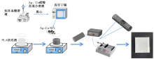
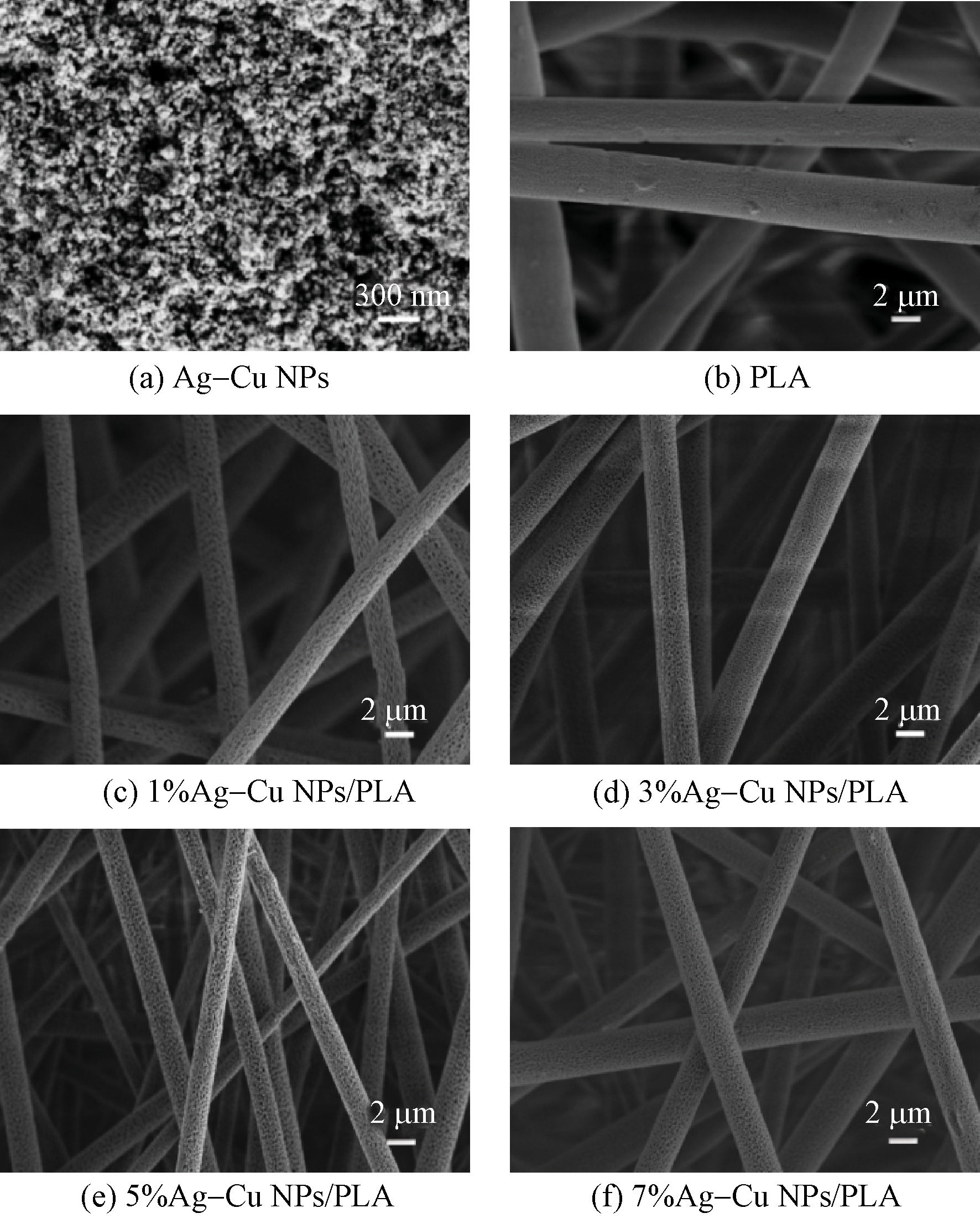
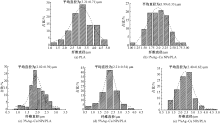
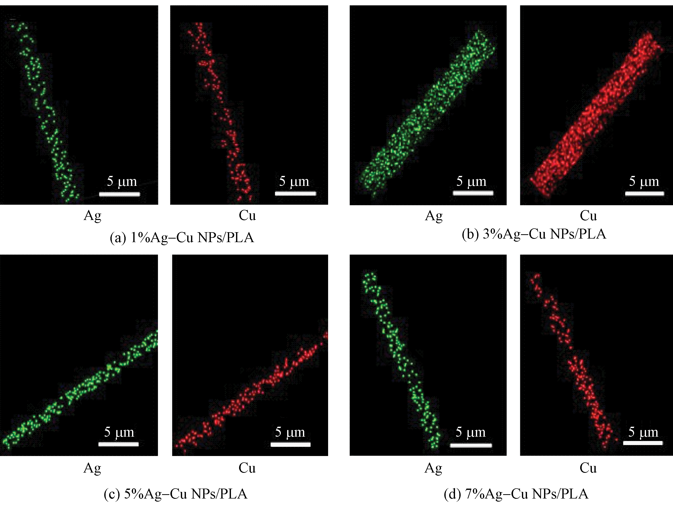
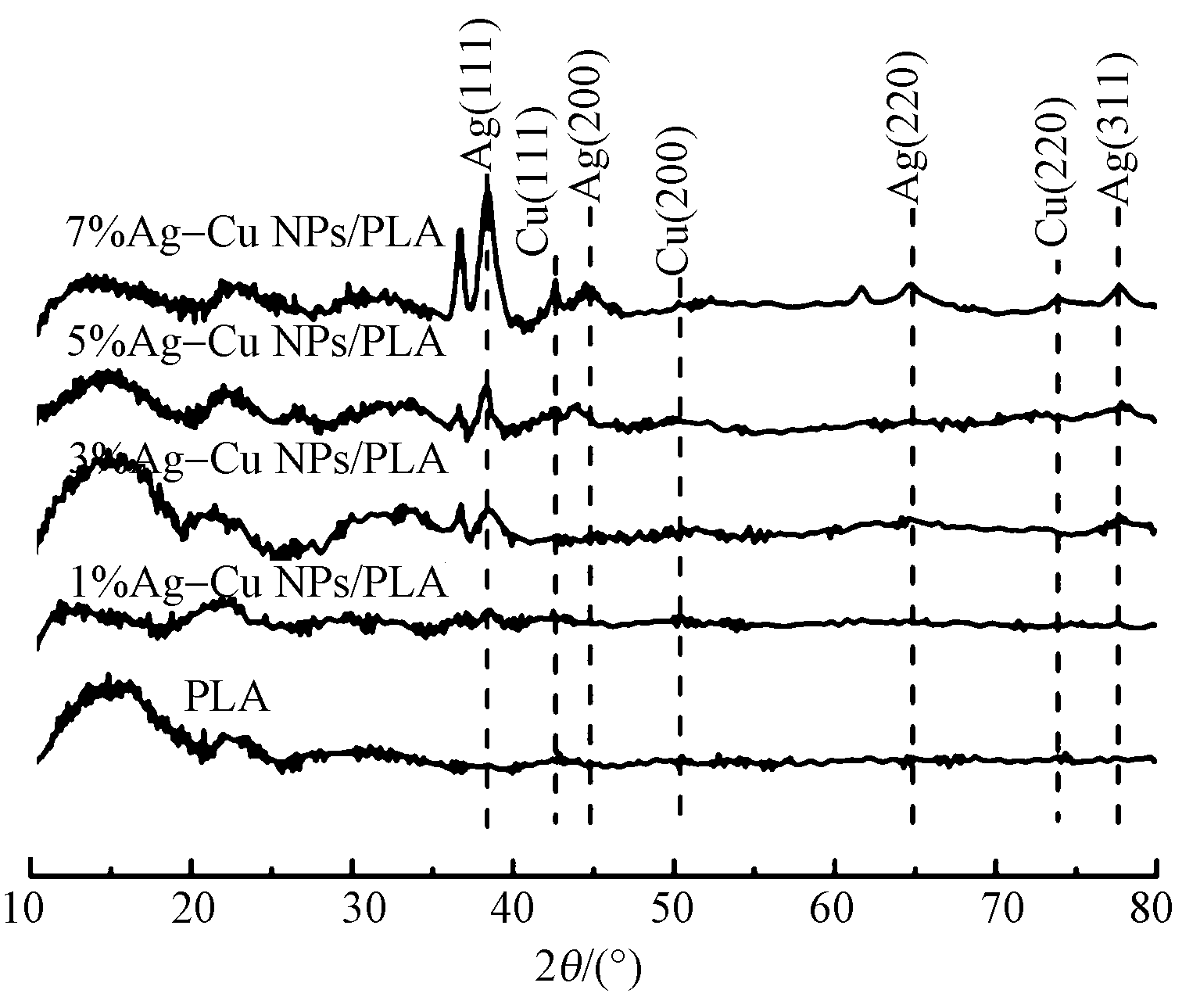
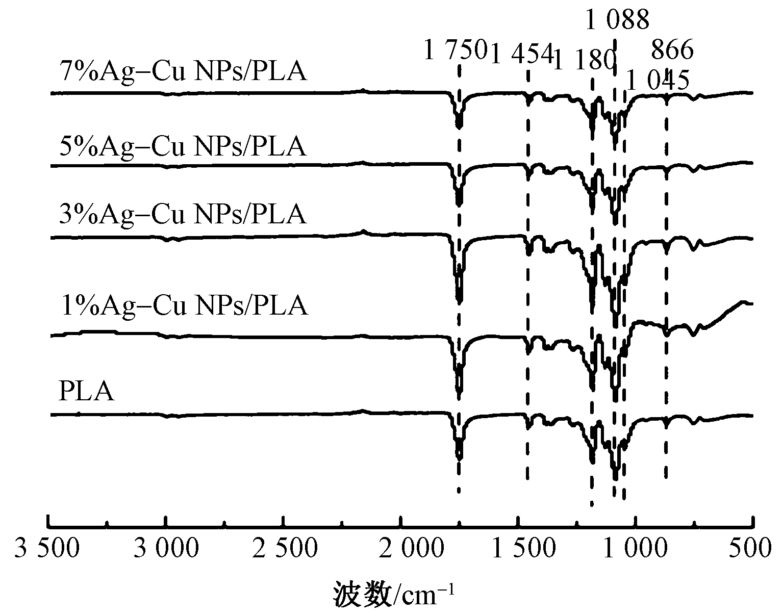
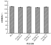
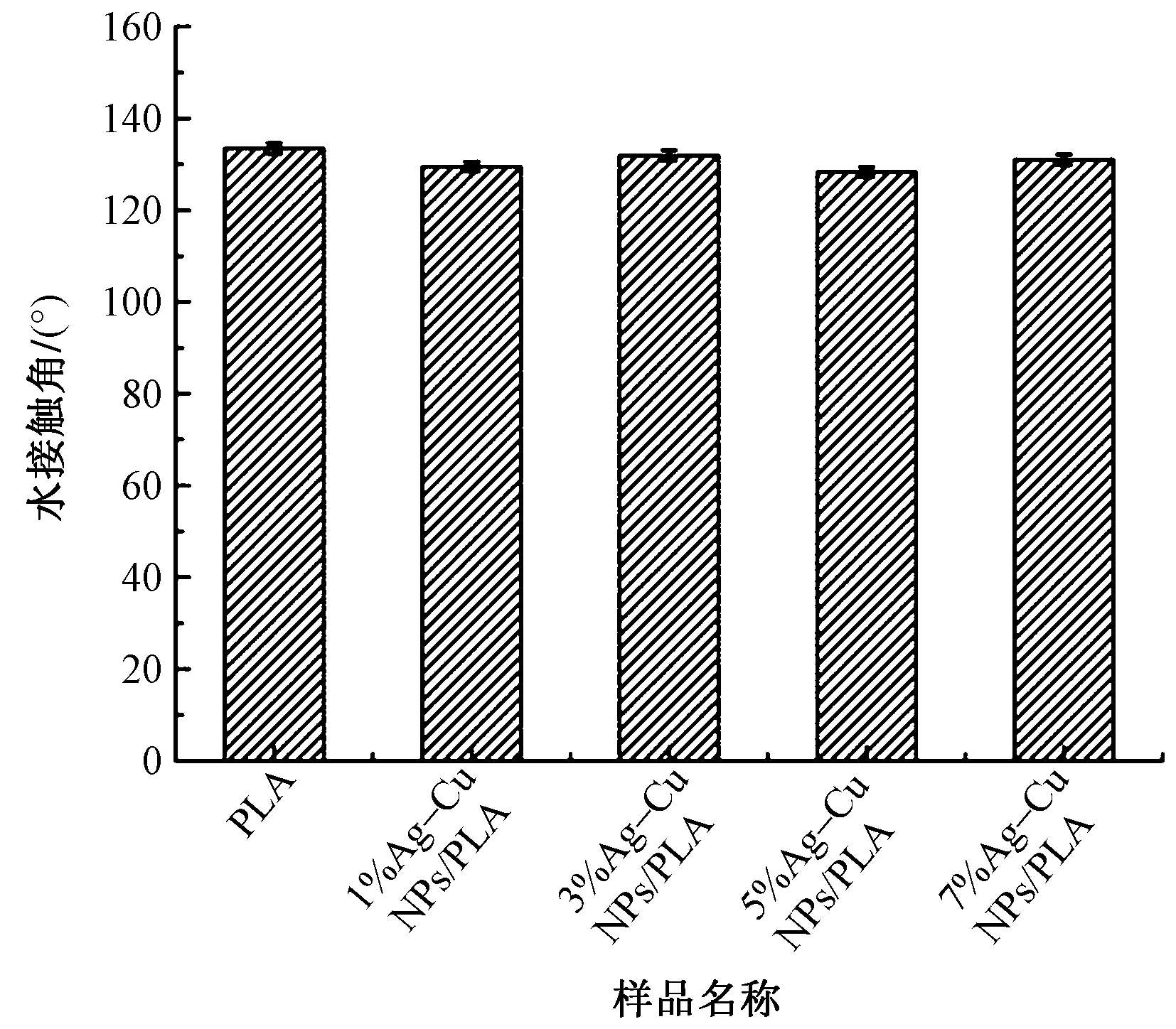
为制备高效抗菌的生物可降解聚乳酸(PLA)静电纺丝纤维膜,首先利用L-抗坏血酸对银和铜的硝酸盐溶液进行化学还原,得到银-铜双金属纳米粒子(Ag-Cu NPs)。然后将Ag-Cu NPs与PLA纺丝液共混,通过静电纺丝技术制备了不同组成的Ag-Cu NPs/PLA复合纳米纤维膜,并对其形貌、结构、亲水性和抗菌性能等进行测试。结果表明:合成的Ag-Cu NPs的粒径约为32 nm,复合纳米纤维膜中Ag-Cu NPs被PLA基体包覆,且沿着纤维径向排列,纤维表面存在大量微小的孔洞;加入Ag-Cu NPs后,Ag-Cu NPs/PLA的水接触角略微降低,亲水性增加,且Ag-Cu NPs和PLA之间仅发生物理作用,未产生明显的化学作用;相比于纯PLA纳米纤维膜,Ag-Cu NPs/PLA的抗菌率明显提高,当纺丝液中Ag-Cu NPs相对于PLA质量为7% 时,复合纳米纤维膜对大肠杆菌和金黄色葡萄球菌的抑菌率均达到99%。
中图分类号:
| [1] | LIU C, SHEN J, YEUNG K W K, et al. Development and antibacterial performance of novel polylactic acid-graphene oxide-silver nanoparticle hybrid nanocomposite mats prepared by electrospinning[J]. ACS Biomaterials Science & Engineering, 2017, 3(3): 471-486. |
| [2] | BALASUBRAMANIAM B, PRATEE K, RANJAN S, et al. Antibacterial and antiviral functional materials: chemistry and biological activity toward tackling COVID-19-like pandemics[J]. ACS Pharmacology & Translational Science, 2020, 4(1): 8-54. |
| [3] |
HUSSAIN Z, KHAN M A, IQBAL F, et al. Electrospun microbial-encapsulated composite-based plasticized seed coat for rhizosphere stabilization and sustainable production of canola (Brassica napus L.)[J]. Journal of Agricultural and Food Chemistry, 2019, 67(18): 5085-5095.
doi: 10.1021/acs.jafc.8b06505 |
| [4] |
SHANG L, YU Y, LIU Y, et al. Spinning and applications of bioinspired fiber systems[J]. ACS Nano, 2019, 13(3): 2749-2772.
doi: 10.1021/acsnano.8b09651 pmid: 30768903 |
| [5] |
YANG H, WANG L, XIANG C, et al. Electrospun porous PLLA and poly (LLA-co-CL) fibers by phase separation[J]. New Journal of Chemistry, 2018, 42(7): 5102-5108.
doi: 10.1039/C7NJ04970F |
| [6] |
ZHANG H, ZHANG T, QIU Q, et al. Quaternary ammonium salt-modified polyacrylonitrile/polycaprolactone electrospun nanofibers with enhanced antibacterial properties[J]. Textile Research Journal, 2021, 91(19/20): 2194-2203.
doi: 10.1177/0040517521997187 |
| [7] | ALTAN E, KARACELEBI Y, SAATCIOGLU E, et al. Fabrication of electrospun Juglans regia (Juglone) loaded poly (lactic acid) scaffolds as a potential wound dressing material[J]. Polymers, 2022. DOI: 10.3390/POLYM14101971. |
| [8] | BACKES E H, PIRES L D N, BEATRICE C A G, et al. Fabrication of biocompatible composites of poly-(lactic acid)/hydroxyapatite envisioning medical applications[J]. Polymer Engineering & Science, 2020, 60(3): 636-644. |
| [9] | NONATO R C, MEI L H I, BONSE B C, et al. Nanocomposites of PLA/ZnO nanofibers for medical applications: antimicrobial effect, thermal, and mechanical behavior under cyclic stress[J]. Polymer Engineering & Science, 2022, 62(4): 1147-1155. |
| [10] |
GODOY-GALLARDO M, ECKHARD U, DELGADO L M, et al. Antibacterial approaches in tissue engineering using metal ions and nanoparticles: from mechanisms to applications[J]. Bioactive Materials, 2021, 6(12): 4470-4490.
doi: 10.1016/j.bioactmat.2021.04.033 |
| [11] |
PUTHALATH A K, HAZEL S, KOTTAPPARA R, et al. Synthesis and antibacterial activity of silver-copper nano-composites formed by microwave assisted chemical reduction[J]. Materials Today: Proceedings, 2021, 41: 525-529.
doi: 10.1016/j.matpr.2020.05.238 |
| [12] |
JUNG H, KING M E, PERSONICK M L. Strategic synergy: advances in the shape control of bimetallic nanoparticles with dilute alloyed surfaces[J]. Current Opinion in Colloid & Interface Science, 2019, 40: 104-117.
doi: 10.1016/j.cocis.2019.02.004 |
| [13] |
NOWAK A, SZADE J, TALIK E, et al. Physicochemical and antibacterial characterization of ionocity Ag/Cu powder nanoparticles[J]. Materials Characterization, 2016, 117: 9-16.
doi: 10.1016/j.matchar.2016.04.013 |
| [14] | VALDEZ-SALAS B, BELTRÁN-PARTIDA E, ZLATEV R, et al. Structure-activity relationship of diameter controlled Ag@Cu nanoparticles in broad-spectrum antibacterial mechanism[J]. Materials Science and Engineering:C, 2020. DOI:10.1016/j.msec.2020.111501. |
| [15] |
ZAIN N M, STAPLEY A G, SHAMA G J. Green synthesis of silver and copper nanoparticles using ascorbic acid and chitosan for antimicrobial appli-cations[J]. Carbohydrate Polymers, 2014, 112:195-202.
doi: 10.1016/j.carbpol.2014.05.081 |
| [16] |
ZAIN N M, STAPLEY A G F, SHAMA G. Green synthesis of silver and copper nanoparticles using ascorbic acid and chitosan for antimicrobial appli-cations[J]. Carbohydrate Polymers, 2014, 112: 195-202.
doi: 10.1016/j.carbpol.2014.05.081 |
| [17] |
YUAN Z, ZHANG K, JIAO X, et al. A controllable local drug delivery system based on porous fibers for synergistic treatment of melanoma and promoting wound healing[J]. Biomaterials science, 2019, 7(12): 5084-5096.
doi: 10.1039/c9bm01045a pmid: 31565707 |
| [18] |
ZHOU Q, XIE J, BAO M, et al. Engineering aligned electrospun PLLA microfibers with nano-porous surface nanotopography for modulating the responses of vascular smooth muscle cells[J]. Journal of Materials Chemistry B, 2015, 3(21): 4439-4450.
doi: 10.1039/c5tb00051c pmid: 32262788 |
| [19] | 唐晓鹏. 载银离子多孔纳米纤维制备及抗菌性能研究[D]. 苏州: 苏州大学, 2015:31-32. |
| TANG Xiaopeng. Preparation and antibacterial properties of porous nanofibers containing silver ions[D]. Suzhou: Soochow University, 2015:31-32. | |
| [20] |
MA G, YANG D, NIE J. Preparation of porous ultrafine polyacrylonitrile (PAN) fibers by electrospinning[J]. Polymers for Advanced Technologies, 2009, 20(2): 147-150.
doi: 10.1002/pat.v20:2 |
| [21] |
ZONG X, KIM K, FANG D, et al. Structure and process relationship of electrospun bioabsorbable nanofiber membranes[J]. Polymer, 2002, 43(16): 4403-4412.
doi: 10.1016/S0032-3861(02)00275-6 |
| [22] | YANG H, ZHANG X, VELU P, et al. Enhanced green mediated synthesis of optimized Ag-Cu bimetallic nanoparticles using Leucas aspera and its application in anti-cancer activity against alveolar cancer[J]. Materials Letters, 2022. DOI:10.1016/j.matlet.2021.131645. |
| [23] |
KEMALA T, BUDIANTO E, SOEGIYONO B. Preparation and characterization of microspheres based on blend of poly (lactic acid) and poly (ε-caprolactone) with poly (vinyl alcohol) as emulsifier[J]. Arabian Journal of Chemistry, 2012, 5(1): 103-108.
doi: 10.1016/j.arabjc.2010.08.003 |
| [24] | WU X, BOURBIGOT S, LI K, et al. Co-pyrolysis characteristics and flammability of polylactic acid and acrylonitrile-butadiene-styrene plastic blend using TG, temperature-dependent FTIR, Py-GC/MS and cone calorimeter analyses[J]. Fire Safety Journal, 2022. DOI: 10.1016/j.firesaf.2022.103543. |
| [25] | DEGHICHE A, HADDAOUI N, ZERRIOUH A, et al. Effect of the stearic acid-modified TiO2 on PLA nanocomposites: morphological and thermal properties at the microscopic scale[J]. Journal of Environmental Chemical Engineering, 2021. DOI: 10.1016/j.jece.2021.106541. |
| [26] |
GONG X, PAN L, TANG C Y, et al. Preparation, optical and thermal properties of CdSe-ZnS/poly (lactic acid)(PLA) nanocomposites[J]. Composites Part B: Engineering, 2014, 66: 494-499.
doi: 10.1016/j.compositesb.2014.06.016 |
| [27] | PETER A, COZMUTA L M, NICULA C, et al. Chemical and organoleptic changes of curd cheese stored in new and reused active packaging systems made of Ag-graphene-TiO2-PLA[J]. Food Chemistry, 2021. DOI: 10.1016/j.foodchem.2021.130341. |
| [28] |
MAMATHA G, SOWMYA P, MADHURI D, et al. Antimicrobial cellulose nanocomposite films with in situ generations of bimetallic (Ag and Cu) nanoparticles using Vitex negundo leaves extract[J]. Journal of Inorganic and Organometallic Polymers and Materials, 2021, 31(2):802-815.
doi: 10.1007/s10904-020-01819-9 |
| [29] | PERDIKAKI A, GALEOU A, PILATOS G, et al. Ag and Cu monometallic and Ag/Cu bimetallic nanoparticle-graphene composites with enhanced antibacterial performance[J]. ACS Applied Materials & Interfaces, 2016. DOI:10.1021/acsami.6b08403. |
| [30] | ZHOU F, ZHU Y, YANG L, et al. Ag NP catalysis of Cu ions in the preparation of Ag Cu NPs and the mechanism of their enhanced antibacterial efficacy[J]. Colloids and Surfaces A: Physicochemical and Engineering Aspects, 2022. DOI: 10.1016/j.colsurfa.2021.127831. |
| [31] |
CRUCES E, ARANCIBIA-MIRANDA N, MANQUIÁN-CERDA K, et al. Copper/silver bimetallic nanoparticles supported on aluminosilicate geomaterials as antibacterial agents[J]. ACS Applied Nano Materials. 2022, 5(1):1472-1483.
doi: 10.1021/acsanm.1c04031 |
| [32] | DOOLOTKELDIEVA T, BOBUSHEVA S, ZHASNAKUNOV Z, et al. Biological activity of Ag and Cu monometallic nanoparticles and Ag-Cu bimetallic nanocomposites against plant pathogens and seeds[J]. Journal of Nanomaterials, 2022. DOI: 10.1155/2022/1190280. |
| [1] | 杨奇, 刘高慧, 黄琪帏, 胡睿, 丁彬, 俞建勇, 王先锋. 熔喷聚乳酸/聚偏氟乙烯电晕驻极空气过滤材料电荷存储与过滤性能相关性研究[J]. 纺织学报, 2024, 45(01): 12-22. |
| [2] | 杨智超, 刘淑强, 吴改红, 贾潞, 张曼, 李甫, 李慧敏. 可吸收手术缝合线研究进展[J]. 纺织学报, 2024, 45(01): 230-239. |
| [3] | 王镕琛, 张恒, 翟倩, 刘瑞焱, 黄鹏宇, 李霞, 甄琪, 崔景强. 聚乳酸超细纤维敷料的熔喷成形工艺及其快速导液特性[J]. 纺织学报, 2024, 45(01): 30-38. |
| [4] | 陈江萍, 郭朝阳, 张琪骏, 吴仁香, 钟鹭斌, 郑煜铭. 静电纺聚酰胺6/聚苯乙烯复合纳米纤维膜制备及其空气过滤性能[J]. 纺织学报, 2024, 45(01): 56-64. |
| [5] | 王鹏, 申佳锟, 陆银辉, 盛红梅, 王宗乾, 李长龙. 石墨相氮化碳/MXene/磷酸银/聚丙烯腈复合纳米纤维膜的制备及其光催化性能[J]. 纺织学报, 2023, 44(12): 10-16. |
| [6] | 孙辉, 崔小港, 彭思伟, 丰江丽, 于斌. 聚乳酸/磁性金属有机框架材料复合熔喷布的制备及其空气过滤性能[J]. 纺织学报, 2023, 44(12): 26-34. |
| [7] | 雷彩虹, 俞林双, 金万慧, 朱海霖, 陈建勇. 丝素蛋白/壳聚糖复合纤维膜的制备与应用[J]. 纺织学报, 2023, 44(11): 19-26. |
| [8] | 徐志豪, 徐丹瑶, 李彦, 王璐. 基于表面增强拉曼光谱的纳米纤维基生物传感器的研究进展[J]. 纺织学报, 2023, 44(11): 216-224. |
| [9] | 王西贤, 郭天光, 王登科, 牛帅, 贾琳. 聚丙烯腈/银复合纳米纤维高效滤膜的制备及其长效性能[J]. 纺织学报, 2023, 44(11): 27-35. |
| [10] | 范梦晶, 吴玲娅, 周歆如, 洪剑寒, 韩潇, 王建. 镀银聚酰胺6/聚酰胺6纳米纤维包芯纱电容传感器的构筑[J]. 纺织学报, 2023, 44(11): 67-73. |
| [11] | 张广知, 杨甫生, 方进, 杨顺. 聚乳酸非织造布植酸/壳聚糖/硼酸一浴法阻燃整理[J]. 纺织学报, 2023, 44(10): 120-126. |
| [12] | 张成成, 刘让同, 李淑静, 李亮, 刘淑萍. 聚左旋乳酸非溶剂挥发诱导成孔机制与纳米多孔纤维膜制备[J]. 纺织学报, 2023, 44(10): 16-23. |
| [13] | 付征, 穆齐锋, 张青松, 张宇晨, 李玉莹, 蔡仲雨. 胶体静电纺微纳米纤维的研究进展[J]. 纺织学报, 2023, 44(10): 196-204. |
| [14] | 姚双双, 付少举, 张佩华, 孙秀丽. 再生丝素蛋白/聚乙烯醇共混取向纳米纤维膜的制备与性能[J]. 纺织学报, 2023, 44(09): 11-19. |
| [15] | 孟鑫, 朱淑芳, 徐英俊, 闫旭. 用于纸质文档保护的原位静电纺废旧聚对苯二甲酸乙二醇酯膜[J]. 纺织学报, 2023, 44(09): 20-26. |
|
||
 京公网安备11010502044800号