纺织学报 ›› 2023, Vol. 44 ›› Issue (11): 19-26.doi: 10.13475/j.fzxb.20220606801
雷彩虹1,2, 俞林双1, 金万慧3, 朱海霖1,2, 陈建勇1( )
)
LEI Caihong1,2, YU Linshuang1, JIN Wanhui3, ZHU Hailin1,2, CHEN Jianyong1()
摘要:
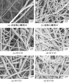
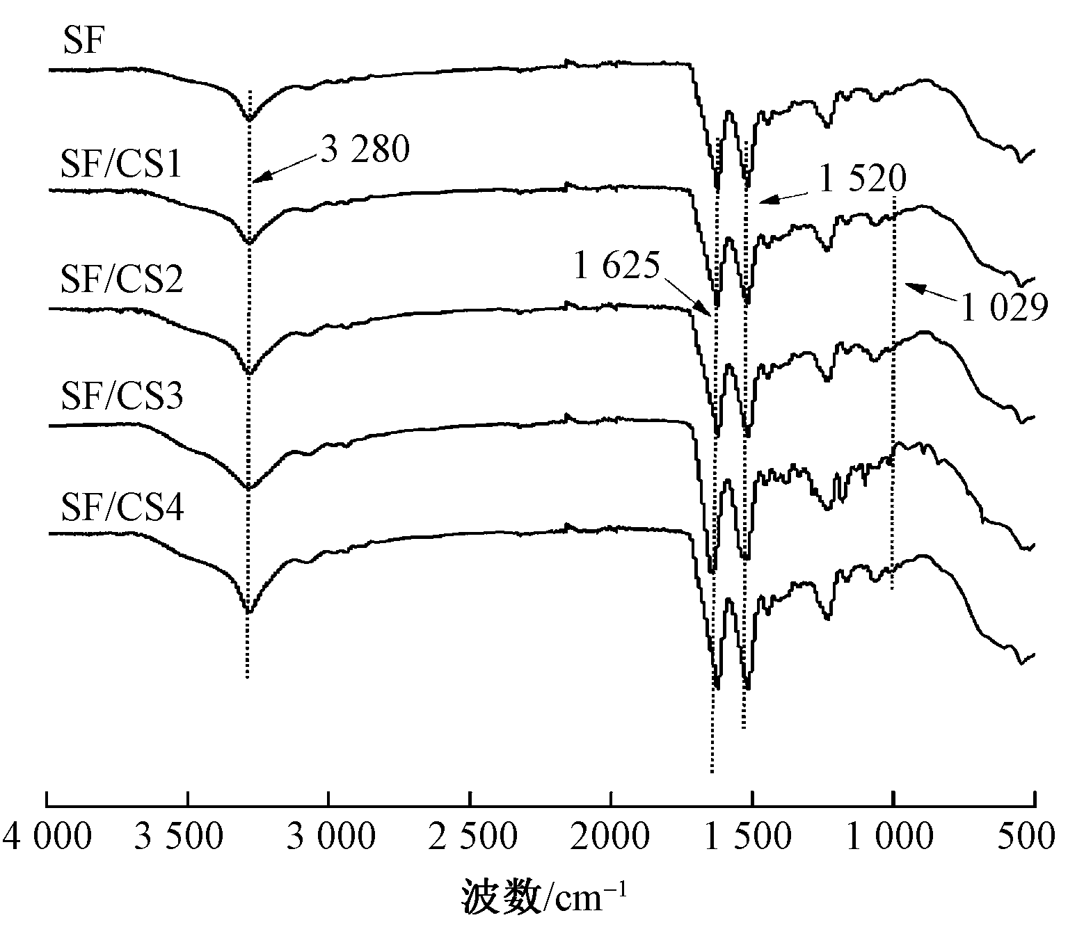
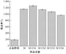
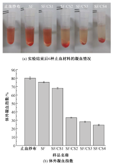
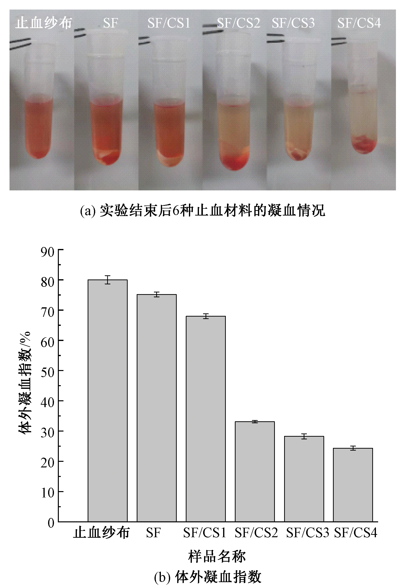
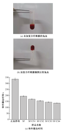
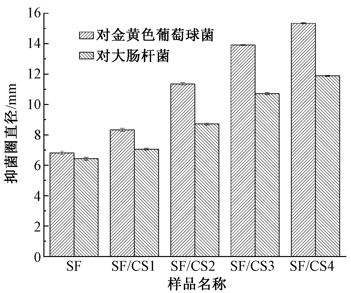
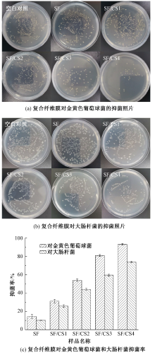
为进一步提升丝素基纤维膜的抗菌性能,以六氟异丙醇和三氟乙酸为溶剂分别溶解丝素蛋白和壳聚糖,制备一定质量比的混合纺丝溶液,然后利用静电纺丝技术制备丝素蛋白/壳聚糖(SF/CS)复合纤维膜。对不同质量比下复合纤维膜的微观形貌、吸水率、止血性能和抗菌性能进行测试与分析。结果表明:SF/CS复合纤维膜中纤维呈光滑致密、无串珠的网状结构;随着壳聚糖添加量的增加,复合纤维膜的抗菌性呈显著增强趋势,当丝素蛋白与壳聚糖质量比为5∶2时,复合纤维膜对大肠杆菌和金黄色葡萄球菌的抑菌圈直径分别为(11.88±0.04)和(15.34±0.04) mm,抑菌率分别达到(73.93±0.85)%和(93.27±0.97)%;同时,在该条件下制备的复合纤维膜具有较高的吸水率((967.59±9.76)%),止血性能优于市售止血纱布。
中图分类号:
| [1] |
CHOUHAN D, MANDAL B B. Silk biomaterials in wound healing and skin regeneration therapeutics: from bench to bedside[J]. Acta Biomaterialia, 2020, 103:24-51.
doi: S1742-7061(19)30800-1 pmid: 31805409 |
| [2] | WEI W, LIU J, PENG Z B, et al. Gellable silk fibroin-polyethylene sponge for hemostasis[J]. Artificial Cells, 2020, 48(1):28-36. |
| [3] |
PATIL P P, REAGAN M R, BOHARA R A. Silk fibroin and silk-based biomaterial derivatives for ideal wound dressings[J]. International Journal of Biological Macromolecules, 2020, 164:4613-4627.
doi: 10.1016/j.ijbiomac.2020.08.041 pmid: 32814099 |
| [4] |
TEUSCHL A H, ZIPPERLE J, HUBER Gries C, et al. Silk fibroin based carrier system for delivery of fibrinogen and thrombin as coagulant supplements[J]. Journal of Biomedical Materials Research: Part A, 2017, 105(3):687-696.
doi: 10.1002/jbm.v105.3 |
| [5] | 熊亮, 陈锦涛, 韦加娜. 丝素蛋白电纺抗菌止血材料的制备及其性能评价[J]. 广东化工, 2018, 45(11):67-69. |
| XIONG Liang, CHEN Jintao, WEI Jiana. Preparation of silk fibroin electrospun antimicrobial hemostatic material and its performance evaluation[J]. Guangdong Chemical Industry, 2018, 45(11):67-69. | |
| [6] | 马烨. 壳聚糖医用敷料的制备及性能研究[D]. 南京: 南京理工大学, 2018:16-18. |
| MA Ye. Research on preparation and performance of chitosan wound dressing[D]. Nanjing: Nanjing University of Science and Technology, 2018:16-18. | |
| [7] | 白雪, 毕华, 张雪峰, 等. 壳聚糖改性及其用于止血海绵的研究进展[J]. 高分子通报, 2021(3):13-19. |
| BAI Xue, BI Hua, ZHANG Xuefeng, et al. Progress of chitosan study in hemostatic sponge[J]. Chinese Polymer Bulletin, 2021(3):13-19. | |
| [8] | 廖硕, 何星. 静电纺丝用于制备止血材料的研究进展[J]. 广东化工, 2018, 45(7):153-154. |
| LIAO Shuo, HE Xing. Research progress in preparation of hemostatic materials by electrospinning[J]. Guangdong Chemical Industry, 2018, 45(7):153-154. | |
| [9] | 张蓓蕾, 沈明武, 史向阳. 静电纺短纤维的制备及其生物医学应用[J]. 纺织学报, 2021, 42(5):1-8. |
|
ZHANG Beilei, SHEN Mingwu, SHI Xiangyang. Preparation and biomedical applications of electrospun short fibers[J]. Journal of Textile Research, 2021, 42(5):1-8.
doi: 10.1177/004051757204200101 |
|
| [10] | 赵新哲, 王绍霞, 高晶, 等. 静电纺胶原/聚环氧乙烷纳米纤维膜的制备及其性能[J]. 纺织学报, 2021, 42(4):33-41. |
|
ZHAO Xinzhe, WANG Shaoxia, GAO Jing, et al. Preparation and properties of electrospun collagen/polyethylene oxide nanofiber membranes[J]. Journal of Textile Research, 2021, 42(4):33-41.
doi: 10.1177/004051757204200107 |
|
| [11] | LI Mengna, LI Na, QIU Weiwang, et al. Nitric oxide-releasing L-tryptophan and L-phenylalanine based poly(ester urea)s electrospun composite mats as antibacterial and antibiofilm dressing for wound healing[J]. Composites Part B: Engineering, 2022. DOI: 10.1016/j.compositesb.2021.109484. |
| [12] |
VARSHNEY N, SAHI A K, PODDAR S, et al. Soy protein isolate supplemented silk fibroin nanofibers for skin tissue regeneration: fabrication and characteriza-tion[J]. International Journal of Biological Macromolecules, 2020, 160:112-127.
doi: 10.1016/j.ijbiomac.2020.05.090 |
| [13] | 范小红, 徐国平, 刘玉, 等. 棉织带丝素蛋白丝质化后处理工艺[J]. 现代纺织技术, 2020, 28(2):76-79. |
| FAN Xiaohong, XU Guoping, LIU Yu, et al. Treatment process of cotton ribbon with silk fibroin[J]. Advanced Textile Technology, 2020, 28(2):76-79. | |
| [14] | 赵新飞, 宋立新, 熊杰. 丝素蛋白/聚己内酯共混复合纳米纤维拉伸性能研究[J]. 现代纺织技术, 2017, 25(2):1-5. |
| ZHAO Xinfei, SONG Lixin, XIONG Jie. Study on tensile property of silk fibroin/polycaprolactone blend composite nanofibers[J]. Advanced Textile Technology, 2017, 25(2):1-5. | |
| [15] | HUANG X, FU Q, DENG Y, et al. Surface roughness of silk fibroin/alginate microspheres for rapid hemostasis in vitro and in vivo[J]. Carbohydrate Polymers, 2021.DOI: 10.1016/j.carbpol.2020.117256. |
| [16] |
CHENG K, TAO X, QI Z, et al. Highly absorbent silk fibroin protein xerogel[J]. ACS Biomaterials Science and Engineering, 2021, 7(8):3594-3607.
doi: 10.1021/acsbiomaterials.1c00467 |
| [17] | WANG Z, HU W, DU Y, et al. Green gas-mediated cross-linking generates biomolecular hydrogels with enhanced strength and excellent hemostasis for wound healing[J]. ACS Applied Materials & Interfaces, 2020, 12(12):13622-13633. |
| [18] | LI T T, ZHONG Y, YAN M, et al. Synergistic effect and characterization of graphene/carbon nanotubes/polyvinyl alcohol/sodium alginate nanofibrous membranes formed using continuous needleless dynamic linear electrospinning[J]. Nanomaterials, 2019.DOI:10.3390/nano9050714. |
| [19] |
QIAO Ziwen, LV X, HE S, et al. A mussel-inspired supramolecular hydrogel with robust tissue anchor for rapid hemostasis of arterial and visceral bleedings[J]. Bioactive Materials, 2021, 6(9):2829-2840.
doi: 10.1016/j.bioactmat.2021.01.039 pmid: 33718665 |
| [20] | ZHANG M, WANG D, JI N, et al. Bioinspired design of sericin/chitosan/Ag@MOF/GO hydrogels for efficiently combating resistant bacteria, rapid hemostasis, and wound healing[J]. Polymers, 2021. DOI: 10.3390/polym13162812. |
| [21] | MA Y, XIN L, TAN H, et al. Chitosan membrane dressings toughened by glycerol to load antibacterial drugs for wound healing[J]. Materials Science & Engineering C: Materials for Biological Applications, 2017, 81:522-531. |
| [22] | 许宗溥. 壳聚糖/丝素微纤维复合材料以及功能性丝素微纤维的研究[D]. 杭州: 浙江大学, 2018: 52-63. |
| XU Zongbo. Study of chitosan/silk microfibers composite materials and functional silk microfibers[D]. Hangzhou: Zhejiang University, 2018: 52-63. | |
| [23] | 黄如翼. 基于壳聚糖的抗菌止血复合敷料[D]. 武汉: 华中师范大学, 2018: 20-28. |
| HUANG Ruyi. Novel hydrogel wound dressing with hemostatic and antimicrobial properties based on chitosan[D]. Wuhan: Central China Normal University, 2018: 20-28. | |
| [24] |
XU Z, GAO Y, LI J, et al. Bio-macromolecules/modified-halloysite composite hydrogel used as multi-functional wound dressing[J]. Smart Materials in Medicine, 2021, 2: 134-144.
doi: 10.1016/j.smaim.2021.03.004 |
| [25] |
XING J, WANG Q, HE T, et al. Polydopamine-assisted immobilization of copper ions onto hemodialysis membranes for anti-microbial[J]. ACS Applied Bio Materials, 2018, 1(5):1236-1243.
doi: 10.1021/acsabm.8b00106 |
| [26] | 成悦, 胡颖捷, 付译鋆, 等. 抗菌止血非织造弹性绷带的制备及其性能[J]. 纺织学报, 2022, 43(3):31-37. |
| CHENG Yue, HU Yingjie, FU Yiyun, et al. Preparation and properties of antibacterial hemostatic nonwoven elastic bandage[J]. Journal of Textile Research, 2022, 43(3):31-37. |
| [1] | 戎成宝, 孙辉, 于斌. 银-铜双金属纳米粒子/聚乳酸复合纳米纤维膜的制备及其抗菌性能[J]. 纺织学报, 2024, 45(01): 48-55. |
| [2] | 陈江萍, 郭朝阳, 张琪骏, 吴仁香, 钟鹭斌, 郑煜铭. 静电纺聚酰胺6/聚苯乙烯复合纳米纤维膜制备及其空气过滤性能[J]. 纺织学报, 2024, 45(01): 56-64. |
| [3] | 王鹏, 申佳锟, 陆银辉, 盛红梅, 王宗乾, 李长龙. 石墨相氮化碳/MXene/磷酸银/聚丙烯腈复合纳米纤维膜的制备及其光催化性能[J]. 纺织学报, 2023, 44(12): 10-16. |
| [4] | 徐志豪, 徐丹瑶, 李彦, 王璐. 基于表面增强拉曼光谱的纳米纤维基生物传感器的研究进展[J]. 纺织学报, 2023, 44(11): 216-224. |
| [5] | 王西贤, 郭天光, 王登科, 牛帅, 贾琳. 聚丙烯腈/银复合纳米纤维高效滤膜的制备及其长效性能[J]. 纺织学报, 2023, 44(11): 27-35. |
| [6] | 范梦晶, 吴玲娅, 周歆如, 洪剑寒, 韩潇, 王建. 镀银聚酰胺6/聚酰胺6纳米纤维包芯纱电容传感器的构筑[J]. 纺织学报, 2023, 44(11): 67-73. |
| [7] | 张广知, 杨甫生, 方进, 杨顺. 聚乳酸非织造布植酸/壳聚糖/硼酸一浴法阻燃整理[J]. 纺织学报, 2023, 44(10): 120-126. |
| [8] | 张成成, 刘让同, 李淑静, 李亮, 刘淑萍. 聚左旋乳酸非溶剂挥发诱导成孔机制与纳米多孔纤维膜制备[J]. 纺织学报, 2023, 44(10): 16-23. |
| [9] | 付征, 穆齐锋, 张青松, 张宇晨, 李玉莹, 蔡仲雨. 胶体静电纺微纳米纤维的研究进展[J]. 纺织学报, 2023, 44(10): 196-204. |
| [10] | 张子凡, 李鹏飞, 王建南, 许建梅. 丝素蛋白载药纳米粒的研究进展[J]. 纺织学报, 2023, 44(10): 205-213. |
| [11] | 杨其亮, 杨海伟, 王邓峰, 李长龙, 张乐乐, 王宗乾. 超疏水弹性丝素蛋白纤维气凝胶的制备及其吸油性能[J]. 纺织学报, 2023, 44(09): 1-10. |
| [12] | 姚双双, 付少举, 张佩华, 孙秀丽. 再生丝素蛋白/聚乙烯醇共混取向纳米纤维膜的制备与性能[J]. 纺织学报, 2023, 44(09): 11-19. |
| [13] | 孟鑫, 朱淑芳, 徐英俊, 闫旭. 用于纸质文档保护的原位静电纺废旧聚对苯二甲酸乙二醇酯膜[J]. 纺织学报, 2023, 44(09): 20-26. |
| [14] | 罗元泽, 戴梦男, 李蒙, 俞杨销, 王建南. 丝素蛋白基药物载体的应用研究进展[J]. 纺织学报, 2023, 44(09): 213-222. |
| [15] | 邵彦峥, 孙将皓, 魏春艳, 吕丽华. 棉秆皮微晶纤维素/改性壳聚糖吸附纤维制备与性能[J]. 纺织学报, 2023, 44(08): 18-25. |
|
||
 京公网安备11010502044800号