纺织学报 ›› 2020, Vol. 41 ›› Issue (11): 102-108.doi: 10.13475/j.fzxb.20191105407
姜兴茂, 刘奇, 郭琳( )
)
JIANG Xingmao, LIU Qi, GUO Lin()
摘要:
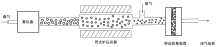
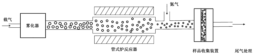
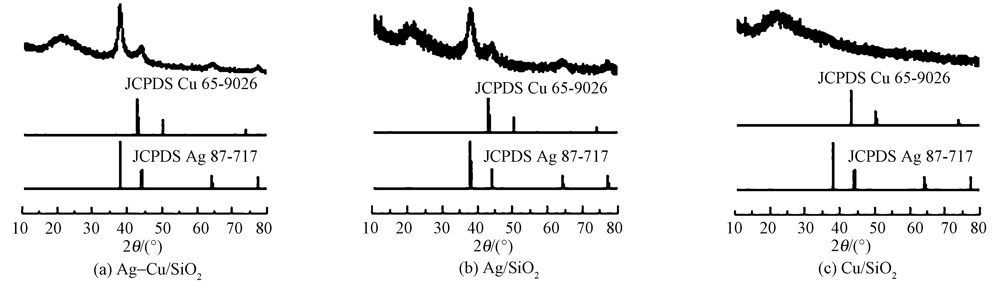
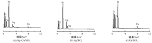
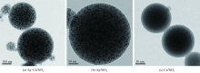
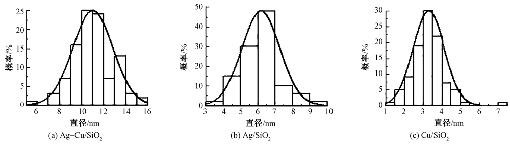
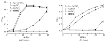
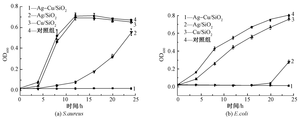
为研究双金属纳米颗粒间协同抗菌作用及防止金属纳米颗粒团聚,利用气溶胶一步法制备了“火龙果”型高负载量(50%)二氧化硅包覆银铜双金属纳米颗粒抗菌剂Ag-Cu/SiO2。借助X射线衍射仪、透射电子显微镜、电子能谱仪对Ag-Cu/SiO2的结构进行研究,并测试该材料对金黄色葡萄球菌和大肠杆菌的最低抑菌浓度(MIC)及细菌的时间与杀菌曲线,研究了2种细菌胞内活性氧的生成情况。结果表明:银铜双金属纳米颗粒均匀分散在球型二氧化硅内部,呈现“火龙果”型结构;与Cu/SiO2和Ag/SiO2相比,相同负载量(50%)的Ag-Cu/SiO2有更好的抗菌效果,对2种细菌的MIC值均为2 μg/mL,在24 h内可以充分抑制细菌生长;Ag-Cu/SiO2生成活性氧的水平明显高于单金属纳米颗粒,致使细菌死亡,从而说明双金属纳米颗粒具有协同抗菌作用。
中图分类号:
| [1] |
HAN S H, BAI J, LIU H M, et al. One-Pot fabrication of hollow and porous Pd-Cu alloy nanospheres and their remarkably improved catalytic performance for hexavalent chromium reduction[J]. ACS Applied Materials & Interfaces, 2016,8(45):30948-30955.
doi: 10.1021/acsami.6b10343 pmid: 27778503 |
| [2] | NOWAK A, SZADE J, TALIK E, et al. Physicochemical and antibacterial characterization of ionocity Ag/Cu powder nanoparticles[J]. Materials Characterization, 2016,117:9-16. |
| [3] |
ASHFAQ M, VERMA N, KHAN S. Copper/zinc bimetal nanoparticles-dispersed carbon nanofibers: a novel potential antibiotic material[J]. Materials Science and Engineering C, 2016,59:938-947.
doi: 10.1016/j.msec.2015.10.079 pmid: 26652451 |
| [4] | 赵兵, 黄小萃, 祁宁, 等. 基于共价结合的纳米银抗菌棉织物研究进展[J]. 纺织学报, 2020,41(3):188-196. |
| ZHAO Bing, HUANG Xiaocui, QI Ning, et al. Research progress of antibacterial cotton fabric treated with silver nanoparticles based on covalent bond[J]. Journal of Textile Research, 2020,41(3):188-196. | |
| [5] |
FENG Q L, WU J, CHEN G Q, et al. A mechanistic study of the antibacterial effect of silver ions on Escherichia coli and Staphylococcus aureus[J]. Journal of Biomedical Materials Research, 2000,52(4):662-668.
doi: 10.1002/1097-4636(20001215)52:4<662::aid-jbm10>3.0.co;2-3 pmid: 11033548 |
| [6] |
CHOI O, HU Z Q. Size dependent and reactive oxygen species related nanosilver toxicity to nitrifying bacteria[J]. Environmental Science & Technology, 2008,42(12):4583-4588.
doi: 10.1021/es703238h pmid: 18605590 |
| [7] |
KIM Y H, LEE D K, CHA H G, et al. Preparation and characterization of the antibacterial Cu nanoparticle formed on the surface of SiO2 nanoparticles[J]. Journal of Physical Chemistry B, 2006,110(49):24923-24928.
doi: 10.1021/jp0656779 |
| [8] |
RUPARELIA J P, CHATTERJEE A K, DUTTAGUPTA S P, et al. Strain specificity in antimicrobial activity of silver and copper nanoparticles[J]. Acta Biomaterialia, 2008,4(3):707-716.
doi: 10.1016/j.actbio.2007.11.006 |
| [9] |
REN G G, HU D W, CHENG E W C, et al. Characterisation of copper oxide nanoparticles for antimicrobial applications[J]. International Journal of Antimicrobial Agents, 2009,33(6):587-590.
doi: 10.1016/j.ijantimicag.2008.12.004 |
| [10] |
ALZAHRANI K E, NIAZY A A, ALSWIELEH A M, et al. Antibacterial activity of trimetal (CuZnFe) oxide nanoparticles[J]. International Journal of Nanomedicine, 2018,13:77-87.
doi: 10.2147/IJN.S154218 pmid: 29317817 |
| [11] | WU W T, ZHAO W J, WU Y H, et al. Antibacterial behaviors of Cu2O particles with controllable morphologies in acrylic coatings[J]. Applied Surface Science, 2019,465:279-287. |
| [12] | VALODKAR M, MODI S, PAL A, et al. Synjournal and anti-bacterial activity of Cu, Ag and Cu-Ag alloy nanoparticles: a green approach[J]. Materials Research Bulletin, 2011,46(3):384-389. |
| [13] | BAGCHI B, DEY S, BHANDARY S, et al. Antimicrobial efficacy and biocompatibility study of copper nanoparticle adsorbed mullite aggregates[J]. Materials Science and Engineering C, 2012,32(7):1897-1905. |
| [14] |
ZAIN N M, STAPLEY A G F, SHAMA G, Green synjournal of silver and copper nanoparticles using ascorbic acid and chitosan for antimicrobial applica-tions[J]. Carbohydrate Polymers, 2014,112:195-202.
doi: 10.1016/j.carbpol.2014.05.081 pmid: 25129735 |
| [15] |
PERDIKAKI A, GALEOU A, PILATOS G, et al. Ag and Cu monometallic and Ag/Cu bimetallic nanoparticle-graphene composites with enhanced antibacterial performance[J]. ACS Applied Materials & Interfaces, 2016,8(41):27498-27510.
doi: 10.1021/acsami.6b08403 pmid: 27680975 |
| [16] |
XU P, LIANG J, CAO X Y, et al. Facile synjournal of monodisperse of hollow mesoporous SiO2 nanoparticles and in-situ growth of Ag nanoparticles for antibac-terial[J]. Journal of Colloid and Interface Science, 2016,474:114-118.
doi: 10.1016/j.jcis.2016.04.009 pmid: 27115332 |
| [17] |
QIN R, LI G A, PAN L P, et al. Preparation of SiO2@Ag composite nanoparticles and their antimicrobial activity[J]. Journal of Nanoscience and Nanotechnology, 2017,17:2305-2311.
doi: 10.1166/jnn.2017.13042 pmid: 29638553 |
| [18] |
ZHANG N C, GAO Y H, ZHANG H, et al. Preparation and characterization of core-shell structure of SiO2@Cu antibacterial agent[J]. Colloids and Surfaces B: Biointerfaces, 2010,81:537-543.
doi: 10.1016/j.colsurfb.2010.07.054 pmid: 20729042 |
| [19] | ISAACS M A, DURNDELL L J, HILTON A C, et al. Tunable Ag@SiO2 core-shell nanocomposites for broad spectrum antibacterial applications[J]. RSC Advances, 2017,7(38):23342-23347. |
| [20] | FANG W J, XU C F, ZHENG J, et al. Fabrication of Cu-Ag bimetal nanotube-based copper silicates for enhancement of antibacterial activities[J]. RSC Advances, 2015,5:39612-39619. |
| [21] |
ZHANG M, WANG P, SUN H Y, et al. Superhydrophobic surface with hierarchical architecture and bimetallic composition for enhanced antibacterial activity[J]. ACS Applied Materials & Interfaces, 2014,6(24):22108-22115.
doi: 10.1021/am505490w pmid: 25418198 |
| [22] | BAKINA O V, GLAZKOVA E A, SVAROVSKAYA N V, et al. 《Janus》-like Cu-Fe bimetallic nanoparticles with high antibacterial activity[J]. Materials Letters, 2019,242:187-190. |
| [23] | SAXENA V, PANDEY L M. Bimetallic assembly of Fe(III) doped ZnO as an effective nanoantibiotic and its ROS independent antibacterial mechanism[J]. Journal of Trace Elements in Medicine and Biology, 2020,57:134-145. |
| [24] |
KOHANSKI M A, DWYER D J, HAYETE B, et al. A common mechanism of cellular death induced by bactericidal antibiotics[J]. Cell, 2007,130(5):797-810.
doi: 10.1016/j.cell.2007.06.049 pmid: 17803904 |
| [1] | 黎俊妤 蒋培清 张文奇 李文斌. 原子层沉积技术对纤维素膜功能化的影响[J]. 纺织学报, 2020, 41(12): 26-30. |
| [2] | 张艳艳, 詹璐瑶, 王培, 耿俊昭, 付飞亚, 刘向东. 用无机纳米粒子制备耐久性抗菌棉织物的研究进展[J]. 纺织学报, 2020, 41(11): 174-180. |
| [3] | 秦益民. 含银海藻酸盐医用敷料的临床应用[J]. 纺织学报, 2020, 41(09): 183-190. |
| [4] | 段红梅, 汪希铭, 黄子欣, 高晶, 王璐. 纤维基介孔SiO2药物载体的构建及其释药性能[J]. 纺织学报, 2020, 41(07): 15-22. |
| [5] | 陈佳颖, 田旭, 彭晶晶, 方彤, 高伟洪. 针织物表面结构色的构建[J]. 纺织学报, 2020, 41(07): 117-121. |
| [6] | 贾琳, 王西贤, 陶文娟, 张海霞, 覃小红. 聚丙烯腈抗菌复合纳米纤维膜的制备及其抗菌性能[J]. 纺织学报, 2020, 41(06): 14-20. |
| [7] | 王婷婷, 刘梁, 曹秀明, 王清清. 竹红菌素-聚( 甲基丙烯酸甲酯-co-甲基丙烯酸)纳米纤维的制备及其光敏抗菌性能[J]. 纺织学报, 2020, 41(05): 1-7. |
| [8] | 刘艳春, 白刚. 小檗碱在聚丙烯腈/ 醋酸纤维素复合纤维染色中的应用[J]. 纺织学报, 2020, 41(05): 94-98. |
| [9] | 王晓菲, 万爱兰. 紫外线辐照聚吡咯/银导电涤纶织物的制备[J]. 纺织学报, 2020, 41(04): 112-116. |
| [10] | 高思梦, 王鸿博, 杜金梅, 王文聪. 甜菜碱聚合物的合成及其在棉织物抗菌整理中的应用[J]. 纺织学报, 2020, 41(02): 89-94. |
| [11] | 吴倩倩, 李珂, 杨立双, 付译鋆, 张瑜, 张海峰. 载药聚偏氟乙烯伤口敷料的制备及其性能 [J]. 纺织学报, 2020, 41(01): 26-31. |
| [12] | 张治斌, 李刚, 毛森贤, 厉巽巽, 陈玉霜, 毛青山, 李翼, 潘志娟, 王晓沁. 丝素蛋白/壳聚糖微球制备及其抗菌性能[J]. 纺织学报, 2019, 40(10): 7-12. |
| [13] | 韩健健, 胡勇杰, 胡敏专. 基于纳滤技术的质检萃取液脱色预处理方法[J]. 纺织学报, 2019, 40(09): 136-142. |
| [14] | 王文聪, 范静静, 丁超, 王鸿博. 多功能复合导电毛织物的制备及其性能[J]. 纺织学报, 2019, 40(08): 117-123. |
| [15] | 吴娇, 于湖生, 万兴云, 田平, 李慧敏, 侯晓欣. 抗菌防螨防霉功能改性粘胶纤维的制备及其性能[J]. 纺织学报, 2019, 40(07): 19-23. |
|
||
 京公网安备11010502044800号