纺织学报 ›› 2025, Vol. 46 ›› Issue (05): 116-124.doi: 10.13475/j.fzxb.20240700101
李鹏飞1, 罗忆心1, 张子凡1, 陆宁1, 陈碧泠1, 许建梅1,2( )
)
LI Pengfei1, LUO Yixin1, ZHANG Zifan1, LU Ning1, CHEN Biling1, XU Jianmei1,2()
摘要:
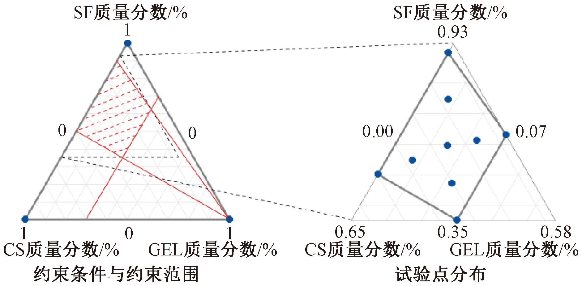
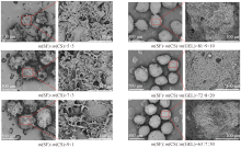
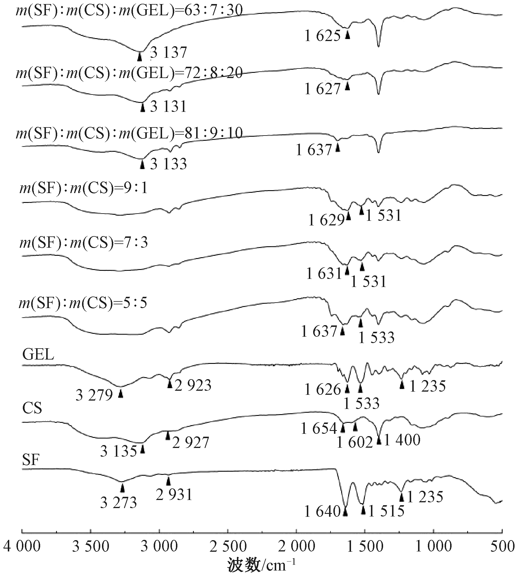
为实现栓塞微球降解行为的可控设计,采用离子凝胶法与乳化交联法联用,利用具有不同降解速度和特性的丝素蛋白(SF)、壳聚糖(CS)、明胶(GEL)材料以不同配比复合制备栓塞微球。经正交试验探究了SF与CS质量比分别为9:1、7:3、5:5时栓塞微球的最优制备工艺条件,并在此基础上利用配方设计中的极端顶点法设计并制备了不同配比的SF/CS/GEL栓塞微球,对栓塞微球的外观形貌、化学组成、热稳定性、降解性能进行了表征。结果表明:在丝素蛋白与壳聚糖不同配比下均可以通过正交试验获得较大尺寸栓塞微球(微球粒径为200~400 μm和550~750 μm)的最优制备工艺条件,制得球形圆整、分散性好的微球;微球中3种材料之间存在氢键、席夫碱等相互作用;获得不同组分配方在3种降解酶体系(蛋白酶ⅩⅣ、溶菌酶或2种酶联用)中的降解率回归方程,拟合优度显著,可实现对不同配方栓塞微球降解性能的预测与设计。
中图分类号:
| [1] | NUZULIA N A, MART T, AHMED I, et al. The use of microspheres for cancer embolization therapy: recent advancements and prospective[J]. ACS Biomaterials Science & Engineering, 2024, 10: 637-656. |
| [2] |
LIU K L, JIN Z C, HU X L, et al. A biodegradable multifunctional porous microsphere composed of carrageenan for promoting imageable trans-arterial chemoembolization[J]. International Journal of Biological Macromolecules, 2020, 142: 866-878.
doi: S0141-8130(19)35185-2 pmid: 31622716 |
| [3] | FUCHS K, DURAN R, DENYS A, et al. Drug-eluting embolic microspheres for local drug delivery-state of the art[J]. Journal of Controlled Release, 2017, 262: 127-138. |
| [4] |
POURSAID A, JENSEN M M, HUO E, et al. Polymeric materials for embolic and chemoembolic applications[J]. Journal of Controlled Release, 2016, 240: 414-433.
doi: S0168-3659(16)30096-7 pmid: 26924353 |
| [5] | HAN S, ZHANG X, LI M. Progress in research and application of PLGA embolic microspheres[J]. Frontiers in Bioscience-Landmark, 2016, 21: 931-940. |
| [6] | MOSCHOVAKI-ZEIGER O, ARKOUDIS N A, GIANNAKIS A, et al. Biodegradable microspheres for transarterial chemoembolization in malignant liver disease[J]. Medicina, 2024. DOI: 10.3390/medicina60040678. |
| [7] | KUNDU B, KURLAND N E, BANO S, et al. Silk proteins for biomedical applications: bioengineering perspectives[J]. Progress in Polymer Science, 2014, 39(2): 251-267. |
| [8] | HONG H, LEE O J, LEE Y J, et al. Cytocompatibility of modified silk fibroin with glycidyl methacrylate for tissue engineering and biomedical applications[J]. Biomolecules, 2020. DOI: 10.3390/biom11010035. |
| [9] | NOSRATI Z, LI N, MICHAUD F, et al. Development of a coflowing device for the size-controlled preparation of magnetic-polymeric microspheres as embolization agents in magnetic resonance navigation technology[J]. ACS Biomaterials Science & Engineering, 2018, 4(3): 1092-1102. |
| [10] |
ZHANG X H, BAUGHMAN C B, KAPLAN D L. In vitro evaluation of electrospun silk fibroin scaffolds for vascular cell growth[J]. Biomaterials, 2008, 29(14): 2217-2227.
doi: 10.1016/j.biomaterials.2008.01.022 pmid: 18279952 |
| [11] | LEE H S, KIM E H, SHAO H P, et al. Synthesis of SPIO-chitosan microspheres for MRI-detectable embolotherapy[J]. Journal of Magnetism and Magnetic Materials, 2005, 293(1): 102-105. |
| [12] |
张子凡, 李鹏飞, 王建南, 等. 丝素蛋白载药纳米粒的研究进展[J]. 纺织学报, 2023, 44(10): 205-213.
doi: 10.13475/j.fzxb.20220607102 |
|
ZHANG Zifan, LI Pengfei, WANG Jiannan, et al. Research progress in silk fibroin drug-loaded nanoparticles[J]. Journal of Textile Research, 2023, 44(10): 205-213.
doi: 10.13475/j.fzxb.20220607102 |
|
| [13] |
ZHAO Z, LI Y, XIE M B. Silk fibroin-based nanoparticles for drug delivery[J]. International Journal of Molecular Sciences, 2015, 16(3): 4880-4903.
doi: 10.3390/ijms16034880 pmid: 25749470 |
| [14] | QIU W, PATIL A, HU F, et al. Hierarchical structure of silk materials versus mechanical performance and mesoscopic engineering principles[J]. Small, 2019. DOI: 10.1002/smll.201903948. |
| [15] | HU J, ALBADAWI H, ZHANG Z, et al. Silk embolic material for catheter-directed endovascular drug deliv-ery[J]. Advanced Materials, 2021. DOI: 1002/adma.202106865. |
| [16] |
LIU Q, LIU H F, FAN Y B. Preparation of silk fibroin carriers for controlled release[J]. Microscopy Research and Technique, 2017, 80(3): 312-320.
doi: 10.1002/jemt.22606 pmid: 26638113 |
| [17] | 王刚, 李晓萍, 王进. 植入壳聚糖体内降解的机理研究[J]. 当代医学, 2015, 21(34): 5-8. |
| WANG Gang, LI Xiaoping, WANG Jin. Mechanism study of degradation in chitosan implanted[J]. Contemporary Medicine, 2015, 21(34): 5-8. | |
| [18] | DOUCET J, KIRI L, O'CONNELL K, et al. Advances in degradable embolic microspheres: a state of the art review[J]. Journal of Functional Biomaterials, 2018. DOI: 10.1016/j.jconrel.2017.07.016. |
| [19] | GAO F, RAFIQ M, CONG H, et al. Current research status and development prospects of embolic microspheres containing biological macromolecules and others[J]. International Journal of Biological Macromolecules, 2024. DOI: 10.1016/j.ijbiomac.2024.131494. |
| [20] |
CHEN G, WEI R, HUANG X, et al. Synthesis and assessment of sodium alginate-modified silk fibroin microspheres as potential hepatic arterial embolization agent[J]. International Journal of Biological Macromolecules, 2020, 155: 1450-1459.
doi: S0141-8130(19)36999-5 pmid: 31734365 |
| [21] |
ZHOU X, KONG M, CHENG X J, et al. In vitro and in vivo evaluation of chitosan microspheres with different deacetylation degree as potential embolic agent[J]. Carbohydrate Polymers, 2014, 113: 304-313.
doi: 10.1016/j.carbpol.2014.06.080 pmid: 25256489 |
| [22] |
KWAK B K, SHIM H J, HAN S M, et al. Chitin-based embolic materials in the renal artery of rabbits: pathologic evaluation of an absorbable particulate agent[J]. Radiology, 2005, 236(1): 151-158.
pmid: 15987971 |
| [23] |
李枫, 杨嘉豪, 赖耿昌, 等. 高分子聚合物栓塞微球的研究进展[J]. 纺织学报, 2021, 42(10): 180-189.
doi: 10.13475/j.fzxb.20200702810 |
|
LI Feng, YANG Jiahao, LAI Gengchang, et al. Research progress of polymer embolic microspheres[J]. Journal of Textile Research, 2021, 42(10): 180-189.
doi: 10.13475/j.fzxb.20200702810 |
|
| [24] | YANG S H, JU X J, DENG C F, et al. In vitro study on effects of physico-chemo-mechanical properties of embolic microspheres on embolization performances[J]. Industrial & Engineering Chemistry Research, 2023, 62: 2636-2648. |
| [25] | JIA G, VAN VALKENBURGH J, CHEN A Z, et al. Recent advances and applications of microspheres and nanoparticles in transarterial chemoembolization for hepatocellular carcinoma[J]. Wiley Interdisciplinary Reviews-Nanomedicine and Nanobiotechnology, 2021. DOI: 10.1002/wnan.1749. |
| [26] |
VAIDYA S, TOZER K, CHEN J. An overview of embolic agents[J]. Seminars in Interventional Radiology, 2008, 25(3): 204-215.
doi: 10.1055/s-0028-1085930 pmid: 21326511 |
| [27] | CHEN C, HUANG X, WANG F, et al. Preparation of a modified silk-based gel/microsphere composite as a potential hepatic arterial embolization agent[J]. Biomaterials Advances, 2023. DOI: 10.1016/j.bioadv.2023.213559. |
| [1] | 于梦菲, 高文丽, 任婧, 曹雷涛, 彭若铉, 凌盛杰. 摩擦纳米发电机用皮芯结构纤维的制备及其性能[J]. 纺织学报, 2025, 46(05): 1-9. |
| [2] | 曹展瑞, 纪灿灿, 赫羴姗, 周丰, 向阳, 高飞, 刘轲, 王栋. 阴离子交换型乙烯-乙烯醇共聚物纳米纤维气凝胶蛋白分离材料[J]. 纺织学报, 2025, 46(04): 29-37. |
| [3] | 董子靖, 吴欣媛, 王瑞霞, 赵华祥, 钱利江, 应城唯, 孙润军. 壳聚糖改性的炭黑导电织物制备及其在人体运动监测中的应用[J]. 纺织学报, 2025, 46(04): 146-153. |
| [4] | 罗欣, 王磊, 王筱悠, 伍韬, 张贞贞, 张一帆. 丝素蛋白多级结构的自组装机制及其重构材料研究进展[J]. 纺织学报, 2025, 46(03): 225-235. |
| [5] | 詹克静, 杨鑫, 张应龙, 张昕, 潘志娟. 自凝聚丝素蛋白微纳米纤维膜的制备及其力学增强[J]. 纺织学报, 2025, 46(02): 10-19. |
| [6] | 杨鑫, 张昕, 潘志娟. 丝素纳米原纤增强再生丝素蛋白/聚乙烯醇纤维的结构与性能[J]. 纺织学报, 2024, 45(11): 1-9. |
| [7] | 李蒙, 戴梦男, 俞杨销, 王建南. 丝素蛋白基骨修复材料的应用研究进展[J]. 纺织学报, 2024, 45(10): 224-231. |
| [8] | 房磊, 刘秀明, 贾娇娇, 蔺志浩, 任燕飞, 侯凯文, 巩继贤, 扈延龄. 高分子量壳聚糖皮芯结构微纳米纤维膜制备[J]. 纺织学报, 2024, 45(09): 1-9. |
| [9] | 王勃翔, 徐航丹, 李佳, 林杰, 程德红, 路艳华. 柞蚕丝素纳米纤维温敏复合膜制备及其生物相容性[J]. 纺织学报, 2024, 45(09): 18-25. |
| [10] | 吕子豪, 徐慧慧, 袁小红, 王清清, 魏取福. 光动力抗菌水刺棉的染整一体化制备及其性能[J]. 纺织学报, 2024, 45(08): 26-34. |
| [11] | 吴雨航, 魏建斐, 顾伟文, 王玉萍, 张安莹, 王锐. 共聚阻燃改性聚对苯二甲酸乙二醇酯的制备及其性能[J]. 纺织学报, 2024, 45(06): 1-10. |
| [12] | 冯颖, 于汉哲, 张宏, 李可心, 马标, 董鑫, 张建伟. 静电纺壳聚糖基纳米纤维的制备及其在水处理中应用研究进展[J]. 纺织学报, 2024, 45(05): 218-227. |
| [13] | 陈锦苗, 李纪伟, 陈萌, 宁新, 崔爱华, 王娜. 壳聚糖微纳米纤维复合抗菌空气滤材的制备及其性能[J]. 纺织学报, 2024, 45(05): 19-26. |
| [14] | 胡自强, 骆晓蕾, 魏璐琳, 刘琳. 植酸/壳聚糖对涤纶/棉混纺织物的协同阻燃整理[J]. 纺织学报, 2024, 45(04): 126-135. |
| [15] | 朱维维, 舒伟, 顾文娟. 负载不同极性药物对粘胶织物结构和性能的影响[J]. 纺织学报, 2024, 45(04): 136-141. |
|
||
 京公网安备11010502044800号