纺织学报 ›› 2022, Vol. 43 ›› Issue (10): 16-23.doi: 10.13475/j.fzxb.20210906708
俞杨销1, 李枫1, 王煜煜1, 王善龙1, 王建南1,2, 许建梅1,2( )
)
YU Yangxiao1, LI Feng1, WANG Yuyu1, WANG Shanlong1, WANG Jiannan1,2, XU Jianmei1,2()
摘要:
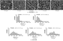
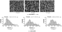
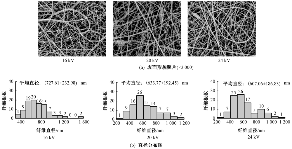
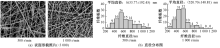
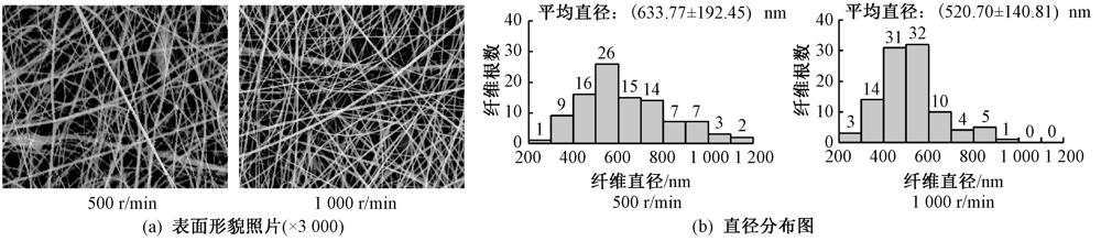
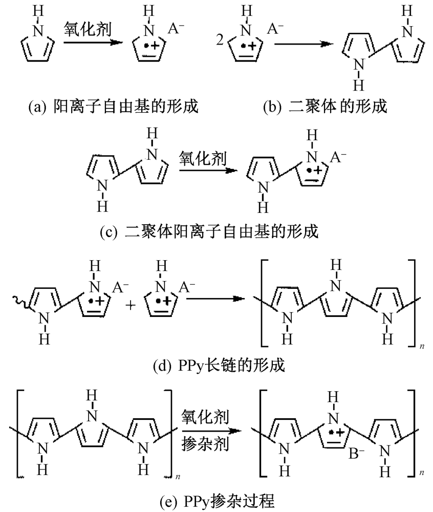
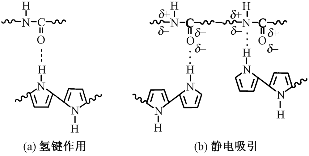
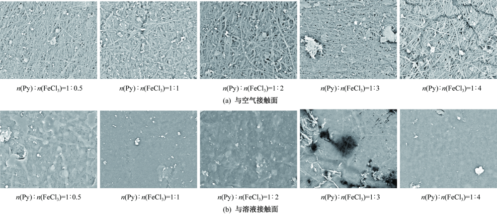
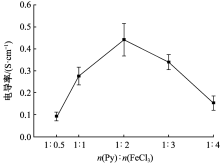
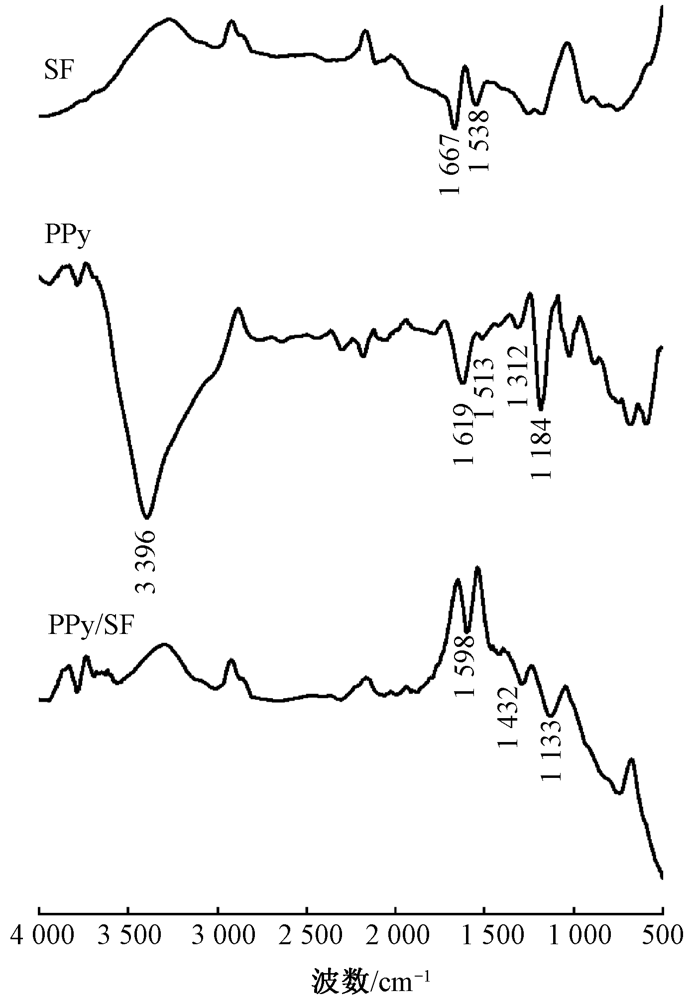
为开发具有一定导电性的组织再生材料,采用静电纺丝法制备了丝素纳米纤维膜,通过原位氧化聚合获得了聚吡咯/丝素导电性纳米纤维膜,探究了纺丝参数对纳米纤维膜表面形貌的影响,利用四探针测试仪测试了纳米纤维膜的导电性,借助红外光谱仪对纳米纤维膜化学结构进行了表征。结果表明:在质量浓度为0.16 g/mL,推注速度为0.2 mL/h,电压为20 kV,滚筒转速为1 000 r/min的条件下,制备的丝素纳米纤维膜表面规整,珠状物少,纤维平均直径为(520.70±140.81) nm;在吡咯单体浓度为0.3 mol/L,掺杂剂浓度为0.3 mol/L,吡咯单体与FeCl3的量比为1∶2,聚合时间为6 h条件下,制备的聚吡咯/丝素导电性纳米纤维膜保留了丝素纳米纤维膜原有的纳米纤维结构,电导率达到(0.44±0.07) S/cm。
中图分类号:
| [1] | ESLAHI N, REZA M, JOGHATAEI M T, et al. The effects of poly L-lactic acid nanofiber scaffold on mouse spermatogonial stem cell culture[J]. International JournaL of Nanomedicine, 2013, 8: 4563-4576. |
| [2] | 贾琳, 陈莉娜, 张海霞, 等. 聚氨酯/胶原蛋白复合纳米纤维支架的性能[J]. 纺织学报, 2016, 37(8): 1-6. |
|
JIA Lin, CHEN Li'na, ZHANG Haixia, et al. Performance of composite polyurethane/collagen nanofiber scaffolds[J]. Journal of Textile Research, 2016, 37(8): 1-6.
doi: 10.1177/004051756703700101 |
|
| [3] |
ALTMAN G H, DIAZ F, JAKUBA C. Silk-based biomaterials[J]. Biomaterials, 2003, 24(3): 401-416.
pmid: 12423595 |
| [4] | 姜雨淋, 王卉, 张克勤. 生物3D打印用丝素蛋白基凝胶墨水的研究进展[J]. 纺织学报, 2021, 42(11): 1-8. |
|
JIANG Yulin, WANG Hui, ZHANG Keqin. Research progress of silk fibroin-based hydrogel bioinks for 3D bio-printing[J]. Journal of Textile Research, 2021, 42(11): 1-8.
doi: 10.1177/004051757204200101 |
|
| [5] |
LI X F, ZHANG Q, LUO Z W, et al. Biofunctionalized silk fibroin nanofibers for directional and long neurite outgrowth[J]. Biointerphases, 2019. DOI: 10.1063/1.5120738.
doi: 10.1063/1.5120738 |
| [6] |
SCHMIDT C E, SHASTRI V R, VACANTI J P, et al. Stimulation of neurite outgrowth using an electrically conducting polymer[J]. Proceedings of the National Academy of Sciences of the United States of America, 1997, 94(17): 8948-8953.
pmid: 9256415 |
| [7] |
LEE M H, PARK Y J, HONG S H, et al. Pulsed electrical stimulation enhances consistency of directional migration of adipose-derived stem cells[J]. Cells, 2021. DOI: 10.3390/cells10112846.
doi: 10.3390/cells10112846 |
| [8] |
LONG Y, LI J, YANG F, et al. Wearable and implantable electroceuticals for therapeutic electrostimulations[J]. Advanced Science, 2020. DOI: 10.1002/advs.202004023.
doi: 10.1002/advs.202004023 |
| [9] |
FUNK R H W. Endogenous electric fields as guiding cue for cell migration[J]. Frontiers in Physiology, 2015. DOI: 10.3389/fphys.2015.00143.
doi: 10.3389/fphys.2015.00143 |
| [10] | VAITKUVIENE A, KASETA V, VORONOVIC J, et al. Evaluation of cytotoxicity of polypyrrole nanoparticles synthesized by oxidative polymerization[J]. Journal of Hazardous Materials, 2013, 250: 167-174. |
| [11] |
KIM S, OH W K, JEONG Y S, et al. Cytotoxicity of, and innate immune response to, size-controlled polypyrrole nanoparticles in mammalian cells[J]. Biomaterials, 2011, 32(9): 2342-2350.
doi: 10.1016/j.biomaterials.2010.11.080 pmid: 21185594 |
| [12] | SUN B B, ZHOU Z F, LI D W, et al. Polypyrrole-coated poly(L-lactic acid-co-epsilon-caprolactone)/silk fibroin nanofibrous nerve guidance conduit induced nerve regeneration in rat[J]. Materials Science & Engineering C-Materials for Biological Applications, 2019, 94: 190-199. |
| [13] |
ZHAO Y H, NIU C M, SHI J Q, et al. Novel conductive polypyrrole/silk fibroin scaffold for neural tissue repair[J]. Neural Regeneration Research, 2018, 13(8): 1455-1464.
doi: 10.4103/1673-5374.235303 |
| [14] | ZHAO Y H, LIANG Y Y, DING S P, et al. Application of conductive PPy/SF composite scaffold and electrical stimulation for neural tissue engineering[J]. Biomaterials, 2020, 255: 120-164. |
| [15] |
LEE J Y, BASHUR C A, GOLDSTEIN A S, et al. Polypyrrole-coated electrospun PLGA nanofibers for neural tissue applications[J]. Biomaterials, 2009, 30(26): 4325-4335.
doi: 10.1016/j.biomaterials.2009.04.042 pmid: 19501901 |
| [16] |
XU Y X, HUANG Z B, PU X M, et al. Fabrication of chitosan/polypyrrole-coated poly(L-lactic acid)/polycaprolactone aligned fibre films for enhancement of neural cell compatibility and neurite growth[J]. Cell Proliferation, 2019. DOI: 10.1111/cpr.12588.
doi: 10.1111/cpr.12588 |
| [17] |
QI F Y, WANG Y Q, MA T, et al. Electrical regulation of olfactory ensheathing cells using conductive polypyrrole/chitosan polymers[J]. Biomaterials, 2013, 34(7): 1799-1809.
doi: 10.1016/j.biomaterials.2012.11.042 pmid: 23228424 |
| [18] |
LIANG Y S, MITRIASHKIN A, LIM T T, et al. Conductive polypyrrole-encapsulated silk fibroin fibers for cardiac tissue engineering[J]. Biomaterials, 2021. DOI: 10.1016/j.biomaterials.2021.12100.
doi: 10.1016/j.biomaterials.2021.12100 |
| [19] | 李永舫. 导电聚吡咯的研究[J]. 高分子通报, 2005(4): 51-57. |
| LI Yongfang. Studies on conducting polypyrrole[J]. Polymer Bulletin, 2005(4): 51-57. | |
| [20] |
XIA Y Y, LU Y. Fabrication and properties of conductive conjugated polymers/silk fibroin composite fibers[J]. Composites Science and Technology, 2008, 68(6): 1471-1479.
doi: 10.1016/j.compscitech.2007.10.044 |
| [21] | 于波, 徐学诚. 聚吡咯结构与导电性能的研究[J]. 华东师范大学学报(自然科学版), 2014(4): 77-87. |
| YU Bo, XU Xuecheng. Structure-conductive property relationship of polypyrrole[J]. Journal of East China Normal University (Natural Science Edition), 2014(4): 77-87. | |
| [22] |
YURTSEVER M, YURTSEVER E. Structural studies of polypyrroles: I: an ab-initio evaluation of bonding through alpha and beta carbons[J]. Synthetic Metals, 1999, 98(3): 221-227.
doi: 10.1016/S0379-6779(98)00195-7 |
| [23] |
HAN F, LIU S, LIU X, et al. Woven silk fabric-reinforced silk nanofibrous scaffolds for regenerating load-bearing soft tissues[J]. Acta Biomaterialia, 2014, 10(2): 921-930.
doi: 10.1016/j.actbio.2013.09.026 pmid: 24090985 |
| [24] |
JIN X, WANG H, LIU Y M, et al. Hydrogen-bonding power interfacial load transfer of carbon fabric/polypyrrole composite pseudosupercapacitor electrode with improved electrochemical stability[J]. Applied Surface Science, 2019, 470: 783-791.
doi: 10.1016/j.apsusc.2018.11.125 |
| [1] | 姚莹, 赵为陶, 张德锁, 林红, 陈宇岳, 魏红. 超支化季铵盐诱导制备树枝状纳米纤维膜及其性能[J]. 纺织学报, 2022, 43(10): 1-9. |
| [2] | 杨吉震, 刘强飞, 何瑞东, 吴韶华, 何宏伟, 宁新, 周蓉, 董湘琳, 齐贵山. 高效低阻空气过滤材料研究进展[J]. 纺织学报, 2022, 43(10): 209-215. |
| [3] | 胡铖烨, 周歆如, 范梦晶, 洪剑寒, 刘永坤, 韩潇, 赵晓曼. 皮芯结构微纳米纤维复合纱线的制备及其性能[J]. 纺织学报, 2022, 43(09): 95-100. |
| [4] | 李伟平, 杨桂霞, 程志强, 赵春莉. 聚乙烯吡咯烷酮/芦荟复合纳米纤维膜的制备及其性能[J]. 纺织学报, 2022, 43(08): 55-59. |
| [5] | 刘蛟, 陈韶娟, 吴韶华. 丝素蛋白/聚左旋乳酸纳米纤维纱线肌腱补片的制备及其性能[J]. 纺织学报, 2022, 43(08): 60-66. |
| [6] | 渠赟, 马维, 刘颖, 任学宏. 可光降解聚羟基丁酸酯/聚己内酯基抗菌纤维膜的制备及其性能[J]. 纺织学报, 2022, 43(06): 29-36. |
| [7] | 李艾元, 施心雨, 岳万福, 游卫云. 丝素蛋白水凝胶支架的制备及其性能[J]. 纺织学报, 2022, 43(06): 44-48. |
| [8] | 欧康康, 祁琳雅, 侯怡君, 范天华, 齐琨, 王宝秀, 王华平. 纳米纤维基单向导湿抗菌敷料的制备及其性能[J]. 纺织学报, 2022, 43(06): 49-56. |
| [9] | 顾张弘, 姚响, 王锦思, 张耀鹏. 具有细胞黏附反差特性的单层平行丝素蛋白纤维图案的制备及其性能[J]. 纺织学报, 2022, 43(05): 1-6. |
| [10] | 李琴, 李兴兴, 解芳芳, 周文龙, 陈恺宜, 刘宇清. 静电纺丝和炭化法制备纳米纤维素储能材料研究进展[J]. 纺织学报, 2022, 43(05): 178-184. |
| [11] | 陈锋, 姬忠礼, 于文瀚, 董伍强, 王倩琳, 王德国. 纳米纤维膜润湿性对三明治结构复合过滤材料气液过滤性能的影响[J]. 纺织学报, 2022, 43(05): 63-69. |
| [12] | 陈明军, 李好义, 杨卫民. 聚合物熔体微分静电纺电场对射流的影响及其物理模型[J]. 纺织学报, 2022, 43(05): 70-76. |
| [13] | 杨科, 闫俊, 肖勇, 徐晶, 陈磊, 刘雍. 电化学沉积锌电池MnOx/碳纳米纤维膜自支撑正极的制备及其电化学特性[J]. 纺织学报, 2022, 43(05): 77-85. |
| [14] | 孙哲茹, 张庆乐, 郝林聪, 程璐, 夏鑫. 仿星型拓扑几何结构聚氨酯/聚二甲基硅氧烷防水透湿膜制备与性能[J]. 纺织学报, 2022, 43(04): 40-46. |
| [15] | 雷彩虹, 俞林双, 朱海霖, 郑涛, 陈建勇. 不同水解方式下蚕丝丝素蛋白材料的止血性能[J]. 纺织学报, 2022, 43(04): 15-19. |
|
||
 京公网安备11010502044800号