纺织学报 ›› 2025, Vol. 46 ›› Issue (03): 236-244.doi: 10.13475/j.fzxb.20240404702
陆宁1, 陈碧泠1, 宋功吉1, 罗忆心1, 王建南1,2, 许建梅1,2( )
)
LU Ning1, CHEN Biling1, SONG Gongji1, LUO Yixin1, WANG Jiannan1,2, XU Jianmei1,2()
摘要:
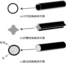
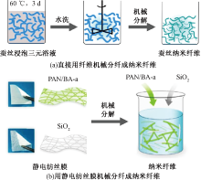
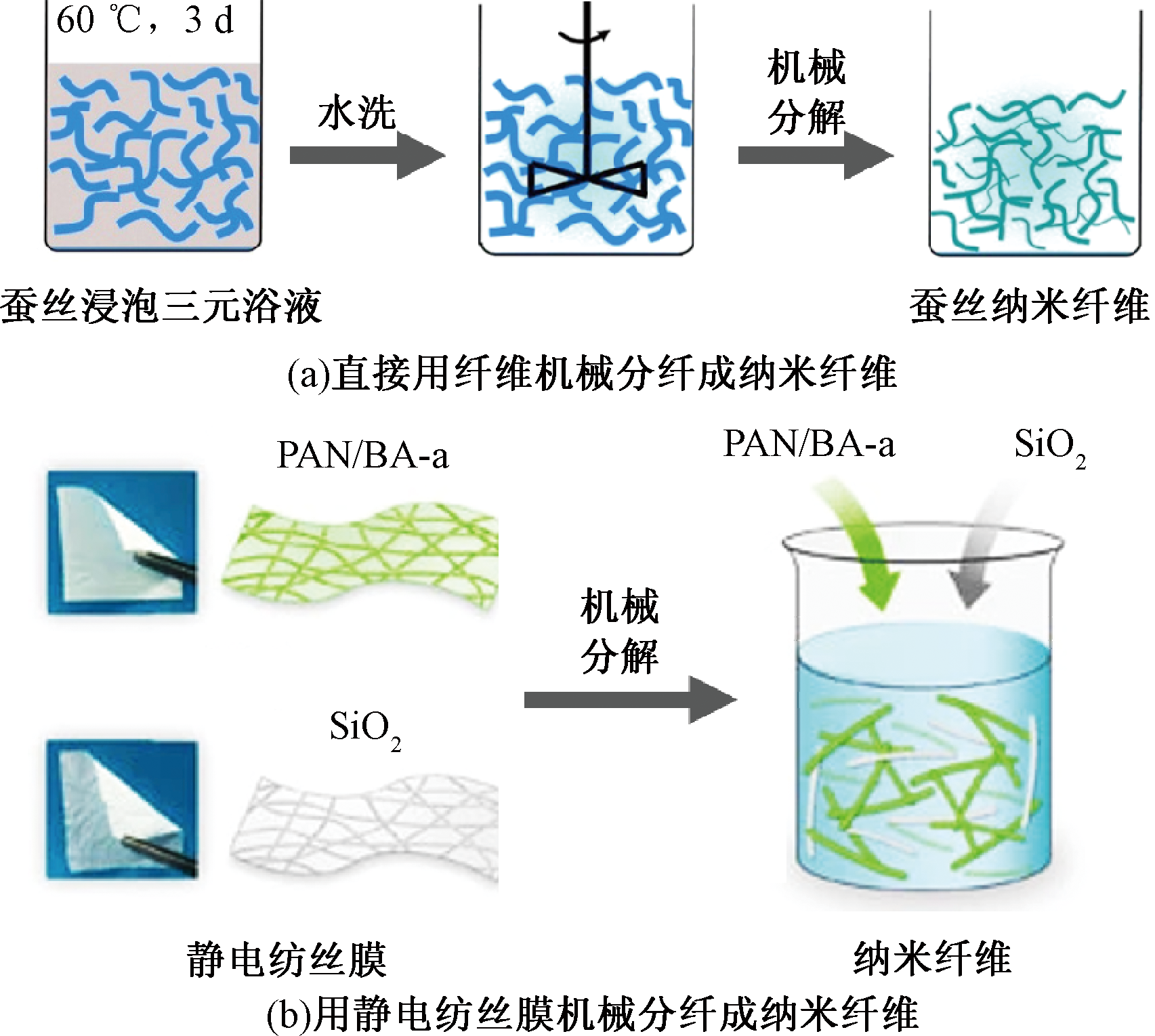
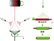
为探讨纳米纤维在神经再生修复中的作用,开发出纳米纤维基神经导管,分析了纳米纤维对周围神经损伤修复的再生机制,探讨了不同材料在纳米纤维基神经导管中的作用与应用,综述了纳米纤维制备的方法及相应的纳米纤维基神经导管的制备方法,讨论了纳米纤维基神经导管的不同内部结构。最后指出:纳米纤维基神经导管制备应考虑宏量化、标准化制备方法;从结构仿生与功能仿生的角度,去构筑组织与材料相接触的纳米仿生界面,包括:仿细胞外基质环境、微图案纳米尺度仿生界面、多尺度分级有序的微纳米仿生界面;将多种修复机制结合,发挥协同增效作用,可实现长距离神经损伤的修复。
中图分类号:
| [1] | TAYLOR C A, BRAZA D, RICE J B, et al. The incidence of peripheral nerve injury in extremity trauma[J]. American Journal of Physical Medicine & Rehabilitation, 2008, 87(5): 381-385. |
| [2] |
MEEK M F, COERT J H. Clinical use of nerve conduits in peripheral-nerve repair: review of the literature[J]. Journal of Reconstructive Microsurgery, 2002, 18(2): 97-109.
doi: 10.1055/s-2002-19889 pmid: 11823940 |
| [3] |
LI A, HOKUGO A, YALOM A, et al. A bioengineered peripheral nerve construct using aligned peptide amphiphile nanofibers[J]. Biomaterials, 2014, 35(31): 8780-8790.
doi: 10.1016/j.biomaterials.2014.06.049 pmid: 25064803 |
| [4] | YANG G, LI X L, HE Y, et al. From nano to micro to macro: Electrospun hierarchically structured polymeric fibers for biomedical applications[J]. Progress in Polymer Science, 2018, 81: 80-113. |
| [5] | KONG Y, XU J W, HAN Q, et al. Electrospinning porcine decellularized nerve matrix scaffold for peripheral nerve regeneration[J]. International Journal of Biological Macromolecules, 2022, 209: 1867-1881. |
| [6] | CLEMENTS I P, KIM Y T, ENGLISH A W, et al. Thin-film enhanced nerve guidance channels for peripheral nerve repair[J]. Biomaterials, 2009, 30(23/24): 3834-3846. |
| [7] | LI S, WU H, HU X D, et al. Preparation of electrospun PLGA-silk fibroin nanofibers-based nerve conduits and evaluation in vivo[J]. Artificial Cells Blood Substitutes and Biotechnology, 2012, 40(1/2): 171-178. |
| [8] |
WANG J, CHENG Y, WANG H Y, et al. Biomimetic and hierarchical nerve conduits from multifunctional nanofibers for guided peripheral nerve regeneration[J]. Acta Biomaterialia, 2020, 117: 180-191.
doi: 10.1016/j.actbio.2020.09.037 pmid: 33007489 |
| [9] | WANG C Y, LIU J J, FAN C Y, et al. The effect of aligned core-shell nanofibres delivering NGF on the promotion of sciatic nerve regeneration[J]. Journal of Biomaterials Science-Polymer Edition, 2012, 23(1/4): 167-184. |
| [10] |
WANG L, WU Y B, HU T L, et al. Aligned conductive core-shell biomimetic scaffolds based on nanofiber yarns/hydrogel for enhanced 3D neurite outgrowth alignment and elongation[J]. Acta Biomaterialia, 2019, 96: 175-187.
doi: S1742-7061(19)30457-X pmid: 31260823 |
| [11] |
XU H X, HOLZWARTH J M, YAN Y H, et al. Conductive PPY/PDLLA conduit for peripheral nerve regeneration[J]. Biomaterials, 2014, 35(1): 225-235.
doi: 10.1016/j.biomaterials.2013.10.002 pmid: 24138830 |
| [12] | JHANG J C, LIN J H, LOU C W, et al. Biodegradable and conductive PVA/CNT nanofibrous membranes used in nerve conduit applications[J]. Journal of Industrial Textiles, 2022. DOI: 10.1177/15280837211032086. |
| [13] |
ZHANG J C, ZHANG Y Q, CHEN L, et al. Ulinastatin promotes regeneration of peripheral nerves after sciatic nerve injury by targeting let-7 microRNAs and enhancing NGF expression[J]. Drug Design Development and Therapy, 2020, 14: 2695-2705.
doi: 10.2147/DDDT.S255158 pmid: 32753848 |
| [14] | 任文乾, 王梓尧, 张艺凡, 等. 周围神经损伤的修复机制[J]. 神经损伤与功能重建, 2022, 17(10): 604-605, 608. |
| REN Wenqian, WANG Zixiao, ZHANG Yifan, et al. Mechanisms of repair of peripheral nerve injury[J]. Neural Injury and Functional Reconstruction, 2022, 17(10): 604-605, 608. | |
| [15] | BETTINGER C J, LANGER R, BORENSTEIN J T. Engineering substrate topography at the micro- and nanoscale to control cell function[J]. Angewandte Chemie-International Edition, 2009, 48(30): 5406-5415. |
| [16] | CARVALHO C R, OLIVEIRA J M, REIS R L. Modern trends for peripheral nerve repair and regeneration: beyond the hollow nerve guidance conduit[J]. Frontiers in Bioengineering and Biotechnology, 2019. DOI: 10.3389/fbioe.2019.00337. |
| [17] | DALAMAGKAS K, TSINTOU M, SEIFALIAN A. Advances in peripheral nervous system regenerative therapeutic strategies: a biomaterials approach[J]. Materials Science & Engineering C-Materials for Biological Applications, 2016, 65: 425-432. |
| [18] |
牛小连, 刘柯君, 廖子明, 等. 基于骨组织工程的静电纺纳米纤维[J]. 化学进展, 2022, 34(2): 342-355.
doi: 10.7536/PC210101 |
|
NIU Xiaolian, LIU Kejun, LIAO Ziming, et al. Electrospinning nanofibers based on bone tissue engineering[J]. Progress in Chemistry, 2022, 34(2): 342-355.
doi: 10.7536/PC210101 |
|
| [19] | STEVENS M M. Exploring and engineering the cell-surface interface[J]. Biophysical Journal, 2011. DOI: 10.1016/j.bpj.2010.12.1248 |
| [20] |
LIM S H, LIU X Y, SONG H J, et al. The effect of nanofiber-guided cell alignment on the preferential differentiation of neural stem cells[J]. Biomaterials, 2010, 31(34): 9031-9039.
doi: 10.1016/j.biomaterials.2010.08.021 pmid: 20797783 |
| [21] |
PARK S Y, KI C S, PARK Y H, et al. Functional recovery guided by an electrospun silk fibroin conduit after sciatic nerve injury in rats[J]. Journal of Tissue Engineering and Regenerative Medicine, 2015, 9(1): 66-76.
doi: 10.1002/term.1615 pmid: 23086833 |
| [22] |
WANG W, ITOH S, KONNO K, et al. Effects of Schwann cell alignment along the oriented electrospun chitosan nanofibers on nerve regeneration[J]. Journal of Biomedical Materials Research Part A, 2009, 91 (4): 994-1005.
doi: 10.1002/jbm.a.32329 pmid: 19097155 |
| [23] |
YANG F, MURUGAN R, WANG S, et al. Electrospinning of nano/micro scale poly(L-lactic acid) aligned fibers and their potential in neural tissue engineering[J]. Biomaterials, 2005, 26(15): 2603-2610.
doi: 10.1016/j.biomaterials.2004.06.051 pmid: 15585263 |
| [24] |
SHAH M B, CHANG W, ZHOU G, et al. Novel spiral structured nerve guidance conduits with multichannels and inner longitudinally aligned nanofibers for peripheral nerve regeneration[J]. Journal of Biomedical Materials Research Part B-Applied Biomaterials, 2019, 107(5): 1410-1419.
doi: 10.1002/jbm.b.34233 pmid: 30265781 |
| [25] | PANSERI S, CUNHA C, LOWERY J, et al. Electrospun micro- and nanofiber tubes for functional nervous regeneration in sciatic nerve transections[J]. BMC Biotechnology, 2008, 8: 1-12. |
| [26] |
SUN C H, JIN X B, HOLZWARTH J M, et al. Development of channeled nanofibrous scaffolds for oriented tissue engineering[J]. Macromolecular Bioscience, 2012, 12(6): 761-769.
doi: 10.1002/mabi.201200004 pmid: 22508530 |
| [27] |
LI D W, PAN X, SUN B, et al. Nerve conduits constructed by electrospun P(LLA-CL) nanofibers and PLLA nanofiber yarns[J]. Journal of Materials Chemistry B, 2015, 3(45): 8823-8831.
doi: 10.1039/c5tb01402f pmid: 32263476 |
| [28] | SUN B B, ZHOU Z F, WU T, et al. Development of nanofiber sponges-containing nerve guidance conduit for peripheral nerve regeneration in vivo[J]. ACS Applied Materials & Interfaces, 2017, 9(32): 26684-26696. |
| [29] |
JIN J, LIMBURG S, JOSHI S K, et al. Peripheral nerve repair in rats using composite hydrogel-filled aligned nanofiber conduits with incorporated nerve growth factor[J]. Tissue Engineering Part A, 2013, 19(19-20): 2138-2146.
doi: 10.1089/ten.TEA.2012.0575 pmid: 23659607 |
| [30] |
RUSZCZAK Z, FRIESS W. Collagen as a carrier for on-site delivery of antibacterial drugs[J]. Advanced Drug Delivery Reviews, 2003, 55(12): 1679-1698.
doi: 10.1016/j.addr.2003.08.007 pmid: 14623407 |
| [31] | CHO G, MOON C, MAHARAJAN N, et al. Effect of pre-induced mesenchymal stem cell-coated cellulose/collagen nanofibrous nerve conduit on regeneration of transected facial nerve[J]. International Journal of Molecular Sciences, 2022. DOI: 10.1016/j.nbd.2024.106650 |
| [32] | EBRAHIMI M, AI J, BIAZAR E, et al. In vivo assessment of a nanofibrous silk tube as nerve guide for sciatic nerve regeneration[J]. Artificial Cells Nanomedicine and Biotechnology, 2018, 46: 394-401. |
| [33] |
YANG Y M, CHEN X M, DING F, et al. Biocompatibility evaluation of silk fibroin with peripheral nerve tissues and cells in vitro[J]. Biomaterials, 2007, 28(9): 1643-1652.
doi: 10.1016/j.biomaterials.2006.12.004 pmid: 17188747 |
| [34] | KANKARIYA Y, CHATTERJEE B. Biomedical application of chitosan and chitosan derivatives: a comprehensive review[J]. Current Pharmaceutical Design, 2023, 29(17): 1311-1325. |
| [35] | DIOMEDE F, GUGLIANDOLO A, CARDELLI P, et al. Three-dimensional printed PLA scaffold and human gingival stem cell-derived extracellular vesicles: a new tool for bone defect repair[J]. Stem Cell Research & Therapy, 2018, 9: 1-2. |
| [36] | DEHNAVI N, PARIVAR K, GOODARZI V, et al. Systematically engineered electrospun conduit based on PGA/collagen/bioglass nanocomposites: the evaluation of morphological, mechanical, and bio-properties[J]. Polymers for Advanced Technologies, 2019, 30(9): 2192-2206. |
| [37] |
MOBASSERI S A, TERENGHI G, DOWNES S. Micro-structural geometry of thin films intended for the inner lumen of nerve conduits affects nerve repair[J]. Journal of Materials Science-Materials in Medicine, 2013, 24(7): 1639-1647.
doi: 10.1007/s10856-013-4922-5 pmid: 23572143 |
| [38] |
MIDDLETON J C, TIPTON A J. Synthetic biodegradable polymers as orthopedic devices[J]. Biomaterials, 2000, 21(23): 2335-2346.
doi: 10.1016/s0142-9612(00)00101-0 pmid: 11055281 |
| [39] |
REID A J, DE LUCA A C, FARONI A, et al. Long term peripheral nerve regeneration using a novel PCL nerve conduit[J]. Neuroscience Letters, 2013, 544: 125-130.
doi: 10.1016/j.neulet.2013.04.001 pmid: 23583695 |
| [40] | VIJAYAVENKATARAMAN S, KANNAN S, CAO T, et al. 3D-printed PCL/PPy conductive scaffolds as three-dimensional porous nerve guide conduits (NGCs) for peripheral nerve injury repair[J]. Frontiers in Bioengineering and Biotechnology, 2019. DOI: 10.3389/fbioe.2019.00266. |
| [41] |
CHEN W M, MA J, ZHU L, et al. Superelastic, superabsorbent and 3D nanofiber-assembled scaffold for tissue engineering[J]. Colloids and Surfaces B-Biointerfaces, 2016, 142: 165-172.
doi: S0927-7765(16)30127-8 pmid: 26954082 |
| [42] | WANG C Y, ZHANG K H, FAN C Y, et al. Aligned natural-synthetic polyblend nanofibers for peripheral nerve regeneration[J]. Acta Biomaterialia, 2011, 7(2): 634-643. |
| [43] |
XIE J W, LIU W Y, MACEWAN M R, et al. Neurite outgrowth on electrospun nanofibers with uniaxial alignment: the effects of fiber density, surface coating, and supporting substrate[J]. ACS Nano, 2014, 8(2): 1878-1885.
doi: 10.1021/nn406363j pmid: 24444076 |
| [44] |
PATEL S, KURPINSKI K, QUIGLEY R, et al. Bioactive nanofibers: synergistic effects of nanotopography and chemical signaling on cell guidance[J]. Nano Letters, 2007, 7(7): 2122-2128.
pmid: 17567179 |
| [45] |
马亮, 时学娟, 张笑笑, 等. 可控核/壳结构聚合物电纺纤维的制备与应用[J]. 化学进展, 2019, 31(9): 1213-1220.
doi: 10.7536/PC190132 |
|
MA Liang, SHI Xuejuan, ZHANG Xiaoxiao, et al. Preparation of the controllable core-shell structured electrospun polymer fibers and their application[J]. Progress in Chemistry, 2019, 31(9): 1213-1220.
doi: 10.7536/PC190132 |
|
| [46] |
LI X Y, WANG X F, YAO D S, et al. Effects of aligned and random fibers with different diameter on cell behaviors[J]. Colloids and Surfaces B-Biointerfaces, 2018, 171: 461-467.
doi: S0927-7765(18)30493-4 pmid: 30077146 |
| [47] | ZHAO H P, FENG X Q, GAO H J. Ultrasonic technique for extracting nanofibers from nature materials[J]. Applied Physics Letters, 2007. DOI: 10.1063/1.2450666. |
| [48] | CHEN M, QIN J Z, LU S J, et al. Robust nanofiber mats exfoliated from tussah silk for potential biomedical applications[J]. Frontiers in Bioengineering and Biotechnology, 2021. DOI: 10.3389/fbioe.2021.746016. |
| [49] | LI L, YANG H, LI X F, et al. Natural silk nanofibrils as reinforcements for the preparation of chitosan-based bionanocomposites[J]. Carbohydrate Polymers, 2021. DOI: 10.1016/j.carbpol.2020.117214. |
| [50] | SI Y, YU J Y, TANG X M, et al. Ultralight nanofibre-assembled cellular aerogels with superelasticity and multifunctionality[J]. Nature Communications, 2014. DOI: 10.1038/ncomms6802. |
| [51] | 刘淑琼, 肖秀峰, 刘榕芳, 等. 热致相分离制备聚乳酸纳米纤维支架[J]. 高等学校化学学报, 2011, 32(2): 372-378. |
| LIU Shuqiong, XIAO Xiufeng, LIU Rongfang, et al. Fabrication of poly(L-lactic acid) nanofibrous scaffolds by thermally induced phase separation[J]. Chemical Journal of Chinese Universities, 2011, 32(2): 372-378. | |
| [52] |
HARTGERINK J D, BENIASH E, STUPP S I. Peptide-amphiphile nanofibers: a versatile scaffold for the preparation of self-assembling materials[J]. Proceedings of the National Academy of Sciences of the United States of America, 2002, 99(8): 5133-5138.
pmid: 11929981 |
| [53] | KENRY, LIM C T. Nanofiber technology: current status and emerging developments[J]. Progress in Polymer Science, 2017, 70: 1-17. |
| [54] | WANG J, XIONG H, ZHU T H, et al. Bioinspired multichannel nerve guidance conduit based on shape memory nanofibers for potential application in peripheral nerve repair[J]. ACS Nano, 2020, 14(10): 12579-12595. |
| [55] | MOHAMMADI M, SAADATABADI A R, MASHAYEKHAN S, et al. Conductive multichannel PCL/gelatin conduit with tunable mechanical and structural properties for peripheral nerve regeneration[J]. Journal of Applied Polymer Science, 2020. DOI: 10.1002/app.49219. |
| [56] | DONG X H, LIU S Y, YANG Y, et al. Aligned microfiber-induced macrophage polarization to guide schwann-cell-enabled peripheral nerve regeneration[J]. Biomaterials, 2021. DOI: 10.1016/j.biomaterials.2021.120767. |
| [57] |
HOPKINS T M, LITTLE K J, VENNEMEYER J, et al. Short and long gap peripheral nerve repair with magnesium metal filaments[J]. Journal of Biomedical Materials Research Part A, 2017, 105(11): 3148-3158.
doi: 10.1002/jbm.a.36176 pmid: 28782170 |
| [58] |
JEZNACH O, KOLBUK D, SAJKIEWICZ P. Injectable hydrogels and nanocomposite hydrogels for cartilage regeneration[J]. Journal of Biomedical Materials Research Part A, 2018, 106(10): 2762-2776.
doi: 10.1002/jbm.a.36449 pmid: 29726104 |
| [59] | XU H, YU Y, ZHANG L, et al. Sustainable release of nerve growth factor for peripheral nerve regeneration using nerve conduits laden with bioconjugated hyaluronic acid-chitosan hydrogel[J]. Composites Part B: Engineering, 2022. DOI: 10.1016/j.compositesb.2021. 109509. |
| [60] |
DU J R, LIU J H, YAO S L, et al. Prompt peripheral nerve regeneration induced by a hierarchically aligned fibrin nanofiber hydrogel[J]. Acta Biomaterialia, 2017, 55: 296-309.
doi: S1742-7061(17)30237-4 pmid: 28412554 |
| [61] | MING J, LI M, HAN Y, et al. Novel two-step method to form silk fibroin fibrous hydrogel[J]. Materials Science and Engineering: C, 2016, 59: 185-192. |
| [62] |
YANG S H, ZHU J J, LU C F, et al. Aligned fibrin/functionalized self-assembling peptide interpenetrating nanofiber hydrogel presenting multi-cues promotes peripheral nerve functional recovery[J]. Bioactive Materials, 2022, 8: 529-544.
doi: 10.1016/j.bioactmat.2021.05.056 pmid: 34541418 |
| [1] | 张惠琴, 吴改红, 刘霞, 刘淑强, 赵恒, 刘涛. 生物可降解聚乳酸防护口罩的开发及性能评估[J]. 纺织学报, 2025, 46(03): 116-122. |
| [2] | 詹克静, 杨鑫, 张应龙, 张昕, 潘志娟. 自凝聚丝素蛋白微纳米纤维膜的制备及其力学增强[J]. 纺织学报, 2025, 46(02): 10-19. |
| [3] | 范梦晶, 岳欣琰, 邵剑波, 陈雨, 洪剑寒, 韩潇. 基于静电纺纤维包芯纱的电容式扭转传感器构建及其传感性能[J]. 纺织学报, 2025, 46(02): 106-112. |
| [4] | 赵超, 金欣, 王闻宇, 朱正涛. 自充电超级电容器用聚丙烯腈纳米纤维隔膜的制备及其性能[J]. 纺织学报, 2025, 46(02): 20-25. |
| [5] | 张鑫伟, 李港华, 李林蔚, 刘红, 田明伟, 王航. 基于聚偏氟乙烯/聚多巴胺/UiO-66纳米纤维的复合质子交换膜制备及其性能[J]. 纺织学报, 2025, 46(02): 35-42. |
| [6] | 朱雪, 钱鑫, 郝梦圆, 张永刚. MXene/碳纳米纤维膜的静电纺丝-电泳沉积复合工艺制备及其电磁屏蔽性能[J]. 纺织学报, 2025, 46(01): 1-8. |
| [7] | 梁雯宇, 季东晓, 覃小红. 微纳米纤维包芯纱制备及其电致发光性能[J]. 纺织学报, 2025, 46(01): 42-51. |
| [8] | 雷福旺, 冯其, 侯奥菡, 赵振鸿, 谭佳兆, 赵景, 王先锋. 聚偏氟乙烯-聚丙烯腈/SiO2单向导湿纤维膜的制备及其性能[J]. 纺织学报, 2024, 45(12): 1-8. |
| [9] | 刘霞, 吴改红, 闫子豪, 王彩柳. 智能相变调温聚乳酸纤维膜的制备及其性能[J]. 纺织学报, 2024, 45(12): 18-24. |
| [10] | 王雅文, 刘娜, 王元非, 吴桐. 静电纺纳米纤维纱线及其对细胞迁移和血管化的调控[J]. 纺织学报, 2024, 45(12): 25-32. |
| [11] | 卢海龙, 于影, 左雨欣, 王浩然, 陈洪立, 汝欣. 取向增强抗CO2腐蚀纤维薄膜的制备及其性能[J]. 纺织学报, 2024, 45(12): 33-40. |
| [12] | 王子傲, 黄朋, 程盼, 刘轲, 向阳, 周丰, 高飞, 王栋. 梯度结构纳米纤维膜的制备及其对啤酒除菌过滤性能[J]. 纺织学报, 2024, 45(11): 29-36. |
| [13] | 李韩, 王海霞, 张旭, 刘丽萍, 刘小琨. 基于聚乙烯醇缩丁醛/聚乙二醇的同轴纳米纤维膜储热织物制备及其热管理性能[J]. 纺织学报, 2024, 45(11): 37-45. |
| [14] | 刘允璞, 刘威, 王黎明, 覃小红. 静电纺三维纳米纤维材料的制备方法与应用进展[J]. 纺织学报, 2024, 45(11): 226-234. |
| [15] | 刘健, 王程皓, 董守骏, 刘泳汝. 半封闭自由表面式静电纺丝喷头设计与优化[J]. 纺织学报, 2024, 45(11): 215-225. |
|
||
 京公网安备11010502044800号