纺织学报 ›› 2021, Vol. 42 ›› Issue (02): 41-46.doi: 10.13475/j.fzxb.20201008606
杨亚1,2, 闫凤祎2, 王卉2, 张克勤2( )
)
YANG Ya1,2, YAN Fengyi2, WANG Hui2, ZHANG Keqin2()
摘要:
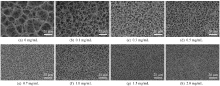
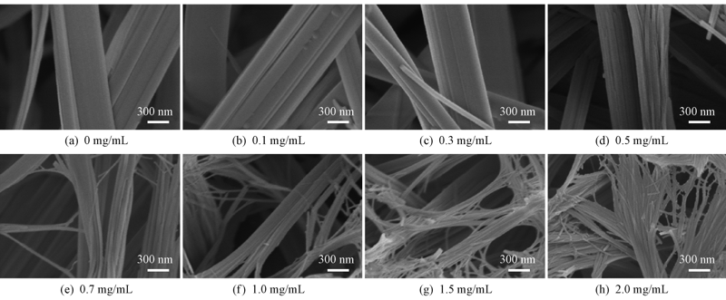
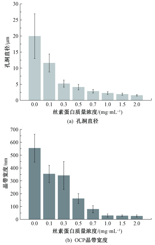
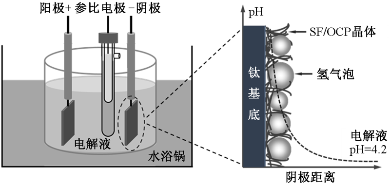
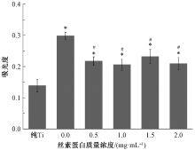
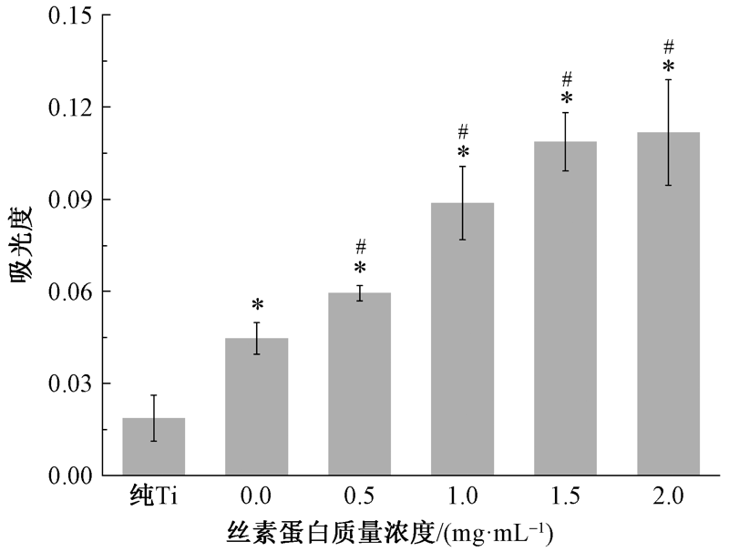
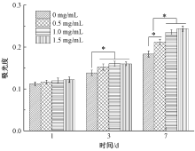
为进一步了解仿生材料结构与功能的关系,为体内骨整合设计一个有利的种植微环境,通过电化学沉积技术调控电解液中丝素蛋白(SF)的质量浓度,制备了具有纳微米多级结构的丝素蛋白/磷酸八钙(SF/OCP)复合涂层。研究了丝素蛋白质量浓度对SF/OCP复合涂层的表面形貌、力学性能、蛋白质吸附以及细胞增殖行为的影响。结果表明:随电解液中丝素蛋白质量浓度的增加,涂层表面孔洞直径由(19.96 ± 6.96) μm减小至(1.56 ± 0.22) μm, OCP晶体宽度减小至纳米级别((26.84 ± 8.2) nm);与纯OCP涂层相比,SF/OCP复合涂层(SF质量浓度为1.0 mg/mL)的弹性模量和硬度分别增加了约1.5和4.3倍;SF/OCP复合涂层选择性地增强了纤连蛋白(Fn)的吸附,经7 d的细胞培养后,SF/OCP复合涂层(SF质量浓度为1.0 mg/mL)表面的细胞活力是纯OCP涂层上的1.28倍。
中图分类号:
| [1] | SAI Y K, SHIWAKU Y K, ANADA T K, et al. Capacity of octacalcium phosphate to promote osteoblastic differentiation toward osteocytes in vitro[J]. Acta Biomaterialia, 2018,69:362-371. |
| [2] |
WANG H, LIU X Y, CHUAH Y J, et al. Design and engineering of silk fibroin scaffolds with biomimetic hierarchical structures[J]. Chemical Communications, 2013,49(14):1431-1433.
doi: 10.1039/c2cc38779d pmid: 23321676 |
| [3] | SHAO Z, VOLLRATH F. Materials: surprising strength of silkworm silk[J]. Nature, 2002,418(6899):741. |
| [4] | RICHERT L, VARIOLA F, ROSEI F, et al. Adsorption of proteins on nanoporous Ti surfaces[J]. Surface Science, 2010,604(17):1445-1451. |
| [5] | SUN M, DENG J, TANG Z, et al. A correlation study of protein adsorption and cell behaviors on substrates with different densities of PEG chains[J]. Colloids & Surfaces B: Biointerfaces, 2014,122(18):134-142. |
| [6] | CARTER D C, HE X M, MUNSON S H, et al. Three-dimensional structure of human serum albumin[J]. Science, 1989,244(4909):1195-1198. |
| [7] | DOCKAL M, CARTER D C, RUKER F. The three recombinant domains of human serum albumin[J]. Journal of Biological Chemistry, 1999,274:29303-29310. |
| [8] | WILSON C J, CLEGG R E, LEAVESLEY D I, et al. Mediation of biomaterial-cell interactions by adsorbed proteins: a review[J]. Tissue Engineering, 2005,11(2):1-18. |
| [9] |
ASURI P, BALE S S, KARAJANAGI S S, et al. The protein-nanomaterial interface[J]. Current Opinion in Biotechnology, 2006,17(6):562-568.
doi: 10.1016/j.copbio.2006.09.002 |
| [10] | MURUGAN R, RAMAKRISHNA S. Development of nanocomposites for bone grafting[J]. Composites Science & Technology, 2005,65(15):2385-2406. |
| [11] |
WEBSTER T J, ERGUN C, DOREMUS R H, et al. Enhanced functions of osteoblasts on nanophase ceramics[J]. Biomaterials, 2000,21(17):1803-1810.
pmid: 10905463 |
| [12] |
LIU H, WEBSTER T J. Nanomedicine for implants: a review of studies and necessary experimental tools[J]. Biomaterials, 2007,28(2):354-369.
doi: 10.1016/j.biomaterials.2006.08.049 pmid: 21898921 |
| [13] |
FELGUEIRAS H P, MURTHY N S, SOMMERFELD S D, et al. Competitive adsorption of plasma proteins using a quartz crystal microbalance[J]. ACS Applied Materials & Interfaces, 2016,8(21):13207-13217.
doi: 10.1021/acsami.5b12600 pmid: 27144779 |
| [14] | KANDORI K, HAMAZAKI H, MATSUZAWA M, et al. Selective adsorption of acidic protein of bovine serum albumin onto sheet-like calcium hydroxyapatite particles produced by microreactor[J]. Advanced Powder Technology, 2014,25(1):354-359. |
| [15] |
SELA M N, BADIHI L, ROSEN G, et al. Adsorption of human plasma proteins to modified titanium surfaces[J]. Clinical Oral Implants Research, 2010,18(5):630-638.
doi: 10.1111/j.1600-0501.2007.01373.x pmid: 17484735 |
| [16] |
GONZÁLEZ-GARCÍA C, SOUSA S R, MORATAL D, et al. Effect of nanoscale topography on fibronectin adsorption, focal adhesion size and matrix organi-zation[J]. Colloids & Surfaces B: Biointerfaces, 2010,77(2):181-190.
pmid: 20185279 |
| [17] |
KHANG D, KIM S Y, LIU-SNYDER P, et al. Enhanced fibronectin adsorption on carbon nanotube/poly(carbonate) urethane: independent role of surface nano-roughness and associated surface energy[J]. Biomaterials, 2007,28(32):4756-4768.
pmid: 17706277 |
| [1] | 宋广州, 涂芳芳, 丁梦瑶, 戴梦男, 殷音, 董凤林, 王建南. 丝素蛋白负电性增强改性及其对降钙素基因相关肽的加载能力[J]. 纺织学报, 2020, 41(12): 7-12. |
| [2] | 王宗乾, 杨海伟, 周剑, 李长龙. 尿素脱胶对丝素蛋白气凝胶力学性能的影响[J]. 纺织学报, 2020, 41(04): 9-14. |
| [3] | 孙广东, 黄益, 邵建中, FAN Qinguo. 光交联丝素蛋白水凝胶的蓝光引发体系[J]. 纺织学报, 2020, 41(04): 64-71. |
| [4] | 钟红荣, 方艳, 包红, 吴婷芳, 张小宁, 徐水, 朱勇. 丝素基双层敷料的制备及其性能[J]. 纺织学报, 2020, 41(02): 13-19. |
| [5] | 张治斌, 李刚, 毛森贤, 厉巽巽, 陈玉霜, 毛青山, 李翼, 潘志娟, 王晓沁. 丝素蛋白/壳聚糖微球制备及其抗菌性能[J]. 纺织学报, 2019, 40(10): 7-12. |
| [6] | 包红, 徐水, 张小宁, 成国涛, 朱勇. 家蚕丝素蛋白阳离子化及其对羊毛性状的影响[J]. 纺织学报, 2019, 40(07): 24-30. |
| [7] | 林永佳, 杨董超, 张佩华, 顾岩. 再生丝素蛋白/脱细胞真皮基质共混纳米纤维膜的制备及其性能[J]. 纺织学报, 2019, 40(07): 13-18. |
| [8] | 王宗乾, 王邓峰, 周杭, 李俊. 超声波辅助对乳化交联工艺制备丝素蛋白微球形貌的影响[J]. 纺织学报, 2019, 40(02): 119-124. |
| [9] | 周倩 袁久刚 李澜 王平 王强. 丝素蛋白的磷酸化及其仿生矿化膜的制备[J]. 纺织学报, 2018, 39(11): 8-13. |
| [10] | 王宗乾 杨海伟 汤立洋 李长龙. 丝素蛋白/聚乙烯醇复合膜的制备及其表征[J]. 纺织学报, 2018, 39(11): 14-19. |
| [11] | 姚勇波 颜志勇 李喆 易洪雷 张玉梅 王华平. 牵伸倍率分配对纤维素/丝素蛋白共混纤维形态结构的影响[J]. 纺织学报, 2018, 39(06): 13-18. |
| [12] | 李鹏飞 邓桦 马军 刘红斌 刘珍珠. 再生丝素蛋白溶液脱盐新工艺及其应用[J]. 纺织学报, 2018, 39(05): 20-24. |
| [13] | 王宗乾 杨海伟 王邓峰. 脱胶对蚕丝纤维的溶解及丝素蛋白性能的影响[J]. 纺织学报, 2018, 39(04): 69-76. |
| [14] | 吴惠英. 脱胶工艺对蚕丝溶解及再生丝素蛋白纤维性能的影响[J]. 纺织学报, 2017, 38(08): 75-80. |
| [15] | 周步光 张宇慧 王平 王强 范雪荣. 丝素/丙烯酰胺/丙烯酸吸水复合材料的紫外光辐照法制备[J]. 纺织学报, 2017, 38(05): 25-30. |
|
||
 京公网安备11010502044800号