纺织学报 ›› 2023, Vol. 44 ›› Issue (10): 205-213.doi: 10.13475/j.fzxb.20220607102
张子凡1, 李鹏飞1, 王建南1,2, 许建梅1,2( )
)
ZHANG Zifan1, LI Pengfei1, WANG Jiannan1,2, XU Jianmei1,2()
摘要:
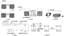
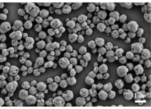
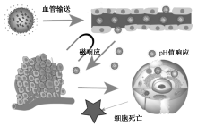
为深入探究丝素蛋白载药纳米粒的制备机制与方法,开发智能响应型丝素载药纳米粒,综述了丝素蛋白的微观结构与性能特点,概述了采用沉淀法、盐析法、电喷雾法、乳液法和脱溶剂法制备丝素蛋白纳米粒的机制与技术特点,分析了丝素蛋白载药方法与药物缓控释实现途径,并重点介绍了pH值响应和磁响应2种靶向释药的机制、方法及其应用。提出:可通过控制纳米粒大小、形状和表面电荷、与药物的化学键结合等方法来提高药物利用效率;通过改变丝素Silk Ⅰ和Silk Ⅱ结晶结构的含量来改变丝素的降解速度,或者通过丝素蛋白与其它不同降解速度的高聚物复合制备纳米粒从而实现药物的可控释放。此外,还可将多种智能响应结合来提高响应效率,最大程度减小药物的全身性毒副作用,实现靶向释药与精准医疗。
中图分类号:
| [1] | AVNESH K, SUDESH K, SUBHASH C. Biodegradable polymeric nanoparticles based drug delivery systems[J]. Colloids and Surfaces B(Biointerfaces), 2009, 75(1): 1-18. |
| [2] |
ZHAO Z, CHEN A, LI Y, et al. Fabrication of silk fibroin nanoparticles for controlled drug delivery[J]. Nanopart Res, 2012, 14(1) : 736-745.
doi: 10.1007/s11051-012-0736-5 |
| [3] | 陈智洋, 叶军, 王洪亮, 等. 基于丝素蛋白的纳米粒药物递送系统研究进展[J]. 药学学报, 2022, 57(6): 1792-1800. |
| CHEN Zhiyang, YE Jun, WANG Hongliang, et al. Research progress of silk fibroin-based nanoparticulate drug delivery systems[J]. Acta Pharmaceutica Sinica, 2022, 57(6): 1792-1800. | |
| [4] | JAHANSHAHI M, BABAEI Z. Protein nanoparticle: a unique system as drug delivery vehicles[J]. African Journal of Biotechnology, 2008, 7(25): 4926-4934. |
| [5] |
LI M, LU S, WU Z, et al. Study on porous silk fibroin materials: I: fine structure of freeze dried silk fib-roin[J]. Journal of Applied Polymer Science, 2015, 79(12): 2185-2191.
doi: 10.1002/(ISSN)1097-4628 |
| [6] |
MOTTAGHITALAB F, FAROKHI M, SHOKRGOZAR M A, et al. Silk fibroin nanoparticle as a novel drug delivery system[J]. Journal of Controlled Release, 2015, 206: 161-176.
doi: 10.1016/j.jconrel.2015.03.020 pmid: 25797561 |
| [7] |
ELAHI M, ALI S, TAHIR H M, et al. Sericin and fibroin nanoparticles-natural product for cancer therapy: a comprehensive review[J]. International Journal of Polymeric Materials, 2021, 70(4): 256-269.
doi: 10.1080/00914037.2019.1706515 |
| [8] |
KUNDU B, RAJKHOWA R, KUNDU S C, et al. Silk fibroin biomaterials for tissue regenerations[J]. Advanced Drug Delivery Reviews, 2013, 65(4): 457-470.
doi: 10.1016/j.addr.2012.09.043 pmid: 23137786 |
| [9] |
WENK E, MERKLE H P, MEINEL L. Silk fibroin as a vehicle for drug delivery applications[J]. Journal of Controlled Release, 2011, 150(2): 128-141.
doi: 10.1016/j.jconrel.2010.11.007 pmid: 21059377 |
| [10] |
SEIB F P, PRITCHARD E M, KAPLAN D L, et al. Self-assembling doxorubicin silk hydrogels for the focal treatment of primary breast cancer[J]. Advanced Functional Materials, 2013, 23(1): 58-65.
pmid: 23646041 |
| [11] | HU Y, QIN Z, YOU R, et al. The relationship between secondary structure and biodegradation behavior of silk fibroin scaffolds[J]. Advances in Materials Science & Engineering, 2013, 2012(6): 15-25. |
| [12] | MIN S, WANG Y, GAO X, et al. Release behavior of a composite of silk fibroin and nano-Ag and its biocompatibility[J]. Journal of Controlled Release, 2013, 172(1): 145-146. |
| [13] | 党婷婷, 陈爱政, 王士斌. 丝素蛋白微球作为药物缓释载体的研究进展[J]. 化工进展, 2012, 31(7): 1587-1591,1596. |
| DANG Tingting, CHEN Aizheng, WANG Shibin. Research progress of silk fibroin microsphere as sustained-release carrier of drug[J]. Chemical Industry and Engineering Progress, 2012, 31(7): 1587-1591,1596. | |
| [14] |
MERCEDES M, JEANNINE C, LOZANO-PÉREZ A, et al. Production of curcumin-loaded silk fibroin nanoparticles for cancer therapy[J]. Nanomaterials, 2018, 8(2): 126.
doi: 10.3390/nano8020126 |
| [15] |
WANG L, PATHAK J L, LIANG D, et al. Fabrication and characterization of strontium-hydroxyapatite/silk fibroin biocomposite nanospheres for bone-tissue engineering applications[J]. International Journal of Biological Macromolecules, 2020, 142: 366-375.
doi: S0141-8130(19)35789-7 pmid: 31593715 |
| [16] |
RADU I C. Silk fibroin nanoparticles reveal efficient delivery of 5-fu in a ht-29 colorectal adenocarcinoma model in vitro[J]. Farmacia, 2021, 69(1): 113-122.
doi: 10.31925/farmacia |
| [17] |
TIAN Y, JIANG X, CHEN X, et al. Doxorubicin-loaded magnetic silk fibroin nanoparticles for targeted therapy of multidrug-resistant cancer[J]. Advanced Materials, 2015, 26(43): 7393-7398.
doi: 10.1002/adma.v26.43 |
| [18] | SONG W, MUTHANA M, MUKHERJEE J, et al. Magnetic-silk core-shell nanoparticles as potential carriers for targeted delivery of curcumin into human breast cancer cells[J]. ACS Biomater, 2017, 3(6): 1027-1038. |
| [19] | TOTTEN J D, WONGPINYOCHIT T, CARROLA J, et al. PEGylation-dependent metabolic rewiring of macrophages with silk fibroin nanoparticles[J]. ACS Applied Materials & Interfaces, 2019, 11(16): 14515-14525. |
| [20] |
OLGA G, ELENI P, CHARALAMBOS S, et al. Silk fibroin nanoparticles for drug delivery: effect of bovine serum albumin and magnetic nanoparticles addition on drug encapsulation and release[J]. Separations, 2018, 5(2): 25-41.
doi: 10.3390/separations5020025 |
| [21] | JING Q, YU L, YU Y, et al. Silk fibroin nanoparticles prepared by electrospray as controlled release carriers of cisplatin[J]. Materials Science and Engineering: C, 2014, 44(1): 166-174. |
| [22] | NIU L, SHI M, FENG Y, et al. The interactions of quantum dot-labeled silk fibroin micro/nanoparticles with cells[J]. Materials, 2020(13): 15-32. |
| [23] |
CAO Y, LIU F, CHEN Y, et al. Drug release from core-shell PVA/silk fibroin nanoparticles fabricated by one-step electrospraying[J]. Scientific Reports, 2017, 7(1): 11913.
doi: 10.1038/s41598-017-12351-1 pmid: 28931908 |
| [24] |
WU P, LIU Q, LI R, et al. Facile preparation of paclitaxel loaded silk fibroin nanoparticles for enhanced antitumor efficacy by locoregional drug delivery[J]. Acs Appl Mater Interfaces, 2013, 5(23): 12638-12645.
doi: 10.1021/am403992b |
| [25] |
SEIB F P, JONES G T, RNJAK-KOVACINA J, et al. pH-dependent anticancer drug release from silk nanoparticles[J]. Advanced Healthcare Materials, 2013, 2(12): 1606-1611.
doi: 10.1002/adhm.201300034 pmid: 23625825 |
| [26] | ZHANG Y Q. Preparation of silk fibroin nanoparticles and enzyme-entrapped silk fibroin nanoparticles[J]. Bio-Protocol, 2018, 8(24): 3113. |
| [27] |
KUNDU J, CHUNG Y, KIM Y H, et al. Silk fibroin nanoparticles for cellular uptake and control release[J]. International Journal of Pharmaceutics, 2010, 388(1/2): 242-250.
doi: 10.1016/j.ijpharm.2009.12.052 |
| [28] |
MYUNG S J, KIM H S, KIM Y, et al. Fluorescent silk fibroin nanoparticles prepared using a reverse microemulsion[J]. Macromolecular Research, 2008, 16(7): 604-608.
doi: 10.1007/BF03218567 |
| [29] |
RAO J P, GECKELER K E. Polymer nanoparticles: preparation techniques and size-control parameters[J]. Progress in Polymer Science, 2011, 36(7): 887-913.
doi: 10.1016/j.progpolymsci.2011.01.001 |
| [30] |
ZHAO Z, LI Y, XIE M B. Silk fibroin-based nanoparticles for drug delivery[J]. International Journal of Molecular Sciences, 2015, 16(3): 4880-4903.
doi: 10.3390/ijms16034880 pmid: 25749470 |
| [31] |
BOCK N, WOODRUFF M A, HUTMACHER D W, et al. Electrospraying, a reproducible method for production of polymeric microspheres for biomedical applications[J]. Polymers, 2011, 3(1): 131-149.
doi: 10.3390/polym3010131 |
| [32] |
YANG Y Y, ZHANG M, LIU Z P, et al. Meletin sustained-release gliadin nanoparticles prepared via solvent surface modification on blending electro-spraying[J]. Applied Surface Science, 2018, 434(15): 1040-1047.
doi: 10.1016/j.apsusc.2017.11.024 |
| [33] |
KIM M K, LEE J Y, OH H J, et al. Effect of shear viscosity on the preparation of sphere-like silk fibroin microparticles by electrospraying[J]. International Journal of Biological Macromolecules, 2015, 79: 988-995.
doi: 10.1016/j.ijbiomac.2015.05.040 pmid: 26027609 |
| [34] |
STURESSON C, CARLFORS J. Incorporation of protein in PLG-microspheres with retention of bioactivity[J]. Journal of Controlled Release, 2000, 67(2/3): 171-178.
doi: 10.1016/S0168-3659(00)00205-4 |
| [35] |
BERNHARD G M, LEUENBERGER H, KISSEL T. Albumin nanospheres as carriers for passive drug targeting: an optimized manufacturing technique[J]. Pharmaceutical Research, 1996, 13(1): 32-37.
pmid: 8668675 |
| [36] |
SRISUWAN Y, SRIHANAM P, BAIMARK Y. Preparation of silk fibroin microspheres and its application to protein adsorption[J]. Journal of Macromolecular Science, 2009, 46(5): 521-525.
doi: 10.1080/00222340701257778 |
| [37] |
LANGER K, ANHORN M G, STEINHAUSER I, et al. Human serum albumin (HSA) nanoparticles: reproducibility of preparation process and kinetics of enzymatic degradation[J]. International Journal of Pharmaceutics, 2008, 347(1/2): 109-117.
doi: 10.1016/j.ijpharm.2007.06.028 |
| [38] |
SUN N, LEI R, XU J, et al. Fabricated porous silk fibroin particles for pH-responsive drug delivery and targeting of tumor cells[J]. Journal of Materials Science, 2019, 54(4): 3319-3330.
doi: 10.1007/s10853-018-3022-9 |
| [39] | CHEN M, SHAO Z, CHEN X. Paclitaxel-loaded silk fibroin nanospheres[J]. Journal of Biomedical Materials Research Part A, 2012. DOI: 10.13475/J.fzkb.10.1002. |
| [40] |
HONG S, CHOI DW, KIM HN, et al. Protein-based nanoparticles as drug delivery systems[J]. Pharmaceutics, 2020, 12(7): 604-633.
doi: 10.3390/pharmaceutics12070604 |
| [41] |
MA X, WANG Y, ZHAO T, et al. Ultra-pH-sensitive nanoprobe library with broad pH tunability and fluorescence emissions[J]. Journal of the American Chemical Society, 2014, 136: 11085-11092.
doi: 10.1021/ja5053158 pmid: 25020134 |
| [42] | 李祥子, 胡平静, 朱振铎, 等. pH响应型纳米药物载体的释药机制及性能研究进展[J]. 无机化学学报, 2018, 34(8): 1399-1412. |
| LI Xiangzi, HU Pingjing, ZHU Zhenduo, et al. Release mechanisms and properties of pH-responsive drug nanocarriers[J]. Chinese Journal of Inorganic Chemistry, 2018, 34(8): 1399-1412. | |
| [43] | 骆强. 荧光磁靶向载药纳米粒的组装及体外抗肿瘤活性研究[D]. 阿拉尔: 塔里木大学, 2021: 6-57. |
| LUO Qiang. Assembly and in vitro antitumor activity of fluorescent magnetic targeting drug loaded nano-particles[D]. Alare: Tarim University, 2021: 6-57. | |
| [44] | 陶灿. 磁性纳米载药体系的构建与抗肿瘤活性研究[D]. 广州: 广东药科大学, 2021: 1-10. |
| TAO Can. Construction of magnetic nanometer drug delivery system and research of antitumor activity[D]. Guangzhou: Guangdong Pharmaceutical University, 2021: 1-10. |
| [1] | 杨其亮, 杨海伟, 王邓峰, 李长龙, 张乐乐, 王宗乾. 超疏水弹性丝素蛋白纤维气凝胶的制备及其吸油性能[J]. 纺织学报, 2023, 44(09): 1-10. |
| [2] | 姚双双, 付少举, 张佩华, 孙秀丽. 再生丝素蛋白/聚乙烯醇共混取向纳米纤维膜的制备与性能[J]. 纺织学报, 2023, 44(09): 11-19. |
| [3] | 罗元泽, 戴梦男, 李蒙, 俞杨销, 王建南. 丝素蛋白基药物载体的应用研究进展[J]. 纺织学报, 2023, 44(09): 213-222. |
| [4] | 狄纯秋, 郭静, 管福成, 相玉龙, 单继成. 双金属离子交联海藻酸盐复合相变纤维的制备与性能[J]. 纺织学报, 2023, 44(05): 54-62. |
| [5] | 陈明星, 张威, 王新亚, 肖长发. 纳米纤维基催化材料的制备及其在环境领域中的应用研究进展[J]. 纺织学报, 2023, 44(01): 209-218. |
| [6] | 曹聪聪, 汤龙世, 刘元军, 赵晓明. 无机抗菌织物的研究进展[J]. 纺织学报, 2022, 43(11): 203-211. |
| [7] | 俞杨销, 李枫, 王煜煜, 王善龙, 王建南, 许建梅. 聚吡咯/丝素导电纳米纤维膜的制备及其性能[J]. 纺织学报, 2022, 43(10): 16-23. |
| [8] | 程宁波, 缪东洋, 王先锋, 王朝晖, 丁彬, 俞建勇. 用于个人热湿舒适管理的功能纺织品研究进展[J]. 纺织学报, 2022, 43(10): 200-208. |
| [9] | 刘蛟, 陈韶娟, 吴韶华. 丝素蛋白/聚左旋乳酸纳米纤维纱线肌腱补片的制备及其性能[J]. 纺织学报, 2022, 43(08): 60-66. |
| [10] | 李艾元, 施心雨, 岳万福, 游卫云. 丝素蛋白水凝胶支架的制备及其性能[J]. 纺织学报, 2022, 43(06): 44-48. |
| [11] | 顾张弘, 姚响, 王锦思, 张耀鹏. 具有细胞黏附反差特性的单层平行丝素蛋白纤维图案的制备及其性能[J]. 纺织学报, 2022, 43(05): 1-6. |
| [12] | 雷彩虹, 俞林双, 朱海霖, 郑涛, 陈建勇. 不同水解方式下蚕丝丝素蛋白材料的止血性能[J]. 纺织学报, 2022, 43(04): 15-19. |
| [13] | 张涛, 王富平, 陈国宝, 吴基玉, 庞亚妮, 陈忠敏. 壳聚糖基抑菌凝胶剂的制备及其性能[J]. 纺织学报, 2022, 43(03): 71-77. |
| [14] | 姚若彤, 赵婧媛, 闫一欣, 段立蓉, 王恬, 严佳, 张淑军, 李刚. 新型可降解编织结构神经再生导管的制备及其性能[J]. 纺织学报, 2022, 43(02): 125-131. |
| [15] | 姜雨淋, 王卉, 张克勤. 生物3D打印用丝素蛋白基凝胶墨水的研究进展[J]. 纺织学报, 2021, 42(11): 1-8. |
|
||
 京公网安备11010502044800号