纺织学报 ›› 2025, Vol. 46 ›› Issue (11): 43-51.doi: 10.13475/j.fzxb.20250204901
曾一洪, 付开秀, 王彦, 罗健, 陈国宝( )
)
ZENG Yihong, FU Kaixiu, WANG Yan, LUO Jian, CHEN Guobao()
摘要:
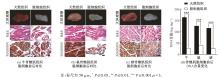
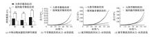
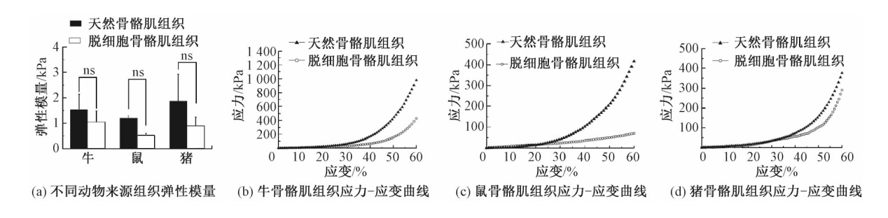
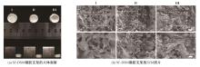
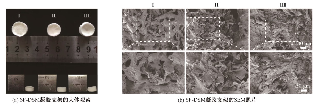
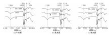
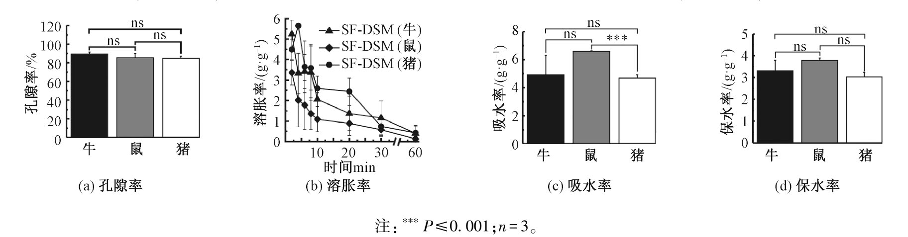
为探究不同物种来源脱细胞基质(dECM)的理化性质差异,以便在组织工程的应用中筛选适宜物种来源的dECM,将牛、鼠、猪不同动物来源的脱细胞骨骼肌(DSM)分别与丝素蛋白(SF)复合,制备SF-DSM支架。对支架的力学性能、亲水性能以及微观结构进行表征,分析不同物种来源的dECM对凝胶支架性能的影响。结果表明:不同动物来源的骨骼肌经过脱细胞处理后,其力学性能无显著性差异;牛、鼠、猪来源SF-DSM支架的弹性模量、亲水性能均无明显差异;红外光谱结果证实,SF和不同来源的DSM成功交联;不同物种来源的骨骼肌组织脱细胞前后的力学性能无明显差异,且物种间也无明显差异;SF-DSM支架的弹性模量、孔隙率、吸水性、保水性以及溶胀率均未因DSM来源不同而产生差异。
中图分类号:
| [1] |
EDOUARD P, REURINK G, MACKEY A L, et al. Traumatic muscle injury[J]. Nat Rev Dis Primers, 2023, 9(1): 56.
doi: 10.1038/s41572-023-00469-8 pmid: 37857686 |
| [2] |
TIDBALL J G. Mechanisms of muscle injury, repair, and regeneration[J]. Comprehensive Physiology, 2011, 1(4): 2029-2062.
doi: 10.1002/cphy.c100092 pmid: 23733696 |
| [3] |
ESTEVES DE LIMA J, BLAVET C, BONNIN M A, et al. Unexpected contribution of fibroblasts to muscle lineage as a mechanism for limb muscle patterning[J]. Nature Communications, 2021, 12(1): 3851.
doi: 10.1038/s41467-021-24157-x pmid: 34158501 |
| [4] | LUO W, ZHANG H L, WAN R W, et al. Biomaterials-based technologies in skeletal muscle tissue engineering[J]. Advanced Healthcare Materials, 2024, 13(18): e2304196. |
| [5] |
SAMANDARI M, QUINT J, RODRÍGUEZ-DELAROSA A, et al. Bioinks and bioprinting strategies for skeletal muscle tissue engineering[J]. Advanced Materials, 2022, 34(12): 2105883.
doi: 10.1002/adma.v34.12 |
| [6] | DURAN P, BOSCOLO SESILLO F, COOK M, et al. Proregenerative extracellular matrix hydrogel mitigates pathological alterations of pelvic skeletal muscles after birth injury[J]. Science Translational Medicine, 2023, 15(707): eabj3138. |
| [7] |
AGMON G, CHRISTMAN K L. Controlling stem cell behavior with decellularized extracellular matrix scaffolds[J]. Current Opinion in Solid State and Materials Science, 2016, 20(4): 193-201.
doi: 10.1016/j.cossms.2016.02.001 |
| [8] | NAKAMURA N, KIMURA T, KISHIDA A. Overview of the development, applications, and future perspectives of decellularized tissues and organs[J]. ACS Biomaterials Science & Engineering, 2017, 3(7): 1236-1244. |
| [9] |
ZHU L Y, YUHAN J Y, YU H, et al. Decellularized extracellular matrix for remodeling bioengineering organoid's microenvironment[J]. Small, 2023, 19(25): 2207752.
doi: 10.1002/smll.v19.25 |
| [10] |
GOLEBIOWSKA A A, INTRAVAIA J T, SATHE V M, et al. Decellularized extracellular matrix biomaterials for regenerative therapies: advances, challenges and clinical prospects[J]. Bioactive Materials, 2024, 32: 98-123.
doi: 10.1016/j.bioactmat.2023.09.017 pmid: 37927899 |
| [11] |
ZHONG S, LAN Y J, LIU J Y, et al. Advances focusing on the application of decellularization methods in tendon-bone healing[J]. Journal of Advanced Research, 2025, 67: 361-372.
doi: 10.1016/j.jare.2024.01.020 |
| [12] |
LIU H T, WANG Y Q, CUI K L, et al. Advances in hydrogels in organoids and organs-on-a-chip[J]. Advanced Materials, 2019, 31(50): 1902042.
doi: 10.1002/adma.v31.50 |
| [13] |
WŁODARCZYK-BIEGUN M K, DEL CAMPO A. 3D bioprinting of structural proteins[J]. Biomaterials, 2017, 134: 180-201.
doi: 10.1016/j.biomaterials.2017.04.019 |
| [14] |
LEAL-EGAÑA A, SCHEIBEL T. Silk-based materials for biomedical applications[J]. Biotechnology and Applied Biochemistry, 2010, 55(3): 155-167.
doi: 10.1042/BA20090229 |
| [15] |
ZHENG H Y, ZUO B Q. Functional silk fibroin hydrogels: preparation, properties and applications[J]. Journal of Materials Chemistry B, 2021, 9(5): 1238-1258.
doi: 10.1039/d0tb02099k pmid: 33406183 |
| [16] |
GRABSKA-ZIELIŃSKA S, SIONKOWSKA A. How to improve physico-chemical properties of silk fibroin materials for biomedical applications: blending and cross-linking of silk fibroin: a review[J]. Materials, 2021, 14(6): 1510.
doi: 10.3390/ma14061510 |
| [17] |
ORAL C B, YETISKIN B, OKAY O. Stretchable silk fibroin hydrogels[J]. International Journal of Biological Macromolecules, 2020, 161: 1371-1380.
doi: S0141-8130(20)34127-1 pmid: 32791264 |
| [18] |
LI X Y, YE M J, GAO Y E, et al. The systematic evaluation of physicochemical and biological properties in vitro and in vivo for natural silk fibroin nano-particles[J]. Advanced Fiber Materials, 2022, 4(5): 1141-1152.
doi: 10.1007/s42765-022-00167-2 |
| [19] |
HE S J, FU X J, WANG L, et al. Self-assemble silk fibroin microcapsules for cartilage regeneration through gene delivery and immune regulation[J]. Small, 2023, 19(40): 2302799.
doi: 10.1002/smll.v19.40 |
| [20] |
GILBERT-HONICK J, GRAYSON W. Vascularized and innervated skeletal muscle tissue engineering[J]. Advanced Healthcare Materials, 2020, 9(1): 1900626.
doi: 10.1002/adhm.v9.1 |
| [21] |
NAIR M, JOHAL R K, HAMAIA S W, et al. Tunable bioactivity and mechanics of collagen-based tissue engineering constructs: a comparison of EDC-NHS, genipin and TG2 crosslinkers[J]. Biomaterials, 2020, 254: 120109.
doi: 10.1016/j.biomaterials.2020.120109 |
| [22] |
CRAPO P M, GILBERT T W, BADYLAK S F. An overview of tissue and whole organ decellularization processes[J]. Biomaterials, 2011, 32(12): 3233-3243.
doi: 10.1016/j.biomaterials.2011.01.057 pmid: 21296410 |
| [23] |
ROTH S P, GLAUCHE S M, PLENGE A, et al. Automated freeze-thaw cycles for decellularization of tendon tissue-a pilot study[J]. BMC Biotechnology, 2017, 17(1): 13.
doi: 10.1186/s12896-017-0329-6 |
| [24] |
MENDIBIL U, RUIZ-HERNANDEZ R, RETEGI-CARRION S, et al. Tissue-specific decellularization methods: rationale and strategies to achieve regenerative compounds[J]. International Journal of Molecular Sciences, 2020, 21(15): 5447.
doi: 10.3390/ijms21155447 |
| [25] |
NEISHABOURI A, SOLTANI KHABOUSHAN A, DAGHIGH F, et al. Decellularization in tissue engineering and regenerative medicine: evaluation, modification, and application methods[J]. Front Bioeng Biotechnol, 2022, 10: 805299.
doi: 10.3389/fbioe.2022.805299 |
| [26] |
GARCIA H, BARROS A S, GONÇALVES C, et al. Characterization of dextrin hydrogels by FT-IR spectroscopy and solid state NMR spectroscopy[J]. European Polymer Journal, 2008, 44(7): 2318-2329.
doi: 10.1016/j.eurpolymj.2008.05.013 |
| [27] |
TAO X S, JIANG F J, CHENG K, et al. Synthesis of pH and glucose responsive silk fibroin hydrogels[J]. International Journal of Molecular Sciences, 2021, 22(13): 7107.
doi: 10.3390/ijms22137107 |
| [28] |
CONZATTI G, FAUCON D, CASTEL M, et al. Alginate/chitosan polyelectrolyte complexes: a comparative study of the influence of the drying step on physicochemical properties[J]. Carbohydrate Polymers, 2017, 172: 142-151.
doi: S0144-8617(17)30530-1 pmid: 28606520 |
| [29] |
SUNTIVICH R, DRACHUK I, CALABRESE R, et al. Inkjet printing of silk nest arrays for cell hosting[J]. Biomacromolecules, 2014, 15(4): 1428-1435.
doi: 10.1021/bm500027c pmid: 24605757 |
| [1] | 曾姚, 吕金凤, 王介平, 刘荣鹏, 周婵. 三维蚕丝蛋白支架的应用研究进展[J]. 纺织学报, 2025, 46(09): 258-267. |
| [2] | 高闻语, 陈诚, 奚晓玮, 邓林红, 刘杨. 改性丝素蛋白纤维增强胶原基角膜修复材料的制备及其性能[J]. 纺织学报, 2025, 46(08): 1-9. |
| [3] | 江淑宁, 杨海伟, 李长龙, 郑天亮, 王宗乾. 低共熔溶剂剥离法制备丝素蛋白纳米原纤及其成膜性能[J]. 纺织学报, 2025, 46(07): 1-9. |
| [4] | 于梦菲, 高文丽, 任婧, 曹雷涛, 彭若铉, 凌盛杰. 摩擦纳米发电机用皮芯结构纤维的制备及其性能[J]. 纺织学报, 2025, 46(05): 1-9. |
| [5] | 罗欣, 王磊, 王筱悠, 伍韬, 张贞贞, 张一帆. 丝素蛋白多级结构的自组装机制及其重构材料研究进展[J]. 纺织学报, 2025, 46(03): 225-235. |
| [6] | 詹克静, 杨鑫, 张应龙, 张昕, 潘志娟. 自凝聚丝素蛋白微纳米纤维膜的制备及其力学增强[J]. 纺织学报, 2025, 46(02): 10-19. |
| [7] | 杨柳, 杜磊, 徐淮中. 熔体近场直写制备组织工程支架的研究进展[J]. 纺织学报, 2025, 46(01): 206-216. |
| [8] | 杨鑫, 张昕, 潘志娟. 丝素纳米原纤增强再生丝素蛋白/聚乙烯醇纤维的结构与性能[J]. 纺织学报, 2024, 45(11): 1-9. |
| [9] | 李蒙, 戴梦男, 俞杨销, 王建南. 丝素蛋白基骨修复材料的应用研究进展[J]. 纺织学报, 2024, 45(10): 224-231. |
| [10] | 王勃翔, 徐航丹, 李佳, 林杰, 程德红, 路艳华. 柞蚕丝素纳米纤维温敏复合膜制备及其生物相容性[J]. 纺织学报, 2024, 45(09): 18-25. |
| [11] | 雷彩虹, 俞林双, 金万慧, 朱海霖, 陈建勇. 丝素蛋白/壳聚糖复合纤维膜的制备与应用[J]. 纺织学报, 2023, 44(11): 19-26. |
| [12] | 张子凡, 李鹏飞, 王建南, 许建梅. 丝素蛋白载药纳米粒的研究进展[J]. 纺织学报, 2023, 44(10): 205-213. |
| [13] | 杨其亮, 杨海伟, 王邓峰, 李长龙, 张乐乐, 王宗乾. 超疏水弹性丝素蛋白纤维气凝胶的制备及其吸油性能[J]. 纺织学报, 2023, 44(09): 1-10. |
| [14] | 姚双双, 付少举, 张佩华, 孙秀丽. 再生丝素蛋白/聚乙烯醇共混取向纳米纤维膜的制备与性能[J]. 纺织学报, 2023, 44(09): 11-19. |
| [15] | 罗元泽, 戴梦男, 李蒙, 俞杨销, 王建南. 丝素蛋白基药物载体的应用研究进展[J]. 纺织学报, 2023, 44(09): 213-222. |
|
||
 京公网安备11010502044800号