纺织学报 ›› 2026, Vol. 47 ›› Issue (04): 43-51.doi: 10.13475/j.fzxb.20250705701
杨鸿杰1, 徐丽亚2, 汪蔚1( )
)
YANG Hongjie1, XU Liya2, WANG Wei1()
摘要:
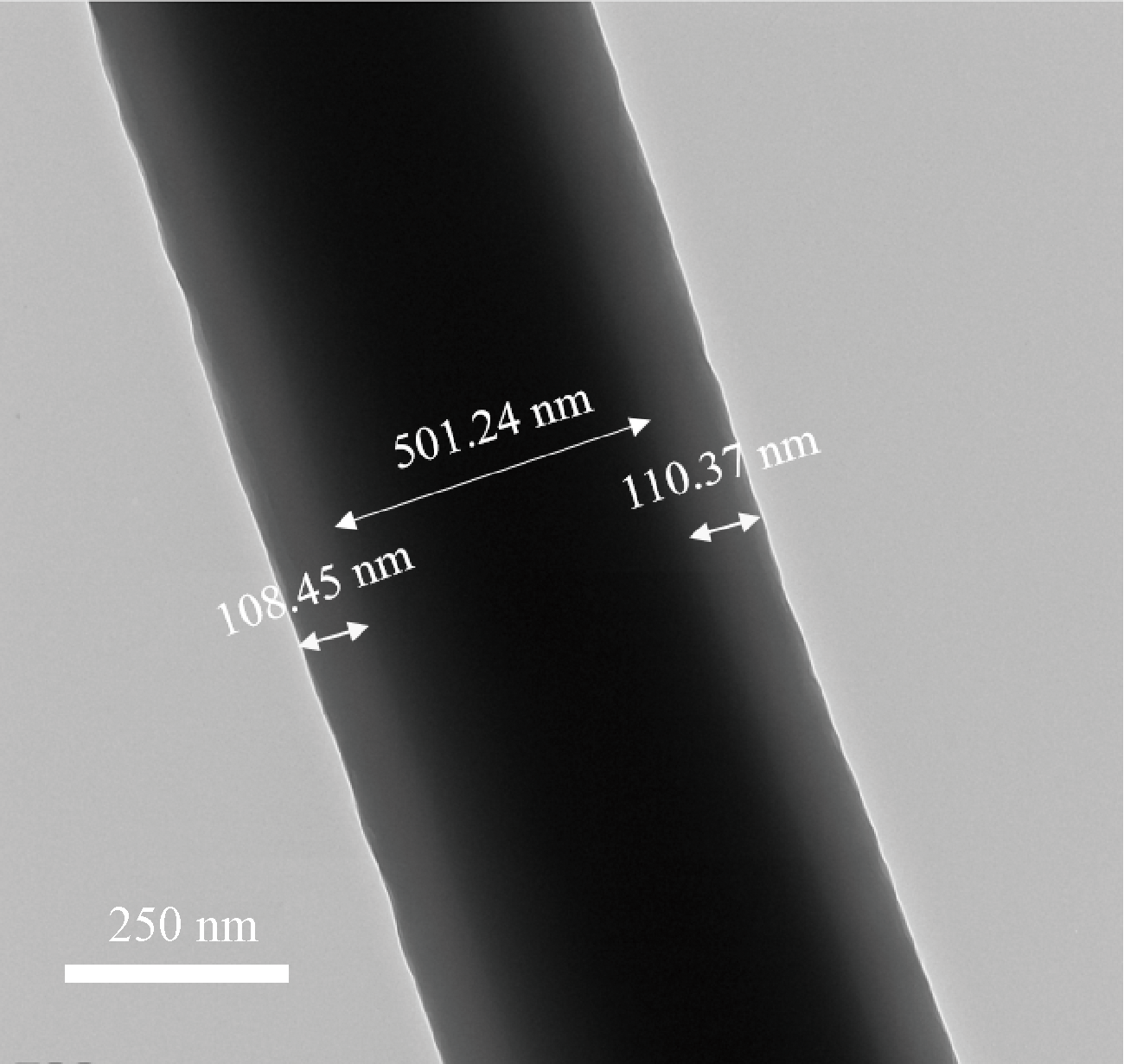
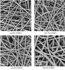
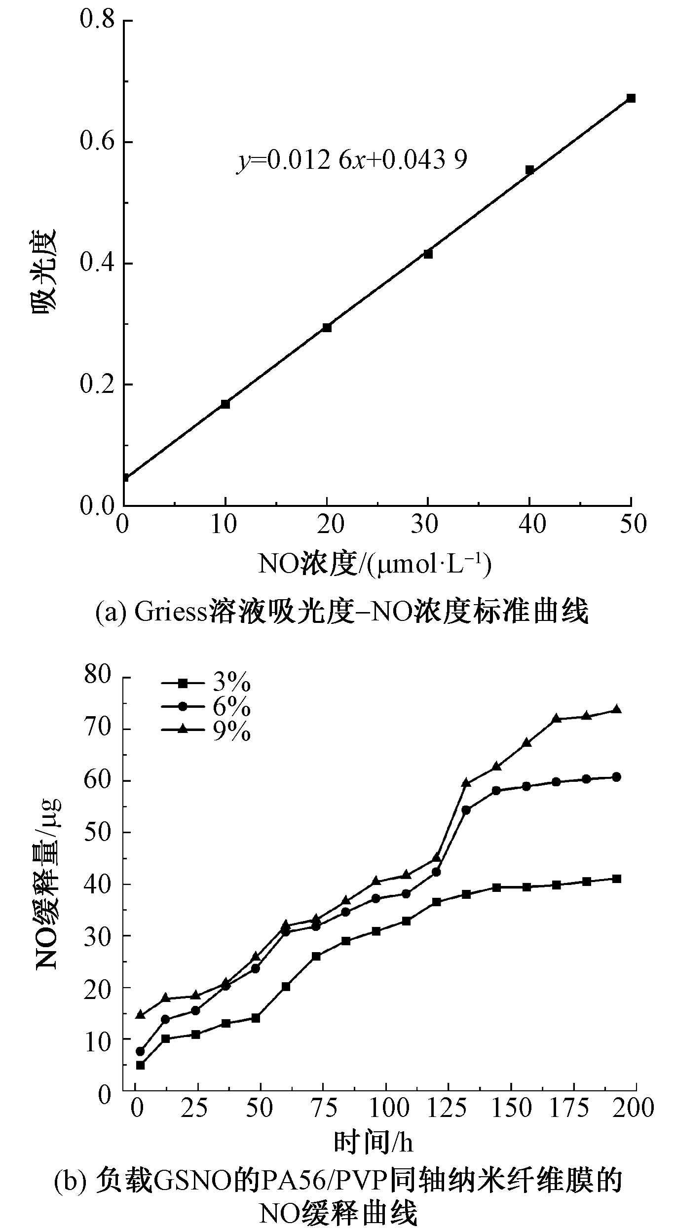
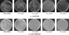
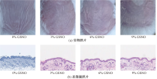
为拓展生物基聚己二酸戊二胺(PA56)材料的功能性和应用领域,以S-亚硝基谷胱甘肽(GSNO)和聚乙烯吡咯烷酮(PVP)共混物为芯,以PA56为壳,采用同轴静电纺丝法制备了纳米纤维膜。通过扫描电子显微镜与透射电子显微镜对纳米纤维膜的同轴结构特征及表面形貌状态进行表征。对GSNO的最小抑菌浓度(MIC)以及负载GSNO的PA56/PVP同轴纳米纤维膜的一氧化氮缓释性能、力学性能、亲水性能、抗菌性能及皮肤刺激性进行测试分析。结果表明,GSNO的MIC值在10.51~21.02 μg/L之间。负载GSNO的PA56/PVP同轴纳米纤维表观形貌规整,具有核-壳结构,但存在少量壳层PA56纤维剥离现象,该现象随着GSNO质量分数的增加而减弱。随着GSNO质量分数的增加,同轴纳米纤维的直径及膜拉伸强度增加,亲水性下降,抗菌性能提高。当载入GSNO质量分数为6%时,同轴纳米纤维膜对金黄色葡萄球菌和大肠埃希菌的抑菌率可达92.18%和84.54%。皮肤刺激性实验表明,负载GSNO的PA56/PVP同轴纳米纤维膜具有优良的皮肤耐受性,可用于功能性敷料。
中图分类号:
| [1] | 张龙贵, 计文希, 李娟. 生物基聚酰胺制备及应用研究进展[J]. 合成树脂及塑料, 2017, 34(2): 86-91, 97. |
| ZHANG Longgui, JI Wenxi, LI Juan. Preparation and application of bio-based PA[J]. China Synthetic Resin and Plastics, 2017, 34(2): 86-91, 97. | |
| [2] | 张腾飞, 石禄丹, 胡红梅, 等. 生物基聚酰胺56低聚物改性聚酯的合成及其表征[J]. 纺织学报, 2019, 40(6): 1-7. |
|
ZHANG Tengfei, SHI Ludan, HU Hongmei, et al. Synthesis and characterization of bio-based polyamide 56 oligomer modified polyester[J]. Journal of Textile Research, 2019, 40(6): 1-7.
doi: 10.1177/004051757004000101 |
|
| [3] | 孙朝续, 刘修才. 生物基聚酰胺56纤维在纺织领域的应用研究进展[J]. 纺织学报, 2021, 42(4): 26-32. |
| SUN Chaoxu, LIU Xiucai. Research progress on applications of bio-based polyamide 56 fibers in textile fields[J]. Journal of Textile Research, 2021, 42(4): 26-32. | |
| [4] |
PERISTERI E, PITSIKAS N. Effects of low doses of different nitric oxide (NO) donors in rat models of obsessive-compulsive disorder (OCD) and posttraumatic stress disorder (PTSD)[J]. Nitric Oxide, 2022, 129: 1-7.
doi: 10.1016/j.niox.2022.09.001 pmid: 36084795 |
| [5] |
HUANG Z J, FU J J, ZHANG Y H. Nitric oxide donor-based cancer therapy: advances and prospects[J]. Journal of Medicinal Chemistry, 2017, 60(18): 7617-7635.
doi: 10.1021/acs.jmedchem.6b01672 pmid: 28505442 |
| [6] |
JIN Y L, LIU S H, WANG X Y, et al. Multifunctional microneedle patches loaded with engineered nitric oxide-releasing nanocarriers for targeted and synergistic chronic wound therapy[J]. Advanced Materials, 2025, 37(5): 2413108.
doi: 10.1002/adma.v37.5 |
| [7] |
PURVIS M E, PICCIOTTI S L, LYNCH A L, et al. Nitric oxide-releasing, hyaluronic acid-coated electrospun fibers as dual-action wound dressings[J]. ACS Applied Bio Materials, 2025, 8(9): 7771-7782.
doi: 10.1021/acsabm.5c00754 pmid: 40794539 |
| [8] | 姜宇, 李妍妍. 一氧化氮供体在抗肿瘤应用中的研究进展[J]. 药学进展, 2024, 48(4): 309-320. |
| JIANG Yu, LI Yanyan. Research progress of nitric oxide donor in antitumor application[J]. Progress in Pharmaceutical Sciences, 2024, 48(4): 309-320. | |
| [9] | 韩林妗. 一氧化氮供体化合物的最新合成方法和研究进展[J]. 科技创新导报, 2020, 17(21): 104-106, 109. |
| HAN Linjin. Recent synthesis methods and research progress of nitric oxide donor compounds[J]. Science and Technology Innovation Herald, 2020, 17(21): 104-106, 109. | |
| [10] |
KUMARI S, SAMMUT I A, GILES G I. The design of nitric oxide donor drugs: s-nitrosothiol tDodSNO is a superior photoactivated donor in comparison to GSNO and SNAP[J]. European Journal of Pharmacology, 2014, 737: 168-176.
doi: 10.1016/j.ejphar.2014.05.012 pmid: 24858367 |
| [11] | 黄连香, 王祥荣, 侯学妮, 等. 茜草色素对生物基聚酰胺56的染色性能[J]. 纺织学报, 2024, 45(7): 94-103. |
| HUANG Lianxiang, WANG Xiangrong, HOU Xueni, et al. Dyeing properties of madder pigment on bio-based polyamide 56[J]. Journal of Textile Research, 2024, 45(7): 94-103. | |
| [12] | 周传兵. S-亚硝基硫醇的合成及生物活性研究[D]. 武汉: 华中科技大学, 2009: 16-20. |
| ZHOU Chuanbing. Synthesis and bioactivities of S-nitrosothiols[D]. Wuhan: Huazhong University of Science and Technology, 2009: 16-20. | |
| [13] |
YAMAKAWA T, MITSUYAMA J, HAYASHI K. In vitro and in vivo antibacterial activity of T-3912, a novel non-fluorinated topical quinolone[J]. The Journal of Antimicrobial Chemotherapy, 2002, 49(3): 455-465.
doi: 10.1093/jac/49.3.455 |
| [14] | 张正斌, 任春艳, 刘春颖, 等. 对一氧化氮作用的新认识及其检测方法[J]. 青岛海洋大学学报(自然科学版), 2003(3): 425-432. |
| ZHANG Zhengbin, REN Chunyan, LIU Chunying, et al. New recognition of the role of nitric oxide and its detection methods[J]. Journal of Ocean University of Qingdao, 2003(3): 425-432. | |
| [15] | 徐丽亚, 汪瑱, 杨鸿杰, 等. 氧化锌-银/生物基聚酰胺56纳米纤维膜的制备及其抗菌性能[J]. 纺织学报, 2025, 46(7):37-45. |
| XU Liya, WANG Zhen, YANG Hongjie, et al. Preparationand antibacterial property of zinc oxide-silver/bio-based polyamide 56 composite nanofiber membranes[J]. Journal of Textile Research, 2025, 46(7):37-45. | |
| [16] |
DAYARAM Y K, TALAUE M T, CONNELL N D, et al. Characterization of a glutathione metabolic mutant of Mycobacterium tuberculosis and its resistance to glutathione and nitrosoglutathione[J]. Journal of Bacteriology, 2006, 188(4): 1364-1372.
pmid: 16452418 |
| [17] | 苏梦茹, 马培培, 李鑫鑫, 等. 9种抗菌药物对大肠埃希菌最小抑菌浓度的测定[J]. 动物医学进展, 2020, 41(3): 52-56. |
| SU Mengru, MA Peipei, LI Xinxin, et al. Determination of minimum inhibitory concentration of nine antibacterial drugs to escherichia coli[J]. Progress in Veterinary Medicine, 2020, 41(3): 52-56. | |
| [18] |
DAN W J, GAO J X, ZHANG J Y, et al. Antibacterial activity and mechanism of action of canthin-6-one against Staphylococcus aureus and its application on beef preservation[J]. Food Control, 2023, 147: 109604.
doi: 10.1016/j.foodcont.2023.109604 |
| [19] |
YU J H, FRIDRIKH S V, RUTLEDGE G C. Production of submicrometer diameter fibers by two-fluid electrospinning[J]. Advanced Materials, 2004, 16(17): 1562-1566.
doi: 10.1002/adma.v16:17 |
| [20] |
李思程, 张岑, 谌迪, 等. 同轴静电纺丝多级结构纳米纤维的研究进展及其在食品领域的应用现状[J]. 食品科学, 2024, 45(13): 300-311.
doi: 10.7506/spkx1002-6630-20230531-285 |
|
LI Sicheng, ZHANG Cen, CHEN Di, et al. Research progress in coaxial electrospinning for the preparation of multi-structure nanofibers and its application in the food industry[J]. Food Science, 2024, 45(13): 300-311.
doi: 10.7506/spkx1002-6630-20230531-285 |
| [1] | 王琎祺, 翟倩, 俞森龙, 朱倩沁, 周哲, 相恒学, 朱美芳. 聚乙烯微细纤维医疗防护应用研究进展[J]. 纺织学报, 2026, 47(03): 156-165. |
| [2] | 邵英海, 朴洪伟, 曹继鹏, 张月, 许兰杰, 于学智, 张明光. 天然彩棉/柞蚕短纤维混纺纱的制备及其抗菌性能[J]. 纺织学报, 2026, 47(03): 240-246. |
| [3] | 刘鹏碧, 任经岗, 张宽祥, 曹东阳, 刘熙, 郭昌盛. 植酸/苯扎氯铵一步共沉积涂层聚丙烯补片的制备及其抗菌性能[J]. 纺织学报, 2026, 47(03): 77-86. |
| [4] | 魏建斐, 魏艳颖, 马超慧, 胡晓鹏, 邴琳涵, 樊瑜, 林彬泽, 董振峰, 朱志国, 王锐. 碳点的宏量制备及其对聚酰胺66阻燃与力学性能协同增强机制[J]. 纺织学报, 2026, 47(02): 37-46. |
| [5] | 王彬, 侯泽明, 徐英俊, 王玉忠. 高阻燃性再生纤维素纤维的制备及其性能[J]. 纺织学报, 2026, 47(02): 47-55. |
| [6] | 王世杰, 孙辉, 于斌. 聚乙烯醇/牡丹皮提取物复合纳米静电纺丝膜的制备及其抗菌性能[J]. 纺织学报, 2026, 47(02): 56-64. |
| [7] | 顾家玉, 张炜栋, 董永春, 孙璇, 徐良军. 银杏叶黄酮对羊毛和蚕丝织物的抗菌整理[J]. 纺织学报, 2026, 47(01): 142-150. |
| [8] | 宋佳怡, 王政驿, 程献伟, 关晋平, 朱亚伟. 液态靛蓝染料的制备及其对棉织物的染色性能[J]. 纺织学报, 2025, 46(12): 133-141. |
| [9] | 候志文, 任泽苹, 王晓宁, 张天骄. 棉织物的壳聚糖/海藻酸盐抗菌阻燃整理及其性能[J]. 纺织学报, 2025, 46(12): 171-180. |
| [10] | 舒祖菊, 袁自钰, 周斐, 黄秀文, 王权, 房显龙, 曹美雪. 载姜黄素核壳结构纳米纤维膜的制备及其缓释性能[J]. 纺织学报, 2025, 46(11): 26-33. |
| [11] | 朱雷, 李晓俊, 程春祖, 徐纪刚, 杜心宇. 四硼酸钠/单宁酸交联对海藻酸钙纤维结构与性能的影响[J]. 纺织学报, 2025, 46(07): 28-36. |
| [12] | 徐丽亚, 汪瑱, 杨鸿杰, 汪蔚. 氧化锌-银/生物基聚酰胺56纳米纤维膜的制备及其抗菌性能[J]. 纺织学报, 2025, 46(07): 37-45. |
| [13] | 张泽祺, 周涛, 周文琪, 范中尧, 杨佳蕾, 陈国印, 潘绍武, 朱美芳. 生理电信号监测用导电纤维及其研究进展[J]. 纺织学报, 2025, 46(05): 70-76. |
| [14] | 郭洁琰, 徐颖雯, 丁放, 任学宏. 卤胺抗菌剂及其改性纤维的应用研究进展[J]. 纺织学报, 2025, 46(04): 255-263. |
| [15] | 欧宗权, 于金超, 潘志娟. 光致变色聚乳酸/聚3-羟基丁酸酯共混纤维的纺制及其结构与性能[J]. 纺织学报, 2024, 45(12): 9-17. |
|
||
 京公网安备11010502044800号