纺织学报 ›› 2025, Vol. 46 ›› Issue (07): 37-45.doi: 10.13475/j.fzxb.20240803101

• 纤维材料 • 上一篇    下一篇

氧化锌-银/生物基聚酰胺56纳米纤维膜的制备及其抗菌性能

徐丽亚1,2, 汪瑱3, 杨鸿杰1, 汪蔚1()   

  1. 1 嘉兴大学 材料与纺织工程学院, 浙江 嘉兴 314001
    2 浙江台华新材料股份有限公司, 浙江 嘉兴 314011
    3 嘉兴大学附属医院 嘉兴市第一医院, 浙江 嘉兴 314001
  • 收稿日期:2024-08-19 修回日期:2025-04-05 出版日期:2025-07-15 发布日期:2025-08-14
  • 通讯作者: 汪蔚(1971—),男,教授,博士。研究方向为功能性纤维及纺织品、聚合物基复合材料。E-mail:zjxuwangwei@163.com
  • 作者简介:徐丽亚(1976—),女,工程师,学士。主要研究方向为功能性纤维及纺织品。
  • 基金资助:
    浙江省“尖兵”“领雁”研发攻关计划项目(2023C01201);浙江省自然科学基金项目(LQ24H300002);嘉兴市科技计划项目(2023AY11012)

Preparation and antibacterial property of zinc oxide-silver/bio-based polyamide 56 composite nanofiber membranes

XU Liya1,2, WANG Zhen3, YANG Hongjie1, WANG Wei1()   

  1. 1 College of Materials and Textile Engineering, Jiaxing University, Jiaxing, Zhejiang 314001, China
    2 Zhejiang Taihua New Materials Co., Ltd., Jiaxing, Zhejiang 314011, China
    3 The First Hospital of Jiaxing, Affiliated Hospital of Jiaxing University, Jiaxing, Zhejiang 314001, China
  • Received:2024-08-19 Revised:2025-04-05 Published:2025-07-15 Online:2025-08-14

摘要:

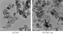
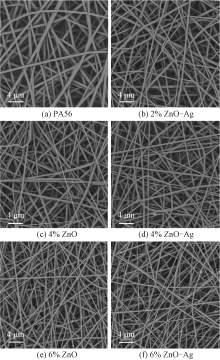
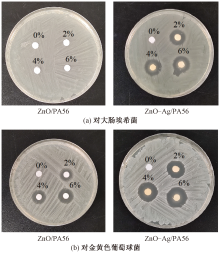
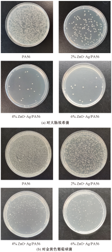
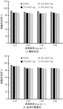
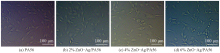
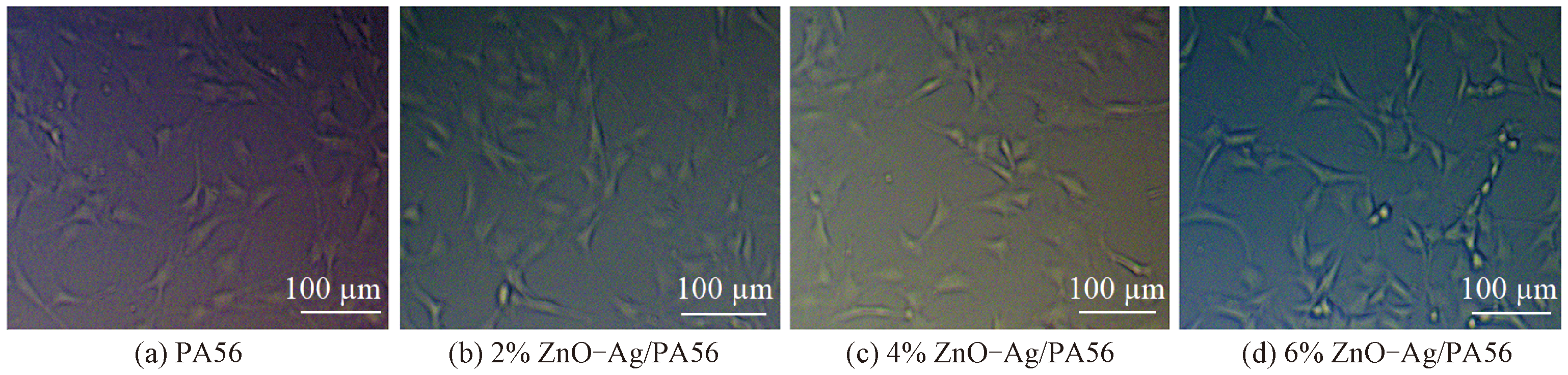
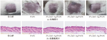
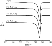
为开发医疗卫生用生物基聚酰胺56(PA56)超细纤维及制品,将纳米氧化锌-银(ZnO-Ag)复合抗菌剂与PA56熔融共混,再经静电纺丝制得ZnO-Ag/PA56纳米纤维膜。采用透射电子显微镜和扫描电子显微镜对ZnO-Ag和ZnO-Ag/PA56复合纳米纤维膜的形貌结构进行表征,并对ZnO-Ag/PA56复合纳米纤维膜的抗菌性能、生物相容性、结晶性能、力学性能及亲水性能进行测试分析。结果表明:ZnO-Ag/PA56复合纳米纤维形态结构圆整,表面ZnO-Ag颗粒分布均匀,无明显团聚现象;随ZnO-Ag质量分数的增加,ZnO-Ag/PA56复合纳米纤维膜的结晶度、拉伸强度及亲水性提高,纤维平均直径减小;当ZnO-Ag质量分数为6%时,ZnO-Ag/PA56复合纳米纤维膜对金黄色葡萄球菌和大肠埃希菌的抑菌率分别达到98.7%和89.3%,且具有良好的生物相容性,有望应用于医疗卫生领域。

关键词: 生物基纤维, 功能纤维, 生物基聚酰胺, 静电纺丝, 纳米纤维, 纳米氧化锌-银, 抗菌性能

Abstract:

Objective Bio-based polypentanediamine adipate (PA56), entirely or partially synthesized from biomass feedstocks, has emerged as environmentally friendly alternative to petroleum-based counterparts and has gained considerable at traction in recent years. PA56 holds significant potential in textiles, food packaging and other fields, by virtue of its good strength and toughness with excellent fatigue resistance. Despite its promising prospects, limited studies have been given to PA56 nanofibers for biomedical applications. Hence, nano ZnO-Ag compound antibacterial agent was prepared and blended with PA56 matrix. ZnO-Ag/PA56 composite nanofiber membranes were then fabricated by electrospinning process for antibacterial applications.

Method The preparation of ZnO-Ag nano particles includes mixing a complex of silver acetate and ammonium hydroxide with nano ZnO dispersions, followed by the addition of formic acid as a reducing agent. The prepared ZnO-Ag particles were blended with PA56 matrix by melt extrusion and ZnO-Ag/PA56 composite nanofiber membranes were then fabricated by electrospinning technology. Morphologies of the ZnO-Ag particles and ZnO-Ag/PA56 nanofiber membranes were characterized with transmission electron microscopy (TEM) and scanning electron microscopy (SEM). The influences of encapsulated ZnO-Ag particles on the antibacterial activity, crystallinity, mechanical and wettability properties of the nanofiber membranes were investigated. Furthermore, the cytotoxicity and skin stimulation of the ZnO-Ag/PA56 nanofiber membranes were analyzed.

Results The average sizes of original ZnO particles are about 20-30 nm and the Ag particles embedded in the prepared ZnO-Ag compound are spherical in shape with an average diameter of 6-10 nm, which have been reported to show higher antimicrobial efficacy. Blank PA56 nanofibers have uniform morphology and smooth surface. ZnO-Ag/PA56 nanofibers have round morphology and ZnO-Ag particels distribute in the nanofibers uniformly without any obvious gathering. The average diameter of ZnO-Ag/PA56 nanofibers decreases with the increase of ZnO-Ag content. This is because that ZnO is a semiconductor and Ag is a conductor, which can increase the charge density of the spinning solution and give a strong elongation force to the ejected polymeric jet. When ZnO-Ag mass fraction is 6%, the average diameter of ZnO-Ag/PA56 nanofiber is 401.18 nm, 54.9% of the average diameter of PA56 nanofiber (731.40 nm). The results of inhibition zone test show that blank PA56 nanofiber membranes do not offer any antibacterial ability. ZnO/PA56 nanofiber membranes have antibacterial activity against Staphylococcus aureus, while have not antibacterial ability against Escherichia coli. ZnO-Ag/PA56 nanofiber membranes exhibit good antibacterial effects against Staphylococcus aureus and Escherichia coli. In order to quantify the antibacterial properties of ZnO-Ag/PA56 nanofibers, a standard plate counting approach that tracked bacterial proliferation was performed. After incubation with the bacterial suspension, Staphylococcus aureus and Escherichia coli in ZnO-Ag/PA56 nanofiber groups show lower viability than those in the PA56 groups, where bacterial colonies proliferate extensively. A sharp decrease in colony formation occurres at a mass fraction of 2% ZnO-Ag and the nanofiber membranes exhibit higher inhibition rate against Staphylococcus aureus (73.5%) than Escherichia coli (51.5%). Increasing the content of ZnO-Ag in the nanofibers significantly enhances their antibacterial capability. The nanofiber membranes containing 4% ZnO-Ag effectively suppress the bacteria growth, and the antibacterial rate is 96.1% for Staphylococcus aureus and 84.2% for Escherichia coli. When the mass fraction of ZnO-Ag are 6%, the antibacterial rate reaches 98.7% and 89.3% against Staphylococcus aureus and Escherichia coli, respectively. The NIH-3T3 cells were incubated with extract solution to check the biosafety of ZnO-Ag/PA56 nanofibers. After 48 h incubation, all the cells have normal morphology and are harvested at viability greater than 85%. In order to evaluate the potential skin inflammation caused by ZnO-Ag/PA56 nanofiber meshes, the samples were placed on the back skin of mice. After patch application for 24 h, no evidence of erythema, edema or other changes is found on the skin surface. Moreover, no conspicuous local inflammation or adverse events occurs in viable epidermis and dermis. These results suggest that the ZnO-Ag/PA56 nanofibers are biocompatible and well tolerated by the skin. The crystalline behavior of ZnO-Ag/PA56 nanofiber was characterized by differential scanning calorimetry (DSC). The addition of ZnO-Ag particles in PA56 nanofibers has no significant influence on melting temperature, while the melting peak area increases, indicating the increased crystallinity of the nanofibers with ZnO-Ag mass fraction. This is because that the growing charge density of the spinning solution promotes the stretching and orientation of the ejected polymeric jet. ZnO-Ag particles can also induce heterogeneous crystallization as nucleating agents and increase the crystallinity of polymer. As well known, a higher degree of crystallinity indicates a greater number of macromolecular chain segments being incorporated into the crystalline region, thereby enhancing deformation resistance. Therefore, the tensile strength of ZnO-Ag/PA56 nanofiber membranes increases with ZnO-Ag mass fraction. The blank PA56 nanofiber membranes present an ultimate breaking strength of 8.81 MPa, and that raises to 13.89 MPa with increasing ZnO-Ag mass fracton to 6%. The wettability of the nanofiber membranes was evaluated through water contact angle (WCA) analysis. The surface WCA of PA56 nanofiber membranes is about 88.37°, and the water droplet immerses through the membrane within 21 s. It is known that PA56 possesses hydrophilicity by virtue of the repeating units with polar amide groups (—CONH—) along the polymer chain. Because ZnO-Ag is a polar substance, the incorporation of ZnO-Ag into PA56 nanofibers improves the hydrophilicity of the membranes. When the mass fraction of ZnO-Ag is 6%, the surface WCA of the membrane is about 58.91°, and the water drop disappears quickly within 3 s.

Conclusion ZnO-Ag composite antibacterial agent containing Ag particles with a diameter of 6-10 nm was prepared and ZnO-Ag/PA56 nanofiber membranes were fabricated by electrospinning process. The ZnO-Ag/PA56 nanofibers have round morphology and ZnO-Ag particles distribute in the nanofibers uniformly without any obvious aggregation. With the increase of ZnO-Ag mass fraction, the crystallinity, tensile strength and hydrophilicity of ZnO-Ag/PA56 nanofibers increase, while the average diameter of the nanofibers decreases. The antibacterial rates of ZnO-Ag/PA56 nanofiber membranes containing 6% ZnO-Ag against Staphylococcus aureus and Escherichia coli are 98.7% and 89.3%, respectively. The ZnO-Ag/PA56 nanofiber membranes also have good biocompatibility and skin tolerability, which offers a great potential in biomedical applications.

Key words: bio-based fiber, functional fiber, bio-based polypentanediamine adipate, electrospinning, nanofiber, nano zinc oxide-silver, antibacterial property

中图分类号: 

  • TS102.6

图1

纳米ZnO和ZnO-Ag的透射电镜照片"

图2

纳米纤维膜的扫描电镜照片"

表1

PA56、ZnO/PA56和ZnO-Ag/PA56纳米纤维的平均直径及直径变异系数"

样品名称 平均直径/nm 直径变异系数/%
PA56 731.40 15.29
2% ZnO/PA56 641.80 11.98
2% ZnO-Ag/PA56 562.61 14.90
4% ZnO/PA56 507.39 12.18
4% ZnO-Ag/PA56 463.43 12.05
6% ZnO/PA56 432.11 16.91
6% ZnO-Ag/PA56 401.18 13.70

图3

PA56、ZnO/PA56和ZnO-Ag/PA56纳米纤维膜对大肠埃希菌和金黄色葡萄球菌的定性检测结果"

表2

PA56、ZnO/PA56和ZnO-Ag/PA56纳米纤维膜的抑菌带宽度"

样品名称 抑菌带宽度/mm
对大肠埃希菌 对金黄色葡萄球菌
PA56 0 0
2% ZnO/PA56 0 3.79
2% ZnO-Ag/PA56 4.48 5.17
4% ZnO/PA56 0 4.24
4% ZnO-Ag/PA56 4.83 5.92
6% ZnO/PA56 0 4.50
6% ZnO-Ag/PA56 5.02 6.58

图4

PA56和ZnO-Ag/PA56纳米纤维膜对大肠埃希菌和金黄色葡萄球菌的定量测试结果"

图5

不同浓度浸提液培养48 h后的细胞存活率"

图6

NIH-3T3细胞经不同样品浸提液培养后的显微镜照片"

图7

小鼠背部皮肤敷用PA56或ZnO-Ag/PA56纳米纤维膜24 h后的实物及显微镜照片"

图8

PA56和ZnO-Ag/PA56纳米纤维膜的升温DSC曲线"

表3

PA56和ZnO-Ag/PA56纳米纤维膜的结晶度及断裂强度"

样品名称 结晶度/% 断裂强度/MPa
PA56 35.82 8.81
2% ZnO-Ag/PA56 37.93 9.68
4% ZnO-Ag/PA56 40.46 11.37
6% ZnO-Ag/PA56 44.68 13.89

表4

PA56和ZnO-Ag/PA56纳米纤维膜的亲水性能"

样品名称 初始接触角/(°) 完全浸润时间/s
PA56 88.37 21
2% ZnO-Ag/PA56 82.04 12
4% ZnO-Ag/PA56 72.45 8
6% ZnO-Ag/PA56 58.91 3
[1] 孙朝续, 刘修才. 生物基聚酰胺56纤维在纺织领域的应用研究进展[J]. 纺织学报, 2021, 42(4): 26-32.
SUN Chaoxu, LIU Xiucai. Research progress on applications of bio-based polyamide 56 fibers in textile fields[J]. Journal of Textile Research, 2021, 42(4): 26-32.
[2] YANG H Y, LIU W T. Bio-based polyamide 56: recent advances in basic and applied research[J]. Polymer Engineering and Science, 2023, 63(8): 2484-2497.
[3] 张施岚. 生物基聚酰胺56织物染色性能研究[D]. 上海: 东华大学, 2020: 5-8.
ZHANG Shilan. Study on dyeing properties of bio-based polyamide 56 fabric[D]. Shanghai: Donghua University, 2002:4-8.
[4] WANG Z, KANG H L, LIN N, et al. Bio-based polyamide 56 fibers by one-step melt-spinning: process, structure and properties[J]. Journal of Applied Polymer Science, 2023. DOI: 10.1002/app.53856.
[5] 黄连香, 王祥荣, 侯学妮, 等. 茜草色素对生物基聚酰胺56的染色性能[J]. 纺织学报, 2024, 45(7): 94-101.
HUANG Lianxiang, WANG Xiangrong, HOU Xueni, et al. Dyeing properties of madder pigment on bio-based polyamide 56[J]. Journal of Textile Research, 2024, 45(7): 94-101.
[6] 丁照轩. 生物基PA56静电纺纳米纤维网形貌调控及其应用[D]. 大连: 大连工业大学, 2021: 52-65.
DING Zhaoxuan. Morphology control and application of bio-based PA56 electro-spun nano-fiber network[D]. Dalian: Dalian Polytechnic University, 2021: 52-65.
[7] XU Yuhan, WANG Jinheng, WANG Zihao, et al. Bio-based polyamide fibers prepared by mussel biomimetic modification of hydroxyapatite[J]. European Polymer Journal, 2023. DOI: 10.1016/j.eurpolymj.2023.111913.
[8] KASTNER J, FAURY T, AUSSERHUBER H M, et al. Silver-based reactive ink for inkjet-printing of conductive lines on textiles[J]. Microelectronic Engineering, 2017, 176: 84-88.
[9] WALKER S B, LEWIS J A. Reactive silver inks for patterning high-conductivity features at mild temperatures[J]. Journal of the American Chemical Society, 2012, 134(3): 1419-1421.
doi: 10.1021/ja209267c pmid: 22220580
[10] MA W, LI L, LIN X H, et al. Novel ZnO/n-halamine-mediated multifunctional dressings as quick antibacterial agent for biomedical applications[J]. ACS Applied Materials & Interfaces, 2019, 11(34): 31411-31420.
[11] 渠赟, 马维, 刘颖, 等. 可光降解聚羟基丁酸酯/聚己内酯基抗菌纤维膜的制备及其性能[J]. 纺织学报, 2022, 43(6): 29-36.
QU Yun, MA Wei, LIU Ying, et al. Antibacterial fiber membrane with photodegradation function based on polyhydroxybutyrate/polycaprolactone[J]. Journal of Textile Research, 2022, 43(6): 29-36.
[12] BHATTARAI R S, BACHU R D, BODDU S H S, et al. Biomedical applications of electrospun nanofibers: drug and nanoparticle delivery[J]. Pharmaceutics, 2019. DOI: 10.3390/pharmaceutics11010005.
[13] ZHENG K Y, SETYAWATI M I, LEONG D T, et al. Antimicrobial silver nanomaterials[J]. Coordination Chemistry Reviews, 2018, 357: 1-17.
[14] XUE C F, HSU K M, CHIU C Y, et al. Fabrication of bio-based polyamide 56 and antibacterial nanofiber membrane from cadaverine[J]. Chemosphere, 2021. DOI: 10.1016/j.chemosphere.2020.128967.
[15] YAN Y R, GOONEIE A, YE H X, et al. Morphology and crystallization of biobased polyamide 56 blended with polyethylene terephthalate[J]. Macromolecular Materials and Engineering, 2018. DOI: 10.1002/mame.201800214.
[16] PUIGGALÍ J, FRANCO L, ALEMÁN C, et al. Crystal structures of nylon 5,6: a model with two hydrogen bond directions for nylons derived from odd diamines[J]. Macromolecules, 1998, 31(24): 8540-8548.
[17] 王宇. 生物基聚酰胺56的共混改性及其结晶性能研究[D]. 上海: 东华大学, 2020: 28-30.
WANG Yu. Study on blending modification and crystallization properties of bio-based polyamide 56[D]. Shanghai: Donghua University, 2020: 28-30.
[18] 王宇, 胡红梅, 朱瑞淑, 等. 纳米粉体改性生物基尼龙56的等温结晶动力学[J]. 东华大学学报(自然科学版), 2020, 46(5): 703-711.
WANG Yu, HU Hongrnei, ZHU Ruishu, et al. Isothermal crystallization kinetics of nano-powder modified bio-based nylon 56[J]. Journal of Donghua University (Natural Science Edition), 2020, 46(5): 703-711.
[19] ELTAHIR Y A, SAEED H A M, XIA Y, et al. Mechanical properties, moisture absorption, and dyeability of polyamide 5,6 fibers[J]. Journal of the Textile Institute, 2015, 107(2): 208-214.
[1] 张利平, 郭羽晴, 丁博, 孙洁. 芳纶纳米纤维/热塑性聚氨酯复合微孔膜与可呼吸覆膜织物制备及其性能[J]. 纺织学报, 2025, 46(07): 19-27.
[2] 朱雷, 李晓俊, 程春祖, 徐纪刚, 杜心宇. 四硼酸钠/单宁酸交联对海藻酸钙纤维结构与性能的影响[J]. 纺织学报, 2025, 46(07): 28-36.
[3] 林玉婷, 许仕林, 胡毅. 多色彩热塑性聚氨酯/聚丙烯腈纳米纤维纱线的制备及其性能[J]. 纺织学报, 2025, 46(07): 78-86.
[4] 贾陈诺瓦, 张勇, 朱威岩, 刘赛, 唐宁. 芯纱种类对聚丙烯腈纳米纤维导电包芯纱性能的影响[J]. 纺织学报, 2025, 46(07): 87-95.
[5] 陈亚娟, 郭瀚宇, 张陈恬, 李欣欣, 张雪萍. 聚乙烯醇/海藻酸钠/锦纶66复合水凝胶包芯纱的制备及其吸湿性能[J]. 纺织学报, 2025, 46(06): 103-110.
[6] 余厚咏, 黄程玲, 陈毅, 高智英. 天然纤维素的多维结构演变及其功能材料研究进展[J]. 纺织学报, 2025, 46(06): 45-55.
[7] 丁振华, 袁开宇, 周敬, 叶冬冬. 面向渗透能收集的纤维素纳米流体系统研究进展[J]. 纺织学报, 2025, 46(06): 56-62.
[8] 王春翔, 李姣, 解开放, 薛宏坤, 徐广标. 天麻多糖/聚乙烯醇静电纺抗菌保鲜膜的制备与性能[J]. 纺织学报, 2025, 46(06): 73-79.
[9] 张嘉诚, 于影, 左雨欣, 顾志清, 汤腾飞, 陈洪立, 吕勇. 聚丙烯腈/二硫化钼纤维薄膜的挠曲电效应与扭转传感特性[J]. 纺织学报, 2025, 46(06): 80-87.
[10] 邱月, 杨询, 李昊, 李海东, 吴国忠, 张彩丹. 聚琥珀酰亚胺纳米纤维膜改性及其染料吸附性能[J]. 纺织学报, 2025, 46(06): 88-95.
[11] 孙洁, 郭羽晴, 屈芸, 张利平. 芳纶纳米纤维/MXene同轴纤维电极制备及其性能[J]. 纺织学报, 2025, 46(05): 125-134.
[12] 王薇, 高建南, 裴笑涵, 陆鑫, 孙银银, 吴建兵. 纤维素/甲基三甲氧基硅烷气凝胶的制备及其油水分离效能[J]. 纺织学报, 2025, 46(05): 135-142.
[13] 时晓聪, 陈莉, 杜迅. 茜素-聚乳酸/胶原蛋白纳米纤维膜的制备及其氨气检测性能[J]. 纺织学报, 2025, 46(05): 143-150.
[14] 闫静, 王亚倩, 刘晶晶, 李好义, 杨卫民, 康卫民, 庄旭品, 程博闻. 熔融静电纺长丝纱的制备及其在摩擦纳米发电机中的应用[J]. 纺织学报, 2025, 46(05): 23-29.
[15] 张泽祺, 周涛, 周文琪, 范中尧, 杨佳蕾, 陈国印, 潘绍武, 朱美芳. 生理电信号监测用导电纤维及其研究进展[J]. 纺织学报, 2025, 46(05): 70-76.
Viewed
Full text


Abstract

Cited

  Shared   
  Discussed   
No Suggested Reading articles found!