纺织学报 ›› 2026, Vol. 47 ›› Issue (05): 65-71.doi: 10.13475/j.fzxb.20250708001
王曙东1,2( ), 严谨3, 沈志高4, 王可1, 马倩1, 戚玉2
WANG Shudong1,2(), YAN Jin3, SHEN Zhigao4, WANG Ke1, MA Qian1, QI Yu2
摘要:
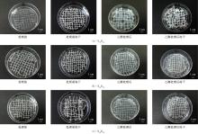
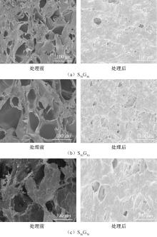
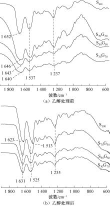
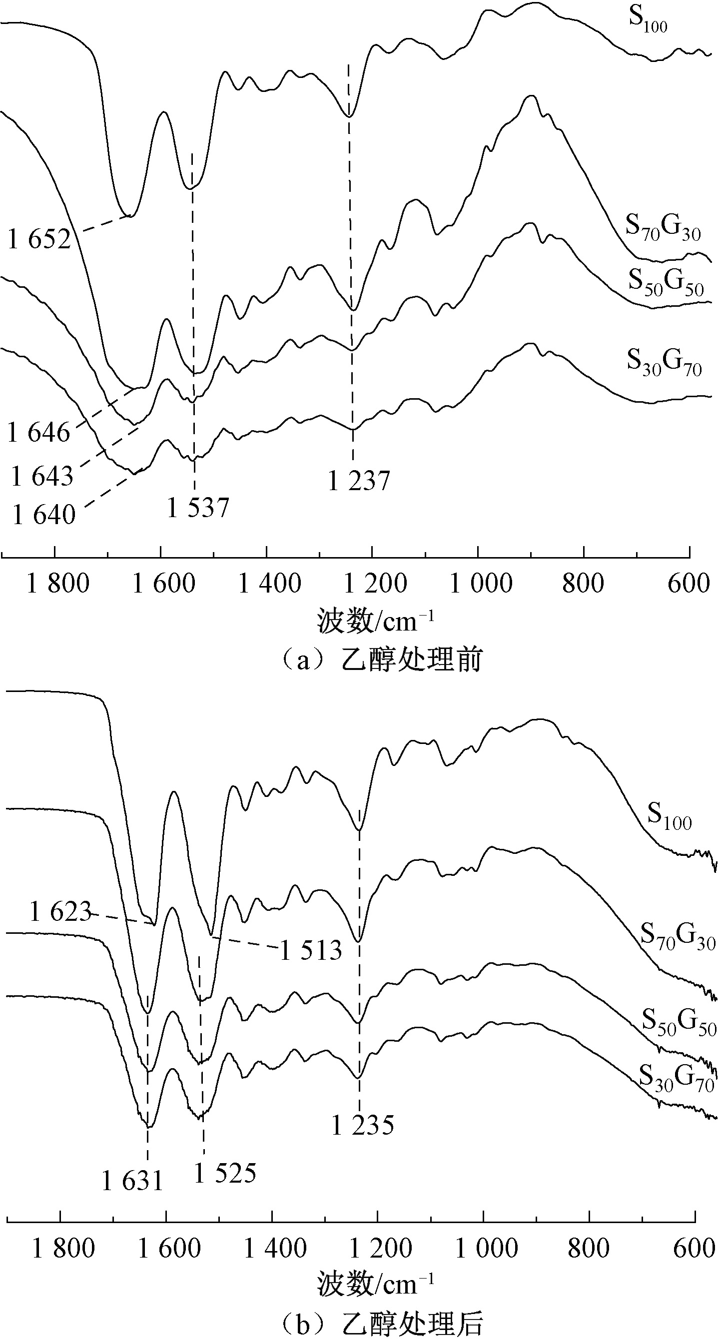
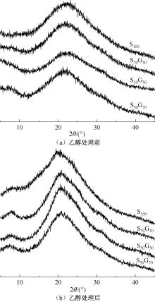
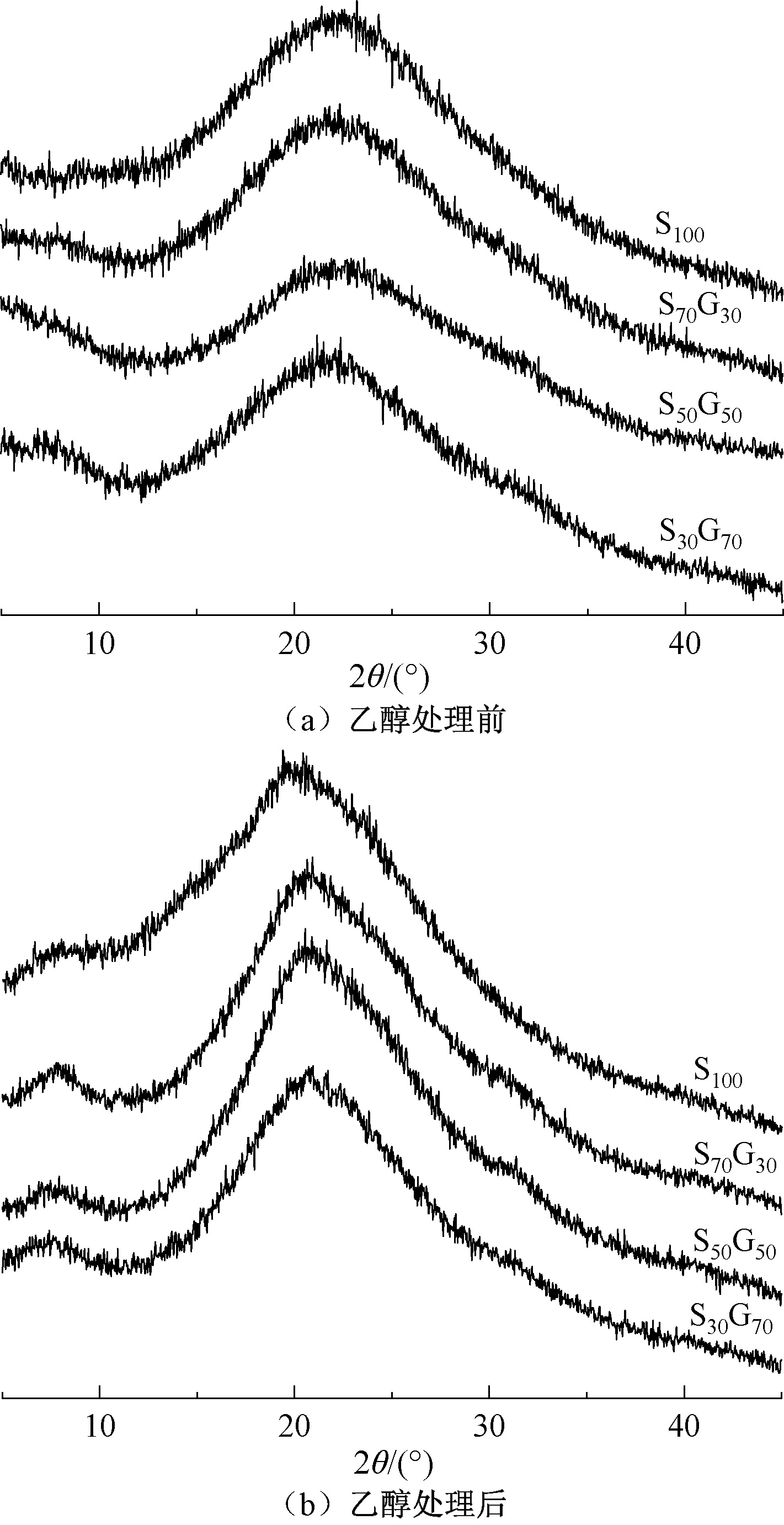
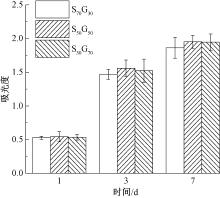
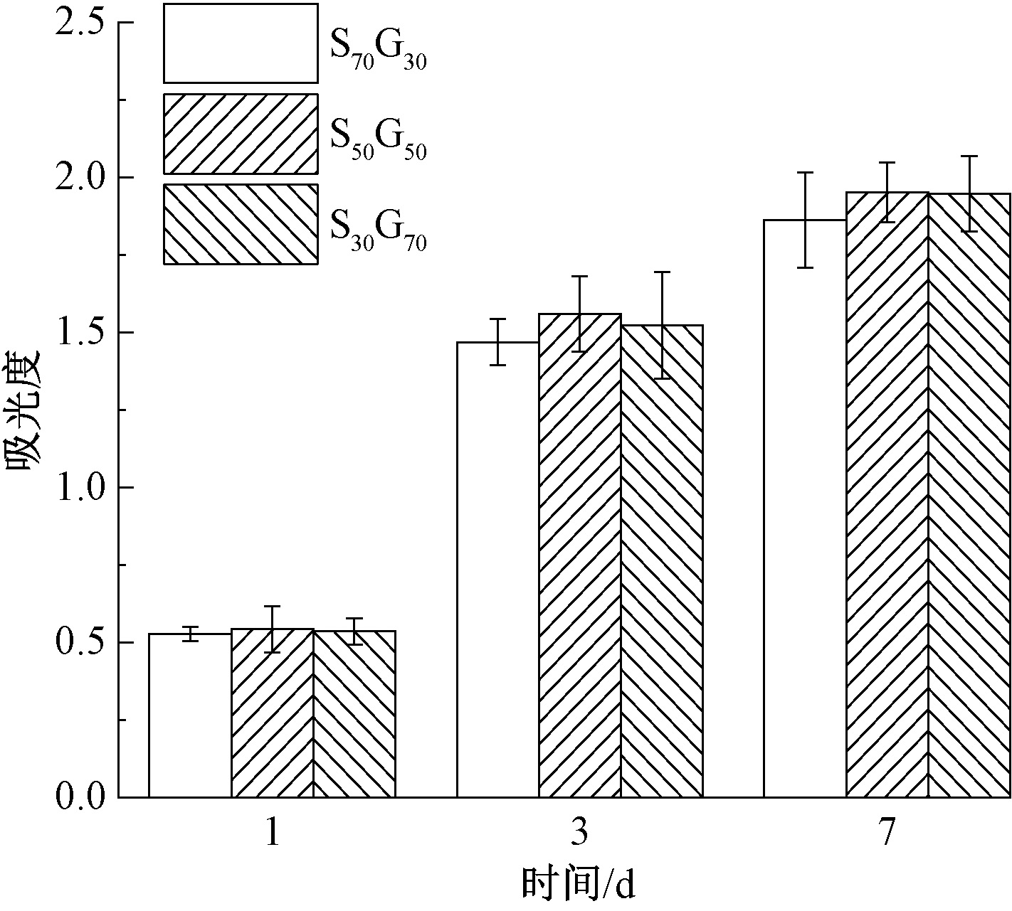
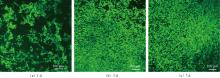
为解决关节软骨损伤修复中传统治疗方法效果有限的问题,采用挤出式3D打印制备丝素蛋白(SF)/明胶(GEL)复合支架,通过优化材料配比和交联工艺提升支架的结构稳定性,并对其微观形貌、化学结构及生物相容性进行表征。结果表明:挤出式3D打印能够成功制备不同质量比(70∶30、50∶50、30∶70)的格栅状SF/GEL支架,其中SF与GEL质量比为50∶50时,所得水凝胶线条连续、结构规整,乙醇处理有利于提高3D打印SF/GEL支架的成型稳定性;SF/GEL支架均呈现蜂窝状多孔结构,经乙醇处理后支架结构更加致密,孔隙数量减少且孔径减小;傅里叶变换红外光谱和X射线衍射分析表明,SF与GEL之间存在氢键相互作用,乙醇处理可诱导SF分子由无规卷曲向β折叠转变,从而提高支架结晶度;细胞实验结果表明,不同质量比的SF/GEL支架均能够支持小鼠胚胎成骨细胞的增殖,培养7 d后细胞呈梭形并沿支架方向排列,表明支架具有良好的生物相容性。该研究为构建具有良好结构稳定性和生物相容性的3D打印软骨组织工程支架提供了实验依据。
中图分类号:
| [1] |
LI C S, XU Y, LI J, et al. Ultramodern natural and synthetic polymer hydrogel scaffolds for articular cartilage repair and regeneration[J]. BioMedical Engineering OnLine, 2025, 24(1): 13.
doi: 10.1186/s12938-025-01342-3 |
| [2] |
LIN X Q, ZHANG Y, LI J R, et al. Biomimetic multizonal scaffolds for the reconstruction of zonal articular cartilage in chondral and osteochondral defects[J]. Bioactive Materials, 2025, 43: 510-549.
doi: 10.1016/j.bioactmat.2024.10.001 pmid: 40115881 |
| [3] |
LIN H, MA M, QI H, et al. 3D-Printed photocatalysts for revolutionizing catalytic conversion of solar to chemical energy[J]. Progress in Materials Science, 2025, 151: 101427.
doi: 10.1016/j.pmatsci.2025.101427 |
| [4] |
WANG K, MA Q, ZHANG Y M, et al. Preparation of bacterial cellulose/silk fibroin double-network hydrogel with high mechanical strength and biocompatibility for artificial cartilage[J]. Cellulose, 2020, 27(4): 1845-1852.
doi: 10.1007/s10570-019-02869-0 |
| [5] |
GHOSH S, PARKER S T, WANG X Y, et al. Direct-write assembly of microperiodic silk fibroin scaffolds for tissue engineering applications[J]. Advanced Functional Materials, 2008, 18(13): 1883-1889.
doi: 10.1002/adfm.v18:13 |
| [6] |
MU X, GONZALEZ-OBESO C, XIA Z Y, et al. 3D printing of monolithic proteinaceous cantilevers using regenerated silk fibroin[J]. Molecules, 2022, 27(7): 2148.
doi: 10.3390/molecules27072148 |
| [7] |
XIE M B, LIAN L M, MU X, et al. Volumetric additive manufacturing of pristine silk-based (bio)inks[J]. Nature Communications, 2023, 14: 210.
doi: 10.1038/s41467-023-35807-7 pmid: 36639727 |
| [8] | 王迪, 甘芳巾, 连小洁, 等. 3D打印双交联丝素/海藻酸钠/矿化胶原骨修复支架材料的构建及性能研究[J]. 中国生物医学工程学报, 2025, 44(3): 325-334. |
| WANG Di, GAN Fangjin, LIAN Xiaojie, et al. The study on fabrication and property of 3D printed double-crosslinked scaffold materials silk fibroin/alginate/mineralized collagen for bone repair[J]. Chinese Journal of Biomedical Engineering, 2025, 44(3): 325-334. | |
| [9] |
ZHENG Z Z, WU J B, LIU M, et al. 3D bioprinting of self-standing silk-based bioink[J]. Advanced Healthcare Materials, 2018, 7(6): 1701026.
doi: 10.1002/adhm.v7.6 |
| [10] | YEON Y K, PARK H S, LEE J M, et al. New concept of 3D printed bone clip (polylactic acid/hydroxyapatite/silk composite) for internal fixation of bone fractures[J]. Journal of Biomaterials Science, Polymer Edition, 2018, 29(7/8/9): 894-906. |
| [11] |
LIU H, QIU L, LIU H, et al. Effects of fiber cross-angle structures on the mechanical property of 3D printed scaffolds and performance of seeded MC3T3-E1 cells[J]. ACS Omega, 2021, 6(49): 33665-33675.
doi: 10.1021/acsomega.1c04672 pmid: 34926914 |
| [12] | PATEL K D, LAMARRA K A, SAWADKAR P, et al. Silk fibroin/GelMA-based hydrogels as a platform for tissue adhesives and tissue engineering[J]. ACS Biomaterials Science & Engineering, 2025, 11(7): 3893-3931. |
| [13] |
CHOI S, LEE K Y, KIM S L, et al. Fibre-infused gel scaffolds guide cardiomyocyte alignment in 3D-printed ventricles[J]. Nature Materials, 2023, 22(8): 1039-1046.
doi: 10.1038/s41563-023-01611-3 pmid: 37500957 |
| [14] |
WANG K, HAZRA R S, Ma Q, et al. Multifunctional silk fibroin/PVA bio-nanocomposite films containing TEMPO-oxidized bacterial cellulose nanofibers and silver nanoparticles[J]. Cellulose, 2022, 29: 1647-1666.
doi: 10.1007/s10570-021-04369-6 |
| [15] |
王曙东, 马倩, 王可, 等. 蚕丝蛋白/明胶复合水凝胶的结构与生物相容性[J]. 纺织学报, 2020, 41(11): 41-47.
doi: 10.13475/j.fzxb.20200301007 |
|
WANG Shudong, MA Qian, WANG Ke, et al. Structure and biocompatibility of silk fibroin/gelatin blended hydrogels[J]. Journal of Textile Research, 2020, 41(11): 41-47.
doi: 10.13475/j.fzxb.20200301007 |
|
| [16] |
WANG S D, MA Q, WANG K, et al. Strong and biocompatible three-dimensional porous silk fibroin/graphene oxide scaffold prepared by phase separation[J]. International Journal of Biological Macromolecules, 2018, 111: 237-246.
doi: 10.1016/j.ijbiomac.2018.01.021 |
| [17] | YANG Y, WANG H, YAN F Y, et al. Bioinspired porous octacalcium phosphate/silk fibroin composite coating materials prepared by electrochemical deposition[J]. ACS Applied Materials & Interfaces, 2015, 7(10): 5634-5642. |
| [18] | 鲍韡韡, 王曙东, 张幼珠, 等. 静电纺再生丝素/明胶纳米纤维的结构与性能[J]. 纺织学报, 2008, 29(3): 1-4, 8. |
| BAO Weiwei, WANG Shudong, ZHANG Youzhu, et al. Microstructure and property of electrospun regenerated silk fibroin/gelatin nanofibers[J]. Journal of Textile Research, 2008, 29(3): 1-4, 8. | |
| [19] |
WANG S D, ZHANG K Q. Electrogelation and rapid prototyping of Bombyx Mori silk fibroin[J]. Materials Letters, 2016, 169: 5-9.
doi: 10.1016/j.matlet.2016.01.079 |
| [1] | 孙哲茹, 李奕萱, 丁泽龙, 王瑜, 惠苗, 夏鑫. 聚氨酯/聚乳酸铰链增强形状记忆织物的制备及其性能[J]. 纺织学报, 2026, 47(05): 114-122. |
| [2] | 周佳丽, 李玉峰, 武会会. 碱凝胶在生丝织物脱胶印花工艺中的应用[J]. 纺织学报, 2026, 47(04): 180-188. |
| [3] | 孔艳辉, 张琳萍, 毛志平, 徐红. 甲基丙烯酰化明胶纤维膜的制备及其止血性能[J]. 纺织学报, 2026, 47(01): 1-10. |
| [4] | 龚维龙, 阳禹辉, 左彪. 基于溶液浓度调控的丝素蛋白构象转变及其动力学[J]. 纺织学报, 2026, 47(01): 29-37. |
| [5] | 史芷丞, 陈凤翔, 王梦云, 白洁, 李娟, 白濛, 伏广伟, 徐卫林. 面向空天应用的高性能无机纤维及制品发展现状及趋势[J]. 纺织学报, 2025, 46(12): 233-242. |
| [6] | 杨孟晓, 邱小雪, 吴芳, 刘琳, 姚菊明. 蚕丝基导电凝胶纤维的制备及其应变传感性能[J]. 纺织学报, 2025, 46(12): 49-56. |
| [7] | 费旌源, 徐乃库, 肖长发. 水可溃散砂质芯模设计及其在中空异形碳纤维复合材料成型中的应用[J]. 纺织学报, 2025, 46(11): 137-146. |
| [8] | 曾一洪, 付开秀, 王彦, 罗健, 陈国宝. 不同动物来源脱细胞骨骼肌基质-丝素蛋白复合支架理化性质比较[J]. 纺织学报, 2025, 46(11): 43-51. |
| [9] | 吴乐然, 吴霓欢, 李林耿, 钟意, 陈鸿鹏, 汤南. 负载厚朴酚的抗菌纳米纤维膜的制备及其性能[J]. 纺织学报, 2025, 46(10): 30-38. |
| [10] | 曾姚, 吕金凤, 王介平, 刘荣鹏, 周婵. 三维蚕丝蛋白支架的应用研究进展[J]. 纺织学报, 2025, 46(09): 258-267. |
| [11] | 高闻语, 陈诚, 奚晓玮, 邓林红, 刘杨. 改性丝素蛋白纤维增强胶原基角膜修复材料的制备及其性能[J]. 纺织学报, 2025, 46(08): 1-9. |
| [12] | 陈晴宇, 陆春红, 张斌, 晋义凯, 黄琪帏, 王超, 丁彬, 俞建勇, 王先锋. 碳纤维增强水泥基灌浆料的制备及其性能[J]. 纺织学报, 2025, 46(08): 120-126. |
| [13] | 江淑宁, 杨海伟, 李长龙, 郑天亮, 王宗乾. 低共熔溶剂剥离法制备丝素蛋白纳米原纤及其成膜性能[J]. 纺织学报, 2025, 46(07): 1-9. |
| [14] | 于梦菲, 高文丽, 任婧, 曹雷涛, 彭若铉, 凌盛杰. 摩擦纳米发电机用皮芯结构纤维的制备及其性能[J]. 纺织学报, 2025, 46(05): 1-9. |
| [15] | 李鹏飞, 罗忆心, 张子凡, 陆宁, 陈碧泠, 许建梅. 丝素/壳聚糖/明胶栓塞微球的制备及其降解性能[J]. 纺织学报, 2025, 46(05): 116-124. |
|
||
 京公网安备11010502044800号